. 2007 Jan;27(1):368-83.
doi: 10.1128/MCB.00814-06. Epub 2006 Oct 30.

BiP internal ribosomal entry site activity is controlled by heat-induced interaction of NSAP1

Affiliations

BiP internal ribosomal entry site activity is controlled by heat-induced interaction of NSAP1

Sungchan Cho et al. Mol Cell Biol. 2007 Jan.

Abstract

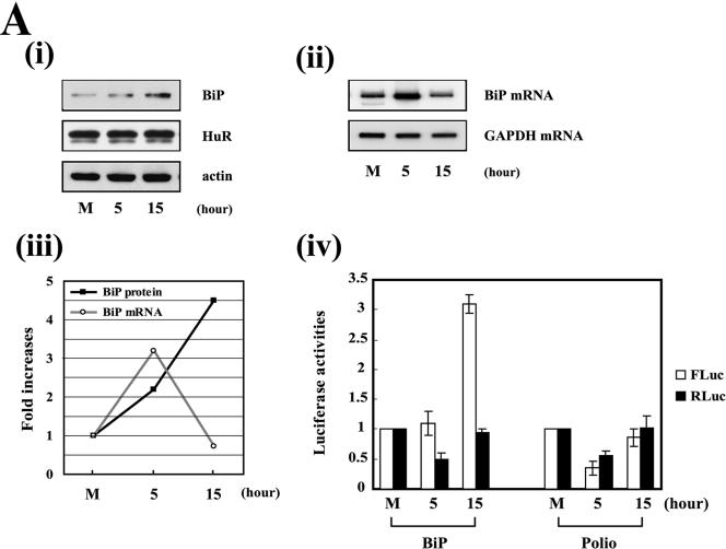
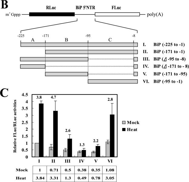
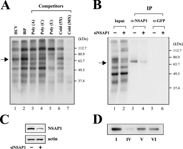
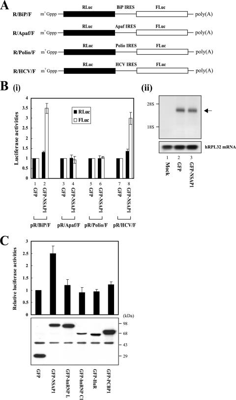
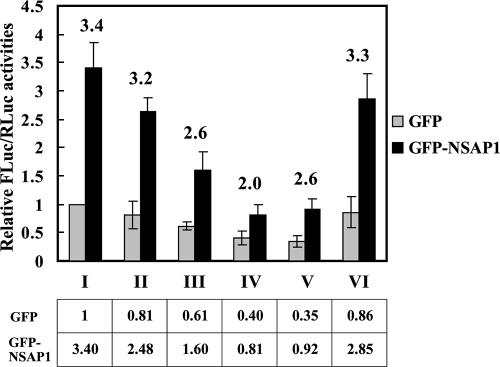
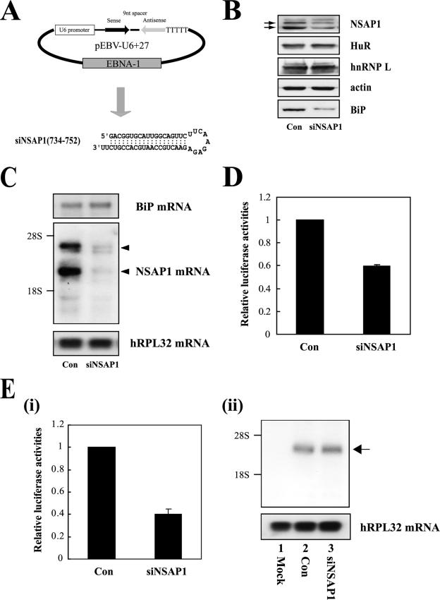
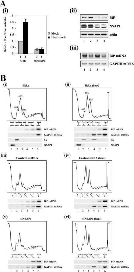
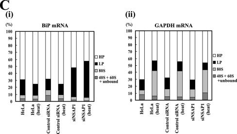
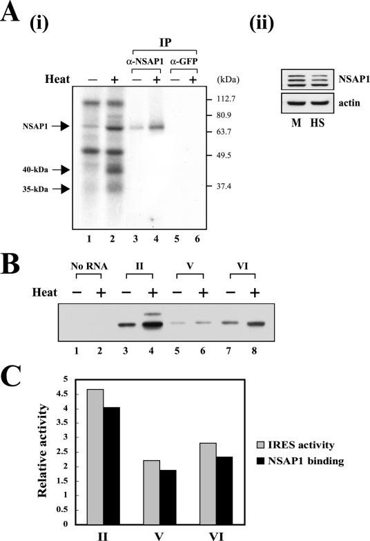
TheBiP protein, a stress response protein, plays an important role in the proper folding and assembly of nascent protein and in the scavenging of misfolded proteins in the endoplasmic reticulum lumen. Translation of BiP is directed by an internal ribosomal entry site (IRES) in the 5' nontranslated region of the BiP mRNA. BiP IRES activity increases when cells are heat stressed. Here we report that NSAP1 specifically enhances the IRES activity of BiP mRNA by interacting with the IRES element. Overexpression of NSAP1 in 293T cells increased the IRES activity of BiP mRNA, whereas knockdown of NSAP1 by small interfering RNA (siRNA) reduced the IRES activity of BiP mRNA. The amount of NSAP1 bound to the BiP IRES increased under heat stress conditions, and the IRES activity of BiP mRNA was increased. Moreover, the increase in BiP IRES activity with heat treatment was not observed in cells lacking NSAP1 after siRNA treatment. BiP mRNAs were redistributed from the heavy polysome to the light polysome in NSAP1 knockdown cells. Together, these data indicate that NSAP1 modulates IRES-dependent translation of BiP mRNA through an RNA-protein interaction under heat stress conditions.


Figures

FIG.1.
FIG.1.
Determination of heat-responsive elements in the BiP 5′NTR. (A, part i) Induction of BiP protein under heat stress conditions. Lysates of HeLa cells grown at 37°C (lane M) and 42°C for 5 and 15 h (lanes 5 and 15) were subjected to immunoblotting with anti-BiP, anti-HuR, and anti-actin antibodies. (ii) Level of BiP mRNA under heat stress conditions. Total RNAs were prepared from HeLa cells grown at 37°C (lane M) and 42°C for 5 and 15 h (lanes 5 and 15) and then subjected to radiolabeling RT-PCR with BiP-specific primers. GAPDH mRNA was used as a control message. (iii) Relative levels of BiP protein and BiP mRNA under heat stress condition. The intensities of BiP protein (i) and BiP mRNA bands (ii) were measured by the Sion Image Analysis Program, and the ratios of the band intensities before and after heat treatment (fold increases) are represented by filled squares and open circles, respectively. The intensities of BiP protein and BiP mRNA in untreated cells were set to 1. (iv) HeLa cells were transfected with dicistronic reporter plasmid pR/BiP/F or pR/Polio/F (5 μg each). These plasmids produce dicistronic mRNAs containing the RLuc and FLuc genes. Translation of RLuc occurs by cap-dependent scanning. On the other hand, translation of FLuc is directed by the BiP IRES or the polioviral IRES in the intercistronic region. The transfected HeLa cells were cultivated at 37°C for 24 h. Thereafter, cells were transferred to an incubator preheated to 42°C and maintained for 5 or 15 h. After heat treatment, the cells were harvested and FLuc and RLuc activities in the cell lysates were measured. The white and black columns depict FLuc and RLuc activities, respectively. The luciferase activities in cell lysate not subjected to heat treatment were set to 1. (B) Schematic diagram of the dicistronic mRNAs used for monitoring of the IRES activities of truncated BiP mRNAs. (C) HeLa cells were transfected with reporter plasmids expressing the dicistronic mRNAs shown in panel B. Twenty-four hours posttransfection, some cells were transferred to an incubator preheated to 42°C and some were left at 37°C. After 15 h of incubation, cells were harvested and luciferase activities were measured. The ratios of FLuc activity to RLuc activity were calculated and are shown in boxes below the graph. The ratio of luciferase activities in cells containing the dicistronic mRNA with the entire BiP IRES (nucleotides −225 to −1; RNA I in panel B) at 37°C was set to 1. The gray and black columns depict relative luciferase activities in mock-treated and heat-treated cells, respectively. The fold increases were calculated by the ratios of heat-stressed to mock-treated cells and are shown above the columns. Experiments were performed at least three times for each experimental set, and standard deviations are shown as error bars.
FIG.1.
FIG.1.
Determination of heat-responsive elements in the BiP 5′NTR. (A, part i) Induction of BiP protein under heat stress conditions. Lysates of HeLa cells grown at 37°C (lane M) and 42°C for 5 and 15 h (lanes 5 and 15) were subjected to immunoblotting with anti-BiP, anti-HuR, and anti-actin antibodies. (ii) Level of BiP mRNA under heat stress conditions. Total RNAs were prepared from HeLa cells grown at 37°C (lane M) and 42°C for 5 and 15 h (lanes 5 and 15) and then subjected to radiolabeling RT-PCR with BiP-specific primers. GAPDH mRNA was used as a control message. (iii) Relative levels of BiP protein and BiP mRNA under heat stress condition. The intensities of BiP protein (i) and BiP mRNA bands (ii) were measured by the Sion Image Analysis Program, and the ratios of the band intensities before and after heat treatment (fold increases) are represented by filled squares and open circles, respectively. The intensities of BiP protein and BiP mRNA in untreated cells were set to 1. (iv) HeLa cells were transfected with dicistronic reporter plasmid pR/BiP/F or pR/Polio/F (5 μg each). These plasmids produce dicistronic mRNAs containing the RLuc and FLuc genes. Translation of RLuc occurs by cap-dependent scanning. On the other hand, translation of FLuc is directed by the BiP IRES or the polioviral IRES in the intercistronic region. The transfected HeLa cells were cultivated at 37°C for 24 h. Thereafter, cells were transferred to an incubator preheated to 42°C and maintained for 5 or 15 h. After heat treatment, the cells were harvested and FLuc and RLuc activities in the cell lysates were measured. The white and black columns depict FLuc and RLuc activities, respectively. The luciferase activities in cell lysate not subjected to heat treatment were set to 1. (B) Schematic diagram of the dicistronic mRNAs used for monitoring of the IRES activities of truncated BiP mRNAs. (C) HeLa cells were transfected with reporter plasmids expressing the dicistronic mRNAs shown in panel B. Twenty-four hours posttransfection, some cells were transferred to an incubator preheated to 42°C and some were left at 37°C. After 15 h of incubation, cells were harvested and luciferase activities were measured. The ratios of FLuc activity to RLuc activity were calculated and are shown in boxes below the graph. The ratio of luciferase activities in cells containing the dicistronic mRNA with the entire BiP IRES (nucleotides −225 to −1; RNA I in panel B) at 37°C was set to 1. The gray and black columns depict relative luciferase activities in mock-treated and heat-treated cells, respectively. The fold increases were calculated by the ratios of heat-stressed to mock-treated cells and are shown above the columns. Experiments were performed at least three times for each experimental set, and standard deviations are shown as error bars.
FIG. 2.
FIG. 2.
Identification of NSAP1 as a cellular protein interacting with the human BiP IRES. (A) UV cross-linking experiments were performed with S10 extracts of HeLa S3 cells (20 μg each) and 32P-labeled RNAs (3 × 10⁵ cpm each) corresponding to nucleotides 18 to 402 of the HCV IRES (lane 1) and nucleotides −225 to +3 of the BiP IRES (lanes 2 to 7). The adenine in the initiation codon of the BiP mRNA was designated nucleotide 1, and the upstream sequences are denoted by minus signs. Competition experiments were carried out with various competitor RNAs, including 50 ng homopolymeric RNAs [poly(A), poly(C), and poly(U)] (lanes 3 to 5) and a 5- or 50-fold molar excess of unlabeled BiP IRES RNA (lanes 6 and 7). After cross-linking reactions, samples were treated with an RNase cocktail and then analyzed by 12% sodium dodecyl sulfate-polyacrylamide gel electrophoresis. The arrow indicates a 65-kDa protein. (B) Immunoprecipitation (IP) of UV cross-linked proteins with the 32P-labeled BiP RNA probe. After UV cross-linking with the cytoplasmic extracts of normal HeLa cells and NSAP1 knockdown cells, samples were precleared with protein G-agarose resin and then reacted with 2 μg of polyclonal anti-NSAP1 antibody (lanes 3 and 4) or an anti-GFP antibody (lanes 5 and 6). Resin-bound proteins were resolved by 10% sodium dodecyl sulfate-polyacrylamide gel electrophoresis. Lanes 1 and 2 show the labeled proteins in normal HeLa cells and NSAP1 knockdown cells, respectively, prior to immunoprecipitation. Lanes 3 and 4 show immunoprecipitated NSAP1. (C) NSAP1 and actin in the lysates used for UV cross-linking were monitored by Western blotting with corresponding antibodies. (D) Determination of the NSAP1 binding site on BiP IRES. UV cross-linking experiments were carried out with purified NSAP1 (100 ng) and 32P-labeled RNAs (3 × 10⁵ cpm each) corresponding to various regions of the BiP IRES shown in Fig. 1B (probes I, IV, V, and VI).
FIG. 3.
FIG. 3.
NSAP1 enhances the IRES activity of BiP mRNA in vivo. (A) Schematic diagram of dicistronic mRNAs used for monitoring the effect of NSAP1 on various IRES activities in vivo. The dicistronic mRNAs containing the BiP, Apaf, poliovirus, and HCV IRES regions are depicted as R/BiP/F, R/Apaf/F, R/Polio/F, and R/HCV/F, respectively. (B) (i) 293T cells were cotransfected with a reporter plasmid (pR/BiP/F, pR/Apaf/F, pR/Polio/F, or pR/HCV/F), an effector plasmid expressing GFP or GFP-NSAP1, and the control plasmid pCMV•SPORT-βgal. Forty-eight hours posttransfection, cells were harvested and luciferase activities were measured. FLuc and RLuc activities were normalized with β-galactosidase activity to adjust for transfection efficiency. Black and white columns depict RLuc and FLuc activities, respectively. Luciferase activities in cells expressing GFP were set to 1 (lanes 1, 3, 5, and 7). Experiments were performed at least three times for each experimental set, and standard deviations are shown as error bars. (ii) Northern blot analysis of the reporter dicistronic mRNA R/BiP/F produced in transfected cells. Three micrograms of poly(A)+ RNA purified from transfected cells was subjected to Northern blotting with a 32P-labeled probe corresponding to the FLuc gene. The positions of the 28S and 18S rRNAs are indicated. The human ribosomal protein large subunit 32 (hRPL32) blot was used as an internal control for poly(A)+ mRNAs. The arrow indicates the position of the reporter mRNA. (C) 293T cells were cotransfected with reporter plasmid pR/BiP/F and an effector plasmid expressing GFP (lane GFP) or expressing ITAFs fused with GFP. Forty-eight hours posttransfection, relative luciferase activities were determined. The ratio of FLuc activity to RLuc activity in cells transfected with effector GFP was set to 1 (upper panel). Lysates were analyzed by Western blotting with a monoclonal anti-GFP antibody (lower panel). The identities of the ITAFs are shown at the bottom. Experiments were performed at least three times for each experimental set, and standard deviations are shown as error bars.
FIG. 4.
FIG. 4.
Determination of NSAP1-responsive elements in the 5′NTR of the BiP mRNA. 293T cells were cotransfected with reporter plasmids expressing one of the dicistronic mRNAs shown in Fig. 1B and an effector plasmid expressing GFP or GFP-NSAP1. Forty-eight hours posttransfection, cells were harvested and luciferase activities were measured. The ratios of FLuc and RLuc activities were calculated, and the mean values are shown in boxes below the graph. Gray and black columns depict the relative luciferase activities in cells transfected with GFP and GFP-NSAP1, respectively. The fold increases were also calculated by the ratio of GFP-NSAP1 to GFP and shown by numbers above each column. The ratio of luciferase activities in cells producing the dicistronic mRNA (RNA I in Fig. 1B) with the entire BiP 5′NTR (nucleotides −225 to −1) and GFP was set to 1. Experiments were performed at least three times for each experimental set, and standard deviations are shown as error bars.
FIG. 5.
FIG. 5.
Effect of NSAP1-specific siRNA on BiP IRES activity. (A) Schematic diagram of siRNA-expressing plasmid pEBV-U6+27 (top) and the predicted secondary structure of siRNAs generated from pEBV-U6+27/NSAP1(734-752) (bottom). (B) Western blot analysis of HeLa cells expressing the siRNA siNSAP1. Stably transformed HeLa cells were generated by transfection of the control (Con) vector and vectors encoding siNSAP1. Cells were harvested, and protein levels were analyzed by immunoblotting with anti-NSAP1, anti-HuR, anti-hnRNP L, anti-actin, and anti-BiP antibodies. Arrows indicate two isoforms of NSAP1, and the uppermost band represents hnRNP R (NSAP1 blot) (C) Northern blot analysis of HeLa cells expressing siRNA against NSAP1. Thirty micrograms of total RNA was resolved on a denaturing gel and immobilized on a nylon membrane. The filter was hybridized consecutively with 32P-labeled probes specific for the BiP, NSAP1, and hRPL32 mRNAs. The arrowheads indicate isoforms of NSAP1 transcripts. (D) The effect of siRNA on BiP IRES function was monitored by transfection of the R/BiP/F dicistronic RNA (shown in Fig. 3A) into the established cell lines. Relative IRES activities were measured 3 h posttransfection. The value of siRNA-lacking control cells was set to 1. Experiments were performed at least three times for each experimental set, and standard deviations are shown as error bars. (E, part i) The effect of siRNA on BiP IRES activity was also monitored by transfection of DNA expressing dicistronic mRNA R/BiP/F (shown in Fig. 3A). Cells with or without siRNA were transfected with plasmid pR/BiP/F, and IRES activity was measured by FLuc activities 48 h posttransfection. DNA transfection efficiency was normalized by RLuc activity directed by cap-dependent translation, and ratios of FLuc activity to RLuc activity were calculated. The value of the control cells was set to 1. Experiments were performed at least three times for each experimental set, and standard deviations are shown as error bars. (ii) Northern blot analysis of dicistronic mRNAs produced from HeLa cells expressing siRNA against NSAP1. Three micrograms of poly(A)+ RNAs prepared from transfected cells was subjected to Northern blotting with a 32P-labeled probe corresponding to the FLuc gene. The positions of the 28S and 18S rRNAs are indicated. The hRPL32 blot was used as an internal control for poly(A)+ mRNAs. The arrow indicates the position of the reporter mRNA.
FIG. 6.
FIG. 6.
The role of NSAP1 in heat-dependent translational activation of BiP mRNA. (A) The IRES activity of BiP mRNA was monitored in cells expressing NSAP1-specific siRNA under heat stress conditions. HeLa cells expressing NSAP1-specific siRNA (siNSAP1) and control (Con) cells were transfected with plasmid pR/BiP/F (3 μg each). Twenty-four hours posttransfection, some of the transfected cells were transferred to an incubator preheated to 42°C (black columns, lanes 2 and 4), while the rest of the transfected cells were maintained at 37°C (gray columns, lanes 1 and 3). After cultivation for 15 h, cells were harvested and FLuc and RLuc activities were measured. The relative luciferase activity in the cell lysate of control cells without siRNA that were incubated at 37°C was set to 1. Experiments were performed at least three times for each experimental set, and standard deviations are shown as error bars. (ii) BiP, NSAP1, and actin protein levels in each cell lysate were monitored by Western blotting with anti-BiP, anti-NSAP1, and anti-actin antibodies. (iii) The levels of BiP mRNAs under heat stress and/or NSAP1 knockdown conditions. Total RNAs were prepared from HeLa cells and then subjected to radiolabeling RT-PCR with BiP-specific primers. GAPDH mRNA was used as a negative control message. (B) Distributions of mRNAs in heat-stressed and/or NSAP1 knockdown cells. HeLa cells were infected with recombinant adenoviruses expressing mutant siRNA (parts iii and iv) or siRNA against NSAP1 (parts v and vi) and then cultivated at 37°C for 24 h. Thereafter, some cells were transferred to an incubator preheated to 42°C and maintained for 15 h (parts ii, iv, and vi) or cultivated at 37°C continuously (parts i, iii, and v). After heat treatment, the cells were treated with cycloheximide (100 μg/ml) for 5 min at 37°C and then harvested. Extracts from heat-treated and/or NSAP1 knockdown HeLa cells were subjected to sucrose gradient centrifugation and then divided into the following six fractions: proteins not associated with ribosomes (fraction 1, unbound proteins), RNAs and proteins at the 40S ribosomal fraction (fraction 2, 40S), RNAs and proteins at the 60S ribosomal fraction (fraction 3, 60S), RNAs and proteins at the monosomal fraction (fraction 4, 80S), RNAs and proteins at the LPs (fraction 5, LP), and RNAs and proteins at the HPs (fraction 6, HP). The distribution of BiP and GAPDH mRNAs across the gradients was analyzed by radiolabeling RT-PCR. In addition, the NSAP1 and S6 proteins were also monitored by Western blotting with corresponding antibodies. Distinct strong peaks appeared right after the heavy polysomal fractions in the heat-treated samples (denoted by asterisks in parts ii, iv, and vi). These rapidly sedimenting entities might be a component of stress granules formed in heat-treated cells. (C) The band intensities of BiP and GAPDH mRNAs shown in panel B were measured with the L process and Image Gauge programs (Fuji Photo Film Co., Ltd.), and the relative proportion of each band was calculated and is depicted in the graphs. The proportions of fractions HP, LP, monosome (80S), and the sum of fractions 40S plus 60S plus unbound proteins are depicted by white, black, light gray, and dark gray bars, respectively.
FIG. 6.
FIG. 6.
The role of NSAP1 in heat-dependent translational activation of BiP mRNA. (A) The IRES activity of BiP mRNA was monitored in cells expressing NSAP1-specific siRNA under heat stress conditions. HeLa cells expressing NSAP1-specific siRNA (siNSAP1) and control (Con) cells were transfected with plasmid pR/BiP/F (3 μg each). Twenty-four hours posttransfection, some of the transfected cells were transferred to an incubator preheated to 42°C (black columns, lanes 2 and 4), while the rest of the transfected cells were maintained at 37°C (gray columns, lanes 1 and 3). After cultivation for 15 h, cells were harvested and FLuc and RLuc activities were measured. The relative luciferase activity in the cell lysate of control cells without siRNA that were incubated at 37°C was set to 1. Experiments were performed at least three times for each experimental set, and standard deviations are shown as error bars. (ii) BiP, NSAP1, and actin protein levels in each cell lysate were monitored by Western blotting with anti-BiP, anti-NSAP1, and anti-actin antibodies. (iii) The levels of BiP mRNAs under heat stress and/or NSAP1 knockdown conditions. Total RNAs were prepared from HeLa cells and then subjected to radiolabeling RT-PCR with BiP-specific primers. GAPDH mRNA was used as a negative control message. (B) Distributions of mRNAs in heat-stressed and/or NSAP1 knockdown cells. HeLa cells were infected with recombinant adenoviruses expressing mutant siRNA (parts iii and iv) or siRNA against NSAP1 (parts v and vi) and then cultivated at 37°C for 24 h. Thereafter, some cells were transferred to an incubator preheated to 42°C and maintained for 15 h (parts ii, iv, and vi) or cultivated at 37°C continuously (parts i, iii, and v). After heat treatment, the cells were treated with cycloheximide (100 μg/ml) for 5 min at 37°C and then harvested. Extracts from heat-treated and/or NSAP1 knockdown HeLa cells were subjected to sucrose gradient centrifugation and then divided into the following six fractions: proteins not associated with ribosomes (fraction 1, unbound proteins), RNAs and proteins at the 40S ribosomal fraction (fraction 2, 40S), RNAs and proteins at the 60S ribosomal fraction (fraction 3, 60S), RNAs and proteins at the monosomal fraction (fraction 4, 80S), RNAs and proteins at the LPs (fraction 5, LP), and RNAs and proteins at the HPs (fraction 6, HP). The distribution of BiP and GAPDH mRNAs across the gradients was analyzed by radiolabeling RT-PCR. In addition, the NSAP1 and S6 proteins were also monitored by Western blotting with corresponding antibodies. Distinct strong peaks appeared right after the heavy polysomal fractions in the heat-treated samples (denoted by asterisks in parts ii, iv, and vi). These rapidly sedimenting entities might be a component of stress granules formed in heat-treated cells. (C) The band intensities of BiP and GAPDH mRNAs shown in panel B were measured with the L process and Image Gauge programs (Fuji Photo Film Co., Ltd.), and the relative proportion of each band was calculated and is depicted in the graphs. The proportions of fractions HP, LP, monosome (80S), and the sum of fractions 40S plus 60S plus unbound proteins are depicted by white, black, light gray, and dark gray bars, respectively.
FIG. 7.
FIG. 7.
Affinity of NSAP1 binding to the BiP IRES and BiP IRES activity. (A) Binding of NSAP1 protein to the BiP IRES before and after heat treatment. (i) UV cross-linking experiments were performed with 32P-labeled BiP IRES RNA and cytoplasmic extract of HeLa cells with (lanes 2, 4, and 6) and without (lanes 1, 3, and 5) heat treatment for 15 h. After UV cross-linking and RNase treatment, samples were precleared with protein G-agarose resin and then reacted with 2 μg of polyclonal anti-NSAP1 antibody (lanes 3 and 4) or anti-GFP antibody (lanes 5 and 6). Resin-bound proteins were resolved by 10% sodium dodecyl sulfate-polyacrylamide gel electrophoresis. Lanes 1 and 2 show the labeled proteins prior to immunoprecipitation (IP). The arrows depict the NSAP1, 40-kDa, and 35-kDa proteins. (ii) NSAP1 and actin in the lysates were identified by Western blotting with corresponding antibodies. M and HS represent cell lysates without and with heat treatment, respectively. (B) Heat-induced enhancement of NSAP1 binding to various regions of the 5′NTR of the BiP mRNA. Extracts of HeLa cells were prepared without (−; lanes 1, 3, 5, and 7) or with (+; lanes 2, 4, 6, and 8) heat treatment for 15 h and subjected to an in vitro RNA-binding assay with biotinylated RNA probes corresponding to different regions of the BiP IRES (II, V, and VI in Fig. 1B). NSAP1 protein bound to each RNA probe was monitored by Western blotting with a polyclonal NSAP1-specific antibody. Negative-control experiments were performed without an RNA probe (lanes 1 and 2). (C) The increase in IRES activity with heat treatment correlates well with the increased binding of NSAP1 to the IRES element. The intensities of NSAP1 bands in panel B were measured by the Sion Image Analysis Program, and the ratios of the band intensities before and after heat treatment were calculated and are depicted by black columns. Fold increases in the IRES activities of the RNAs caused by heat treatment are depicted by gray columns. Names of RNAs (II, V, and VI in Fig. 1C) are shown at the bottom.

Similar articles

Cited by

References

    1. Back, S. H., Y. K. Kim, W. J. Kim, S. Cho, H. R. Oh, J.-E. Kim, and S. K. Jang. 2002. Translation of polioviral mRNA is inhibited by cleavage of polypyrimidine tract-binding proteins executed by polioviral 3C9pro. J. Virol. 76:2529-2542. - PMC - PubMed
    1. Bannai, H., K. Fukatsu, A. Mizutani, T. Natsume, S. I. Iemura, T. Ikegami, T. Inoue, and K. Mikoshiba. 2004. An RNA-interacting protein, SYNCRIP (hnRNP Q1/NSAP1) is a component of mRNA granule transported with inositol 1,4,5-trisphosphate receptor type 1 mRNA in neuronal dendrites. J. Biol. Chem. 279:53427-53434. - PubMed
    1. Bole, D. G., L. M. Hendershot, and J. F. Kearney. 1986. Posttranslational association of immunoglobulin heavy chain binding protein with nascent heavy chains in nonsecreting and secreting hybridomas. J. Cell Biol. 102:1558-1566. - PMC - PubMed
    1. Duncan, R. F. 1996. Translational control during heat shock, p. 271-293. In J. W. B. Hershey, M. B. Mathews, and N. Sonenberg (ed.), Translational control. Cold Spring Harbor Laboratory Press, Cold Spring Harbor, N.Y.
    1. Gabai, V. L., and M. Y. Sherman. 2002. Invited review: interplay between molecular chaperones and signaling pathways in survival of heat shock. J. Appl. Physiol. 92:1743-1748. - PubMed

Publication types

MeSH terms

Substances

LinkOut - more resources