. 2007 Jan;27(1):253-66.
doi: 10.1128/MCB.01071-06. Epub 2006 Oct 30.

Dynamic interplay of transcriptional machinery and chromatin regulates "late" expression of the chemokine RANTES in T lymphocytes

Affiliations

Dynamic interplay of transcriptional machinery and chromatin regulates "late" expression of the chemokine RANTES in T lymphocytes

Yong-Tae Ahn et al. Mol Cell Biol. 2007 Jan.

Abstract

The chemokine RANTES (regulated upon activation normal T cell expressed and secreted) is expressed "late" (3 to 5 days) after activation in T lymphocytes. In order to understand the molecular events that accompany changes in gene expression, a detailed analysis of the interplay between transcriptional machinery and chromatin on the RANTES promoter over time was undertaken. Krüppel-like factor 13 (KLF13), a sequence-specific DNA binding transcription factor, orchestrates the induction of RANTES expression in T lymphocytes by ordered recruitment of effector molecules, including Nemo-like kinase, p300/cyclic AMP response element binding protein (CBP), p300/CBP-associated factor, and Brahma-related gene 1, that initiate sequential changes in phosphorylation and acetylation of histones and ATP-dependent chromatin remodeling near the TATA box of the RANTES promoter. These events recruit RNA polymerase II to the RANTES promoter and are responsible for late expression of RANTES in T lymphocytes. Therefore, KLF13 is a key regulator of late RANTES expression in T lymphocytes.


Figures

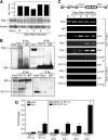
FIG. 1.
FIG. 1.
Kinetics of RANTES expression and the effect of KLF13 knock-down on RANTES expression in T lymphocytes. (A) Time course of RANTES mRNA expression by real-time quantitative PCR in resting (R) and activated T lymphocytes. Data represent the mean ± standard deviation (SD) of three independent experiments. (B) Kinetics of RANTES protein expression by ELISA in resting (R) and activated T lymphocytes. Results represent the mean ± SD of three independent experiments. (C) The rate of RANTES transcription after activation was measured by a nuclear run-on assay. Each column is an individual hybridization blot from resting and activated T lymphocytes at the indicated times. (D) Diagram of regulatory regions in the human RANTES promoter and KLF13 binding site. (E) Knockdown of KLF13 by siRNA reduces RANTES expression. Levels of KLF13, RANTES, and IFNG mRNA (left panel) and protein levels of KLF13 and RANTES (right panel) were measured. All values were normalized to the nonsilencing control. Data represent the mean ± SD of three triplicates.
FIG. 2.
FIG. 2.
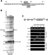
In vivo kinetics of protein-DNA interactions of Pol II, KLF13, and p50 at the RANTES promoter. (A) DNA sequence of the RANTES promoter near the A site. Consensus binding sites for KLF13 and p50 overlap and are represented by bars above and below the sequence. (B) In vivo DMS footprinting of DNA from resting (R) and PHA-activated T lymphocytes. Numbers indicate the distance from the transcription start site at +1. The TATA box at position −32 to −27 and the core KLF13 binding site (GGGGAG) from −58 to −63 are also marked. Naked genomic DNA treated with DMS in vitro was analyzed as a control. (C) ChIP assays of Pol II, KLF13, and p50 at the RANTES promoter in resting (R) and PHA-activated T lymphocytes. Specific DNA-protein complexes were immunoprecipitated from cross-linked chromatin using antibodies specific to Pol II, KLF13, and p50, and eluted DNA was PCR amplified using two RANTES promoter primer sets as indicated.
FIG. 3.
FIG. 3.
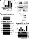
NLK interaction with KLF13 and phosphorylation of histone proteins at the RANTES promoter. (A) Western blot analysis of NLK. HeLa cells were used as a positive control. Actinin was used as a protein loading control with the graph showing the ratio of NLK/actinin expression. (B) In vivo coimmunoprecipitation of NLK and KLF13 with anti-KLF13 and anti-NLK antibodies. NLK (upper panel) and KLF13 (lower panel) were identified in unprecipitated (input) or immunoprecipitated extracts by Western blotting using the indicated antibodies. (C) ChIP and re-ChIP assay of the KLF13/NLK complex on the RANTES promoter in resting (R) and PHA-activated T lymphocytes. Immunoprecipitating antibodies used in each ChIP and re-ChIP reaction are indicated as well as the primer sets used for amplification. (D) NLK phosphorylates histone H3 in vitro. Phosphorylated histone H3 substrates were detected either by autoradiography (upper panel; [32P]H3) or by Western blotting using an antibody specific to phosphorylated serine 10 of histone H3 (middle panel). The blot was stripped and reprobed with anti-histone H3 antibody to control for protein loading (bottom panel). (E) ChIP assay of phosphorylated histone H3 (serine 10) at the RANTES promoter in resting and PHA-activated T lymphocytes. Specific protein-DNA complexes were immunoprecipitated using a P-H3 (S10) antibody and amplified using two primer sets, as indicated. The graph represents the mean ± standard deviation of triplicate determinations, while the images are representative of one of the three replicates.
FIG. 4.
FIG. 4.
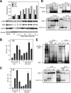
T lymphocytes express HAT proteins that interact with KLF13. (A) Western blot of HAT proteins using anti-p300, anti-CBP, and anti-PCAF antibodies. HeLa cells were used as a positive control. Actinin was used as a protein loading control with the graph showing the ratio of HAT/actinin expression. (B and C) Histone H3 (B) and histone H4 HAT (C) activity assays of resting (R) and PHA-activated T lymphocytes. Increasing amounts of preacetylated histone H3 or histone H4 peptides were included as standards. The control was nuclear extract from HeLa cells. Data represent the mean ± standard deviation of three independent experiments. (D and E) In vivo coimmunoprecipitation of PCAF and KLF13 (D) and p300/CBP and KLF13 (E) were identified in unprecipitated (input) or immunoprecipitated extracts by Western blotting using the indicated antibodies.
FIG. 5.
FIG. 5.
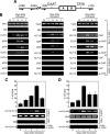
ChIP and re-ChIP assays of KLF13/HAT complexes and acetylation of histone proteins at or near the TATA box of the RANTES promoter. (A) Diagram of the RANTES promoter, indicating positions of the two primer sets used in ChIP and re-ChIP assays. (B) ChIP and re-ChIP assays of KLF13/p300 (left panel), KLF13/CBP (middle panel), and KLF13/PCAF (right panel) complexes on the RANTES promoter in resting (R) and PHA-activated T lymphocytes. Immunoprecipitating antibodies used in each ChIP and re-ChIP reaction are indicated as well as the primer sets used for amplification. DNA eluted from unprecipitated chromatin was used as input. (C and D) ChIP assay of acetylated histone H3 (lysine 14) (C) and acetylated histone H4 (lysine 8) (D) at the RANTES promoter in resting and PHA-activated T lymphocytes, using primers as indicated. The graphs represent the mean ± standard deviation of triplicate determinations, while the images are representative of one of the three replicates.
FIG. 6.
FIG. 6.
KLF13 and Brg-1 are involved in ATP-dependent chromatin remodeling at the RANTES promoter. (A) Western blot analysis of Brg-1. HeLa cells were used as a positive control. Actinin was used as a protein loading control with the graph showing the ratio of Brg-1/actinin expression. (B) In vivo coimmunoprecipitation of Brg-1 and KLF13. Brg-1 (upper panel) and KLF13 (lower panel) were identified in unprecipitated (input) or immunoprecipitated extracts by Western blotting using the indicated antibodies. (C) ChIP and re-ChIP assays of the KLF13/Brg-1 complex on the RANTES promoter in resting (R) and PHA-activated T lymphocytes. Immunoprecipitating antibodies used in each ChIP and re-ChIP reaction are indicated as well as the primer sets used for amplification. DNA eluted from unprecipitated chromatin was used as input. (D) Activation of the RANTES promoter by Brg-1 requires proper chromatin structure and is augmented by synergy with KLF13. (Left panel) Brg-1-deficient SW13 cells were cotransfected with Brg-1 expression plasmid or empty vector (pBJ5) and RANTES reporter plasmids constructed in either pGL3 (pGL3-RP-Luc) or pREP4 (pREP4-RP-Luc). (Right panel) SW13 cells were cotransfected with combinations of the indicated expression plasmids (Brg-1 and KLF13) and empty vector (pcDNA and pBJ5) and either pREP4-RP-Luc or pREP4-ΔA-RP-Luc, which contains a mutated A site. Renilla was used as an internal control to normalize for transfection efficiency, and data are expressed as change relative to transfection with Brg-1 alone. Data represent the mean ± standard deviation of three independent experiments.
FIG. 7.
FIG. 7.
Schematic model of the mechanism of late RANTES expression in T lymphocytes. In resting T lymphocytes, RANTES is not expressed. After activation of T lymphocytes, KLF13 binds to a core binding element on the RANTES promoter (day 1). At this time, NLK binds to KLF13 at the RANTES promoter and phosphorylates serine 10 on histone H3 (histone tails shown in red). KLF13 recruits p300/CBP and PCAF to the RANTES promoter (days 3 to 7). The phosphorylation of serine 10 on histone H3 by a MAP kinase at day 1 is required for p300/CBP or PCAF to acetylate histone H3 at lysine 14. This acetylation results in the recruitment of factors required for transcriptional initiation on the RANTES promoter. In addition, p300/CBP or PCAF also acetylates lysine 8 of histone 4, which allows Brg-1 to be recruited to the RANTES promoter to form a complex with KLF13. This ATP-dependent chromatin remodeling complex at the A site of the RANTES promoter can twist or deform the chromatin structure, exposing the adjacent TATA box to which polymerase II binds, leading to transcriptional initiation.

References

    1. Agalioti, T., G. Chen, and D. Thanos. 2002. Deciphering the transcriptional histone acetylation code for a human gene. Cell 111:381-392. - PubMed
    1. Asano, H., X. S. Li, and G. Stamatoyannopoulos. 2000. FKLF-2: a novel Kruppel-like transcriptional factor that activates globin and other erythroid lineage genes. Blood 95:3578-3584. - PubMed
    1. Barker, N., A. Hurlstone, H. Musisi, A. Miles, M. Bienz, and H. Clevers. 2001. The chromatin remodelling factor Brg-1 interacts with beta-catenin to promote target gene activation. EMBO J. 20:4935-4943. - PMC - PubMed
    1. Berger, S. L. 2002. Histone modifications in transcriptional regulation. Curr. Opin. Genet. Dev. 12:142-148. - PubMed
    1. Beverly, L. J., and A. J. Capobianco. 2004. Targeting promiscuous signaling pathways in cancer: another Notch in the bedpost. Trends Mol. Med. 10:591-598. - PubMed

Publication types

MeSH terms

LinkOut - more resources