. 2007 Jan;75(1):429-42.
doi: 10.1128/IAI.01287-06. Epub 2006 Oct 30.

Intranasal inoculation of mice with Yersinia pseudotuberculosis causes a lethal lung infection that is dependent on Yersinia outer proteins and PhoP

Affiliations

Intranasal inoculation of mice with Yersinia pseudotuberculosis causes a lethal lung infection that is dependent on Yersinia outer proteins and PhoP

Michael L Fisher et al. Infect Immun. 2007 Jan.

Abstract

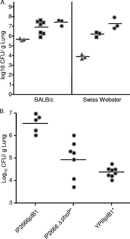
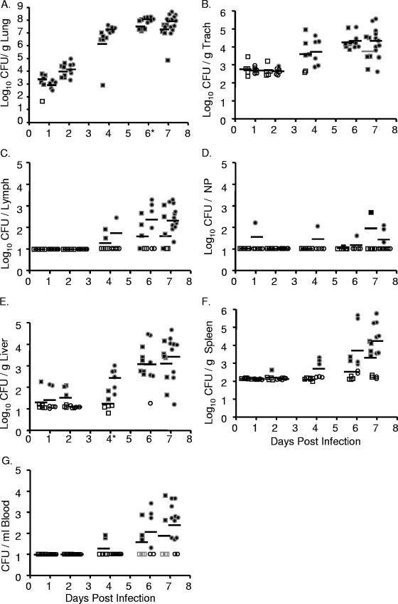
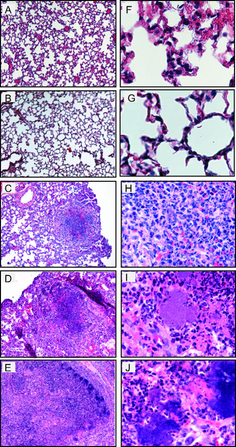
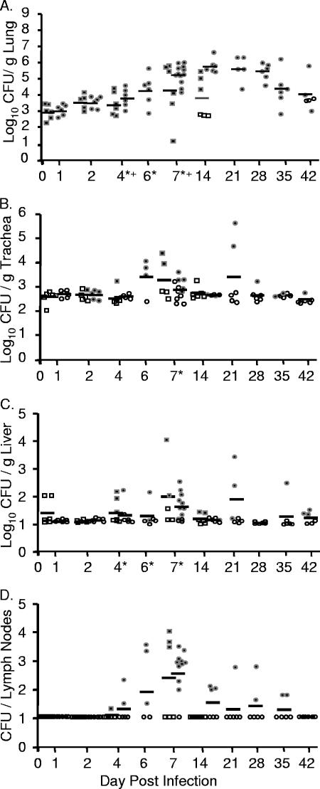
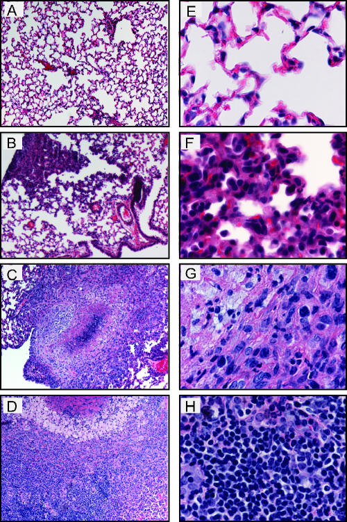
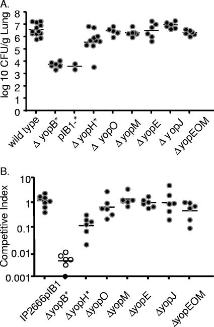
Yersinia pseudotuberculosis infects many mammals and birds including humans, livestock, and wild rodents and can be recovered from the lungs of infected animals. To determine the Y. pseudotuberculosis factors important for growth during lung infection, we developed an intranasal model of infection in mice. Following intranasal inoculation, we monitored both bacterial growth in lungs and dissemination to systemic tissues. Intranasal inoculation with as few as 18 CFU of Y. pseudotuberculosis caused a lethal lung infection in some mice. Over the course of 7 days, wild-type Y. pseudotuberculosis replicated to nearly 1 x 10(8) CFU/g of lung in BALB/c mice, induced histopathology in lungs consistent with pneumonia, but disseminated sporadically to other tissues. In contrast, a Delta yopB deletion strain was attenuated in this model, indicating that translocation of Yersinia outer proteins (Yops) is essential for virulence. Additionally, a Delta yopH null mutant failed to grow to wild-type levels by 4 days postintranasal inoculation, but deletions of any other single effector YOP did not attenuate lung colonization 4 days postinfection. Strains with deletions in yopH and any one of the other known effector yop genes were more attenuated that the Delta yopH strain, indicating a unique role for yopH in lungs. In summary, we have characterized the progression of a lung infection with an enteric Yersinia pathogen and shown that YopB and YopH are important in lung colonization and dissemination. Furthermore, this lung infection model with Y. pseudotuberculosis can be used to test potential therapeutics against Yersinia and other gram-negative infections in lungs.


Figures

FIG. 1.
FIG. 1.
Y. pseudotuberculosis colonization of lungs occurs at low doses and is dependent on phoP. (A) In vivo growth of Y. pseudotuberculosis in BALB/c or Swiss Webster mice. Mice were inoculated intranasally with wild-type Y. pseudotuberculosis with 7.8 × 101 CFU (▴), 8.4 × 10² (▪), or 8.4 × 10³ (•). Four days postinoculation lungs were harvested, weighed, homogenized, and plated for CFU. (B) Strains lacking PhoP are attenuated in colonization of lungs. BALB/c mice were intranasally inoculated with 8.0 × 10² CFU of IP2666pIB1, 6.0 × 10² CFU of IP2666pIB1ΔphoP, or 2.5 × 10³ CFU of YPIIIpIB1. Four days postinoculation, lungs were weighed, homogenized, and plated for CFU. Each dot represents output from one mouse. Bars represent the geometric mean. P values were determined by a two-tailed Student's t test. *, P < 0.01.
FIG. 2.
FIG. 2.
Intranasal inoculation with Y. pseudotuberculosis results in a lethal lung infection. BALB/c mice were inoculated with 300 to 500 CFU of Y. pseudotuberculosis grown at 26°C (•) or 37°C (▪). Mice were sacrificed 1, 2, 4, 6, and 7 days postinoculation and lungs (A), trachea (B), lymph nodes (C), nasopharynx (D), liver (E), spleen (F), and blood (G) were harvested, homogenized, and plated for CFU. Each dot represents data from one mouse. Open symbols indicate that bacteria were below the limit of detection (note, the limit of detection per gram of trachea recovered is generally higher than in the lungs, liver, and spleen because the weight of the trachea is smaller). Bars represent the geometric mean. *, P < 0.01 (Student's t test). NP, nasopharynx; Trach, trachea.
FIG. 3.
FIG. 3.
Histological analysis of lungs from BALB/c mice inoculated with wild-type Y. pseudotuberculosis. Mice were inoculated with 300 to 500 CFU of Y. pseudotuberculosis, and lungs were harvested 1 (A and F), 2 (B and G), 4 (C and H), 6 (D and I), and 7 (E and J) days postinoculation, sectioned, and stained with hematoxylin and eosin. Slides are shown at a magnification of ×10 (A to E) or ×60 (F to J).
FIG. 4.
FIG. 4.
Immunohistochemical analysis of lung sections. Sections from mice 4 days postinoculation with PBS (A and C) or with 300 to 500 CFU of Y. pseudotuberculosis grown at 26°C (B and D) were stained with anti-Y. pseudotuberculosis antibodies. Goat anti-mouse HRP-conjugated antibody was used as a secondary stain. Brown areas represent areas of HRP activity; lighter brown/gray areas are red blood cells. Samples shown are at a magnification of ×10 (A and B) and ×60 (C and D). Arrow (D) indicates an area of colocalization of antibody with bacterial colonies. Boxed sections in panels A and B indicate the areas magnified in panels C and D, respectively.
FIG. 5.
FIG. 5.
YopB is essential for high levels of colonization and dissemination. BALB/c mice were infected with 300 to 500 CFU of Y. pseudotuberculosis ΔyopB grown at 26°C (•) or 37°C (▪). Mice inoculated with the ΔyopB mutant grown at 26°C were sacrificed at 1, 2, 4, 6, 7, 14, 21, 28, 35, and 42 days postinoculation, and mice inoculated with ΔyopB strain grown at 37°C were sacrificed at 1, 2, 4, 7, and 14 days postinoculation. Lungs (A), trachea (B), liver (C), and lymph nodes (D) were harvested, homogenized, and plated for CFU. Each circle represents the CFU from a mouse inoculated with the ΔyopB strain grown at 26°C, and each square represents CFU from a mouse inoculated with the ΔyopB strain grown at 37°C. Open symbols indicate that no bacteria were recovered at that limit of detection. Bars represent the geometric mean. P values were determined between the levels of the ΔyopB strain recovered as shown here and levels of wild-type Y. pseudotuberculosis shown in Fig. 2 using a two-tailed Student's t test. *, P < 0.01 between wild type and yopB mutant grown at 26°C; +, P < 0.01 between wild type and yopB mutant grown at 37°C.
FIG. 6.
FIG. 6.
Histological analysis of lungs from BALB/c mice inoculated with ΔyopB Y. pseudotuberculosis. Mice were inoculated with 300 to 500 CFU of Y. pseudotuberculosis, and lungs were harvested 1 (A and E), 7 (B and F), 28 (C and G), and 42 (D and H) days postinoculation. Lungs were sectioned and stained with hematoxylin and eosin. Slides show lungs at a magnification of ×10 (A to D) or ×60 (E to H).
FIG. 7.
FIG. 7.
Colonization of lungs of BALB/c mice with yop mutant strains 4 days after intranasal inoculation in single-strain and competition infections. (A) Mice were inoculated with ∼8 × 10² CFU wild-type Y. pseudotuberculosis or an isogenic mutant of yopB, yopH, yopO, yopM, yopE, yopJ, or a yopEOM triple mutant or a strain of IP2666 lacking the virulence plasmid (pIB1). Four days postinoculation lungs were harvested, homogenized, and plated for CFU. (B) BALB/c mice were inoculated with ∼8 × 10² CFU of an equal mixture of MLF-13 and either IP2666pIB1 or a yopB, yopH, yopO, yopM, yopE, or yopJ mutant or a yopEOM triple mutant. Four days postinoculation lungs were harvested, homogenized, and plated on L plates containing X-Gal, and the ratios of wild type to mutants were determined. Each dot represents data from one mouse. Open circles indicate that no bacteria were recovered at the limit of detection. Bars represent the geometric mean. P values were calculated by a two-tailed Student's t test. *, P < 0.05 for the number of CFU of wild-type Y. pseudotuberculosis recovered versus the number of CFU of a mutant strain.
FIG. 8.
FIG. 8.
Colonization of Y. pseudotuberculosis yopH mutants in competition with wild-type Y. pseudotuberculosis 4 days postinoculation. BALB/c mice were inoculated with ∼8 × 10² CFU of an equal mixture of MLF-13 and either the MLF-9, yopH(R409A), yopH(K342A), yopH(V31G), yopH(Q11R), or yopHΔ223-226 strain. Bacteria from lungs were grown on L plates containing X-Gal, and the competitive index was calculated as described in Materials and Methods. Black circles represent the CI from individual mice, open circles indicate that no mutants were detected, and bars represent the geometric mean. P values were determined by a two-tailed Student's t test. *, P < 0.05.
FIG. 9.
FIG. 9.
Colonization of Y. pseudotuberculosis yopH double mutants. Mice were intranasally inoculated with ∼8 × 10² CFU of wild-type Y. pseudotuberculosis or mutants of the yopHE, yopHO, yopHM, yopHJ, or yopEOMJ strains. Four days postinoculation lungs were weighed, homogenized, and plated for CFU. Black circles represent the CFU recovered from individual mice, open circles indicate that the number of CFU was below the limit of detection, and bars represent the geometric mean. P values were determined by a two-tailed Student's t test. *, P < 0.05.

References

    1. Achtman, M., K. Zurth, G. Morelli, G. Torrea, A. Guiyoule, and E. Carniel. 1999. Yersinia pestis, the cause of plague, is a recently emerged clone of Yersinia pseudotuberculosis. Proc. Natl. Acad. Sci. USA 96:14043-14048. - PMC - PubMed
    1. Angelichio, M. J., D. S. Merrell, and A. Camilli. 2004. Spatiotemporal analysis of acid adaptation-mediated Vibrio cholerae hyperinfectivity. Infect. Immun. 72:2405-2407. - PMC - PubMed
    1. Anisimov, A. P., S. V. Dentovskaya, G. M. Titareva, I. V. Bakhteeva, R. Z. Shaikhutdinova, S. V. Balakhonov, B. Lindner, N. A. Kocharova, S. y. N. Senchenkova, O. Holst, G. B. Pier, and Y. A. Knirel. 2005. Intraspecies and temperature-dependent variations in susceptibility of Yersinia pestis to the bactericidal action of serum and to polymyxin B. Infect. Immun. 73:7324-7331. - PMC - PubMed
    1. Balada, J. M., and J. Mecsas. 2006. Yersinia has a tropism for B and T cell zones of lymph node that is independent of the type III secretion system. PLOS Pathog, in press. - PMC - PubMed
    1. Barz, C., T. N. Abahji, K. Trulzsch, and J. Heesemann. 2000. The Yersinia Ser/Thr protein kinase YpkA/YopO directly interacts with the small GTPases RhoA and Rac-1. FEBS Lett. 482:139-143. - PubMed

Publication types

MeSH terms

Substances

LinkOut - more resources