. 2006 Nov 27;203(12):2577-87.
doi: 10.1084/jem.20060244. Epub 2006 Oct 30.

IL-23 stimulates epidermal hyperplasia via TNF and IL-20R2-dependent mechanisms with implications for psoriasis pathogenesis

Affiliations

IL-23 stimulates epidermal hyperplasia via TNF and IL-20R2-dependent mechanisms with implications for psoriasis pathogenesis

Jason R Chan et al. J Exp Med. .

Abstract

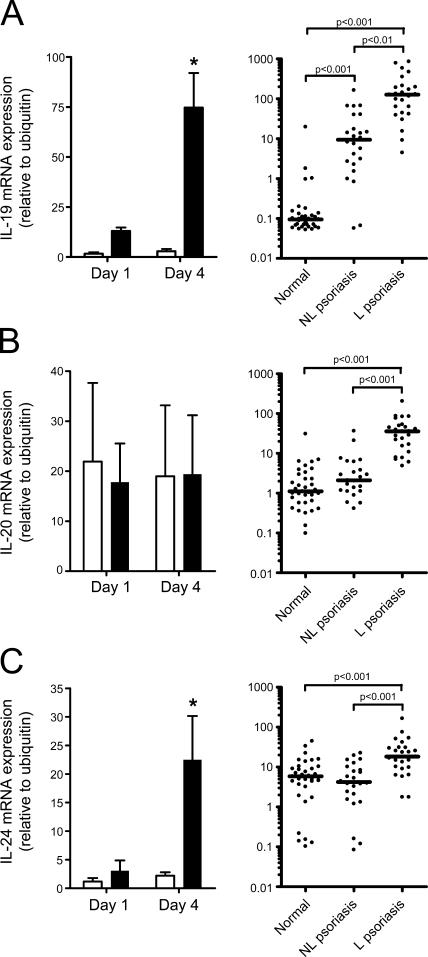
Aberrant cytokine expression has been proposed as an underlying cause of psoriasis, although it is unclear which cytokines play critical roles. Interleukin (IL)-23 is expressed in human psoriasis and may be a master regulator cytokine. Direct intradermal administration of IL-23 in mouse skin, but not IL-12, initiates a tumor necrosis factor-dependent, but IL-17A-independent, cascade of events resulting in erythema, mixed dermal infiltrate, and epidermal hyperplasia associated with parakeratosis. IL-23 induced IL-19 and IL-24 expression in mouse skin, and both genes were also elevated in human psoriasis. IL-23-dependent epidermal hyperplasia was observed in IL-19-/- and IL-24-/- mice, but was inhibited in IL-20R2-/- mice. These data implicate IL-23 in the pathogenesis of psoriasis and support IL-20R2 as a novel therapeutic target.


Figures

Figure 1.
Figure 1.
IL-23 is associated with human psoriasis and drives epidermal hyperplasia in mice. Expression of IL-23p19, IL-12p40, and IL-12p35 mRNA in normal, nonlesional (NL), and lesional (L) human psoriatic skin. Bar represents median (A). Representative photographs (B) of 129SvEv mice at day 4 treated with daily injections of saline (left) or 1 μg IL-23 (right). Representative frozen sections stained for CD31. Samples were taken at day 4 from 129SvEv mice treated daily with saline (top) or 1 μg IL-23 (bottom). Red arrows indicate CD31 staining in the dermal papillary. Bar, 100 μm (C). Representative histological sections at day 4 of littermate wild-type C57BL/6 mice treated daily with saline (left) or 1 μg IL-23 (middle), and IL-23R−/− mice treated daily with 1 μg IL-23 (right). Red arrows indicate parakeratosis. Green arrows indicate neutrophilic exudates. Bar, 100 μm (D). Epidermal thickness was measured in 129SvEv mice treated daily with saline (◯) or 1 μg IL-23 (•). Data was pooled together from six independent experiments performed in quadruplicate. Mean ± SD is shown. *, P < 0.001 (E). Epidermal thickness was measured at day 4 in 129SvEv mice treated daily with saline, 1 μg IL-23, 1 μg IL-12, or 1 μg TNF. Data was pooled together from two independent experiments (F).
Figure 2.
Figure 2.
IL-23–stimulated epidermal thickening is due to keratinocyte hyperplasia in the spinous layer and altered granular layer differentiation. Low magnification (bar, 8.4 μm) of skin illustrating the major ultrastructural features of IL-23–induced epidermal hyperplasia, including hypertrophy and hyperplasia of the spinous layer, with spongiosis and retained nuclei as well as compacted keratin filaments in the keratin layer (A). Transmission electron photomicrograph of skin from an IL-23–treated mouse showing retained nuclei (single arrows) within the stratum corneum. Note the division between the cornified and granular layers (double arrow). Bar, 9.7 μm (B).
Figure 3.
Figure 3.
IL-17A is not required for IL-23–dependent epidermal hyperplasia. Expression of IL-17A (A) or TNF (B) mRNA in human psoriatic skin. Expression of IL-17A (C) or TNF (D) mRNA in mice treated daily with saline (open bars) or 1 μg IL-23 (filled bars). Data are mean ± SEM of three independent experiments using 129SvEv, C57BL/6, or B6 × 129 mice, where mRNA was pooled together from four mice per group. *, P < 0.05. Epidermal thickness at day 4 in 129SvEv mice treated daily with saline or 1 μg IL-23 in the presence of anti–IL-17A (E) or anti-TNF (F) blocking monoclonal antibody or isotype control. Sum of two independent experiments for each antibody is shown. Expression of G-CSF (G) at day 4 in 129SvEv mice (n = 4, mRNA was pooled together) treated daily with saline or IL-23 in the presence of blocking anti–IL-17A monoclonal antibody (filled bars) or isotype control (open bars). One of two representative experiments is shown.
Figure 4.
Figure 4.
IL-23 stimulates keratinocyte proliferation in mice. Representative immunohistochemical staining for Ki67 in mice at day 1 (A) after treatment with saline (top) or 1 μg IL-23 (bottom). Bar, 20 μm. Expression of K16 mRNA (B) in mice treated daily with saline (open bars) or 1 μg IL-23 (filled bars). Data are mean ± SEM of three independent experiments using 129SvEv, C57BL/6, or B6 × 129 mice, where mRNA was pooled together from four mice per group. *, P < 0.05. K16 mRNA expression in mice at day 4 treated daily with saline, 1 μg IL-23, 1 μg IL-12, or 1 μg TNF. Data are mean ± STD, n = 4. One of two representative experiments is shown (C). mRNA expression at day 1 for a panel of keratinocyte growth factors (D) in mice treated with saline (open bars) or IL-23 (filled bars). Data are mean ± SEM of three independent experiments using 129SvEv, C57BL/6, or B6 × 129 mice, where mRNA was pooled together from four mice per group. Keratinocyte proliferation assay (E) in which normal human keratinocytes were treated in triplicate with vehicle or IL-23 (, , or 100 ng/ml). 10 ng/ml KGF was used as a positive control. One of three representative experiments is shown.
Figure 5.
Figure 5.
IL-19 family members are expressed in human psoriatic skin and stimulated by IL-23 in mouse skin. IL-19 (A), IL-20 (B), and IL-24 (C) mRNA expression was measured in mice (left) treated daily with saline (open bars) or 1 μg IL-23 (filled bars), and in human psoriatic skin (right). Mouse data are mean ± SEM of three independent experiments using 129SvEv, C57BL/6, or B6 × 129 mice, where mRNA was pooled together from four mice per group. *, P < 0.05. Bar represents median for human samples.
Figure 6.
Figure 6.
Role of IL-20R2 in IL-23–dependent epidermal hyperplasia. Epidermal thickness was measured at day 4 in wild-type, IL-20R2−/−, IL-19−/−, or IL-24−/− mice treated daily with saline (open symbols) or 1 μg IL-23 (filled symbols). Data from different wild-type strains were pooled together because no differences in IL-23–stimulated epidermal thickening were observed. Data are the aggregate of seven independent experiments (each strain of mouse was used in at least two separate experiments) (A). IL-19 and IL-24 gene expression in IL-20R2−/− mice at day 4 (B). IL-24 gene expression in IL-19−/− mice at day 4 (C). IL-19 gene expression in IL-24−/− mice at day 4 (D). Data are representative of two independent experiments where mRNA (n = 4) was pooled together. Representative immunohistochemical staining for CD4+ T cells (E), CD11c+ dendritic cells (F), F4/80+ macrophages (G), and neutrophils (H) at day 4 in IL-20R2−/− mice treated daily with saline (left) or 1 μg IL-23 (middle), or littermate wild-type mice at day 4 treated daily with 1 μg IL-23 (right). Bar, 100 μm.

Similar articles

Cited by

References

    1. Schon, M.P., and W.H. Boehncke. 2005. Psoriasis. N. Engl. J. Med. 352:1899–1912. - PubMed
    1. Nickoloff, B.J., and F.O. Nestle. 2004. Recent insights into the immunopathogenesis of psoriasis provide new therapeutic opportunities. J. Clin. Invest. 113:1664–1675. - PMC - PubMed
    1. Bos, J.D., M.A. de Rie, M.B. Teunissen, and G. Piskin. 2005. Psoriasis: dysregulation of innate immunity. Br. J. Dermatol. 152:1098–1107. - PubMed
    1. Nestle, F.O., C. Conrad, A. Tun-Kyi, B. Homey, M. Gombert, O. Boyman, G. Burg, Y.J. Liu, and M. Gilliet. 2005. Plasmacytoid predendritic cells initiate psoriasis through interferon-α production. J. Exp. Med. 202:135–143. - PMC - PubMed
    1. Nickoloff, B.J., B. Bonish, B.B. Huang, and S.A. Porcelli. 2000. Characterization of a T cell line bearing natural killer receptors and capable of creating psoriasis in a SCID mouse model system. J. Dermatol. Sci. 24:212–225. - PubMed

MeSH terms