. 2006 Nov;128(5):583-91.
doi: 10.1085/jgp.200609653.

Mechanisms of two modulatory actions of the channel-binding protein Slob on the Drosophila Slowpoke calcium-dependent potassium channel

Affiliations

Mechanisms of two modulatory actions of the channel-binding protein Slob on the Drosophila Slowpoke calcium-dependent potassium channel

Haoyu Zeng et al. J Gen Physiol. 2006 Nov.

Abstract

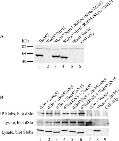
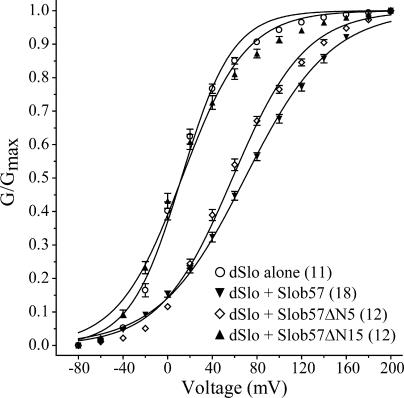
Slob57 is an ion channel auxiliary protein that binds to and modulates the Drosophila Slowpoke calcium-dependent potassium channel (dSlo). We reported recently that residues 1-39 of Slob57 comprise the key domain that both causes dSlo inactivation and shifts its voltage dependence of activation to more depolarized voltages. In the present study we show that removal of residues 2-6 from Slob57 abolishes the inactivation, but the ability of Slob57 to rightward shift the voltage dependence of activation of dSlo remains. A synthetic peptide corresponding in sequence to residues 1-6 of Slob57 blocks dSlo in a voltage- and dose-dependent manner. Two Phe residues and at least one Lys residue in this peptide are required for the blocking action. These data indicate that the amino terminus of Slob57 directly blocks dSlo, thereby leading to channel inactivation. Further truncation to residue Arg(16) eliminates the modulation of voltage dependence of activation. Thus these two modulatory actions of Slob57 are independent. Mutation within the calcium bowl of dSlo greatly reduces its calcium sensitivity (Bian, S., I. Favre, and E. Moczydlowski. 2001. Proc. Natl. Acad. Sci. USA. 98:4776-4781). We found that Slob57 still causes inactivation of this mutant channel, but does not shift its voltage dependence of activation. This result confirms further the independence of the inactivation and the voltage shift produced by Slob57. It also suggests that the voltage shift requires high affinity Ca(2+) binding to an intact calcium bowl. Furthermore, Slob57 inhibits the shift in the voltage dependence of activation of dSlo evoked by Ca(2+), and this inhibition by Slob57 is greater at higher free Ca(2+) concentrations. These results implicate distinct calcium-dependent and -independent mechanisms in the modulation of dSlo by Slob.


Figures

Figure 1.
Figure 1.
Expression of Slob57ΔN5 and Slob57ΔN15 and coimmunoprecipitation with dSlo or dSloD5N5. (A) Slobs were transfected into CHO cells. Slob expression was analyzed in cell lysates with anti-Slob antibody. Lane 1, wild-type Slob57; lane 2, Slob57 with Met1 to Leu mutation; lane 3, Slob57 with Met1 to Leu and Lys6 to Met mutations (Slob57ΔN5); lane 4, Slob57 with Met1 to Leu and Arg16 to Met mutations (Slob57ΔN15); lane 5, cells transfected with blank vector; lane 6, nontransfected cells. (B) Different Slobs were cotransfected into CHO cells together with dSlo or dSloD5N5. Cell lysates were then incubated with anti-Slob antibody and immunoprecipitated by protein A/G agarose beads. Coimmunoprecipitated proteins were assayed by Western blot with anti-dSlo antibody (top). Slobs and dSlo in cell lysates were analyzed for expression level by Western blot with anti-Slob or anti-dSlo antibody (bottom two panels). dSlo cotransfected with Slob57 (lane 1), with Slob57ΔN5 (lane 2), or with Slob57ΔN15 (lane 3). dSloD5N5 cotransfected with Slob57 (lane 4), with Slob57ΔN5 (lane 5), or with Slob57ΔN15 (lane 6). Lane 7, dSlo cotransfected with blank vector used for Slobs; lane 8, Slob57 cotransfected with blank vector used for dSlo channels; lane 9, nontransfected CHO cells.
Figure 2.
Figure 2.
The first six amino-terminal residues of Slob57 are essential for the inactivation of dSlo. Whole cell currents in cells transfected with dSlo and Slob57 or its mutants were evoked, in the presence of 110 μM free Ca2+, by a 350-ms test pulse to different voltages from a holding potential of −80 mV, followed by hyperpolarization to −120 mV. (A) dSlo alone, (B) coexpression of dSlo and Slob57, (C) coexpression of dSlo and Slob57ΔN5, (D) coexpression of dSlo and Slob57ΔN15, (E) pulse protocol.
Figure 3.
Figure 3.
The peptide MFKFNK blocks dSlo. (A) dSlo current traces from an inside-out patch before and during application of 100 μM MFKFNK (note the increase in noise in the current trace), and after wash as indicated in the figure. (B) MFKFNK blocks dSlo in a dose-dependent manner. Current amplitudes in the presence of different concentrations of MFKFNK were normalized to that in its absence. The data were fit with the Hill equation as described in Materials and methods. IC50 is 31 μM, and the Hill coefficient is 0.84 (n = 3). Error bars illustrate SEM.
Figure 4.
Figure 4.
MFKFNK inhibits dSlo in a voltage-dependent manner, and independent of internal Mg2+ or external K+ concentrations. (A) The dSlo current amplitude in the presence of 100 μM MFKFNK was normalized to that in the same patch before addition of the peptide. The normalized current was plotted against voltage and analyzed using the Woodhull model as described in Materials and methods. The x axis represents the voltage to which the cell was stepped from the holding potential of −80 mV. The Kd at 0 mV is 162 μM and the effective electrical distance δ is 0.17 (n = 7). (B) Normalized current amplitude of dSlo at +140 mV in the presence of 100 μM MFKFNK in control solutions, 0 Mg2+ internal solution, or 5 mM K+ external solution are shown (n = 3).
Figure 5.
Figure 5.
A specific sequence is required for peptide block of dSlo. Current amplitude of dSlo at +140 mV with 100 μM MFKFNK or its variants (as indicated in the figure) was normalized to control current in the same patch before peptide application (n = 5–6). *, significantly different from control (100%, dashed line), P < 0.05; #, significantly different from wild type MFKFNK, P < 0.05.
Figure 6.
Figure 6.
Conductance–voltage relationships for dSlo in the presence of Slob57 or its N-terminal truncation mutants. The conductance–voltage relationships for dSlo transfected alone (◯) or together with Slob57 (▾), Slob57ΔN5 (⋄), or Slob57ΔN15 (▴) were measured from tail currents with 110 μM free Ca2+. To prevent dSlo inactivation caused by Slob57 in longer test pulses, a 100-ms test pulse was used (see Results). The conductance–voltage relationship was generated from peak tail currents as described in Materials and methods. The number of experiments is shown in parentheses, and the error bars illustrate SEM. Some of the error bars are so small that they are masked by the symbols.
Figure 7.
Figure 7.
Slob57 causes inactivation of dSloD5N5. Same as Fig. 2, except that the mutated channel dSloD5N5 was used instead of wild-type dSlo. (A) dSloD5N5 alone, (B) dSloD5N5 together with Slob57, (C) dSloD5N5 together with Slob57ΔN5, (D) dSloD5N5 together with Slob57ΔN15, (E) pulse protocol.
Figure 8.
Figure 8.
Conductance–voltage relationships for dSloD5N5. Same protocol as Fig. 6, except that the mutated channel dSloD5N5 was used in the presence (circles) or absence (squares) of Slob57, and the concentration of free Ca2+ was either 110 μM (filled symbols) or 300 μM (open symbols).
Figure 9.
Figure 9.
Effect of calcium on dSlo voltage dependence of activation in the absence or presence of Slob57. Same as Fig. 6, except that the measurements of dSlo expressed alone (A) or together with Slob57 (B) were performed in the presence of different concentrations of free Ca2+: 20 μM (diamonds), 40 μM (inverted triangles), 80 μM (triangles), 110 μM (squares), and 300 μM (circles).
Figure 10.
Figure 10.
Interaction between Slob57 and calcium. The V1/2 (A) of the conductance–voltage relationship for dSlo expressed alone (◯) or together with Slob57 (▪), taken from Fig. 9, and the difference in V1/2 (ΔV1/2) evoked by Slob57 (B) are plotted as a function of the concentration of free Ca2+.

Similar articles

Cited by

References

    1. Adelman, J.P., K.-Z. Shen, M.P. Kavanaugh, R.A. Warren, Y.-N. Wu, A. Lagrutta, C.T. Bond, and R.A. North. 1992. Calcium-activated potassium channels expressed from cloned complementary DNAs. Neuron. 9:209–216. - PubMed
    1. Atkinson, N.S., G.A. Robertson, and B. Ganetzky. 1991. A component of calcium-activated potassium channels encoded by the Drosophila slo locus. Science. 253:551–555. - PubMed
    1. Atkinson, N.S., R. Brenner, W. Chang, J. Wilbur, J.L. Larimer, and J. Yu. 2000. Molecular separation of two behavioral phenotypes by a mutation affecting the promoters of a Ca-activated K channel. J. Neurosci. 20:2988–2993. - PMC - PubMed
    1. Bao, L., A.M. Rapin, E.C. Holmstrand, and D.H. Cox. 2002. Elimination of the BK(Ca) channel's high-affinity Ca2+ sensitivity. J. Gen. Physiol. 120:173–189. - PMC - PubMed
    1. Bao, L., C. Kaldany, E.C. Holmstrand, and D.H. Cox. 2004. Mapping the BKCa channel's “Ca2+ bowl”: side-chains essential for Ca2+ sensing. J. Gen. Physiol. 123:475–489. - PMC - PubMed

Publication types

MeSH terms