Comparative Study
. 2006 Nov;133(22):4561-72.
doi: 10.1242/dev.02641.

Two separate molecular systems, Dachsous/Fat and Starry night/Frizzled, act independently to confer planar cell polarity

Affiliations
Comparative Study

Two separate molecular systems, Dachsous/Fat and Starry night/Frizzled, act independently to confer planar cell polarity

José Casal et al. Development. 2006 Nov.

Abstract

Planar polarity is a fundamental property of epithelia in animals and plants. In Drosophila it depends on at least two sets of genes: one set, the Ds system, encodes the cadherins Dachsous (Ds) and Fat (Ft), as well as the Golgi protein Four-jointed. The other set, the Stan system, encodes Starry night (Stan or Flamingo) and Frizzled. The prevailing view is that the Ds system acts via the Stan system to orient cells. However, using the Drosophila abdomen, we find instead that the two systems operate independently: each confers and propagates polarity, and can do so in the absence of the other. We ask how the Ds system acts; we find that either Ds or Ft is required in cells that send information and we show that both Ds and Ft are required in the responding cells. We consider how polarity may be propagated by Ds-Ft heterodimers acting as bridges between cells.


Figures

Figure 1
Figure 1. A summary of polarising gradients in the abdomen
On the left, the pattern of the cuticle is shown with the types of cuticle in the A (a1-a6) and in the P compartment (blue, p3-p1). The compartments are patterned by gradients; Hh in A and Wg in P (Struhl et al., 1997a; Lawrence et al., 2002). The U-shaped Hh gradient sets up the Ds gradients and also the activity gradients of Fj and Ft that are shown in the next three columns (Casal et al., 2002). Clones that lack or overexpress a gene affect the polarity of wildtype cells around as shown (arrows). For example, the leftmost column shows ptc en clones, which constituitively activate the Hh transduction pathway and produce reversal of the wildtype cells behind the clones (but only when they are in the middle of the A compartment, where they cause a discrepancy in the Hh transduction pathway between the clone and the surround). The next column shows the gradient of Ds: loss of ds reverses the polarity of cells in front of clones at the back of the A compartment (where the level of Ds activity is high) but has no effect when the clones are located at the front of the A compartment (where Ds activity is low). Overexpression of Ds has the opposite effects: repolarising only at the front of the A compartment. The effects of clones involving Fj and Ft are as shown. By contrast to the other genes, clones involving Fz have similar effects wherever they are situated. We imagine there is an alteration in Fz activity that spreads out from the clones as the surrounding wildtype cells readjust their levels of Fz activity by an averaging process (Lawrence et al., 2004); this is symbolised by the haloes. This difference of clonal behaviour points again to a distinction between the Ds and Stan systems.
Figure 2
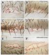
Figure 2. The Ds and Stan systems are different and independent
This figure compares the effects of driving Fz, Ft and ectoDs (a particularly potent signalling form of Ds) in clones in flies lacking either the Ds or the Stan systems. Clones overexpressing fz (UAS.fz) reverse the polarity of wildtype cells over a short range (Lawrence et al., 2004) but they reverse polarity of ds cells over a longer range (2A). UAS.fz clones have no effect in stan flies (2B). UAS.ft clones reverse the polarity of wildtype cells in front of the clone (Fig 3A), but have no effect in ds flies (2C); the same clones reverse polarity of stan flies (2D) Clones overexpressing ectoDs reverse the polarity of wildtype cells behind the clone (Fig 3C), but have no effect in ds flies (2E). These UAS.ectoDs clones reverse polarity of stan flies (2F). Clones marked with pwn (A-D), and pwn sha (E, F). As in all the figures (except Fig 7), anterior is towards the top, red lines outline the clone and red arrows indicate imposed polarity.
Figure 3
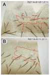
Figure 3. The range of repolarisations due to the Ds system is increased in fj flies
This figure compares the effects of UAS.ft clones (reversing polarity in front of the clone in the A compartment) and UAS.ectoDs clones (reversing polarity behind) in wildtype flies (3A, 3C) with the same types of clones in fj flies. The range in fj flies is increased (3B, 3D). Clones marked with pwn (A, B, D) and with pwn sha (C).
Figure 4
Figure 4. Cells respond more to the Stan system in the absence of the Ds system
A twofold increase in the dose of the fz gene (between clone and surround) has no effect in wildtype flies (not shown) but, in ds flies, reverses polarity in front of the clone and imposes normal polarity behind the clone (4A). Compare with the small effect, indicated by a yellow arrowhead, of similar clones in a ds+/ds fly (4B). Clones marked with trc.
Figure 5
Figure 5. The loss of one or both systems leads to different larval and adult phenotypes
ds tergites have a whorly central area but the bristle pattern is near normal (5A) while stan tergites are dishevelled at the front and back in the A compartment, but near normal elsewhere (5C). In ds stan tergites both the hairs and bristles are dishevelled everywhere (5B). In the 3rd instar larvae, ds have disturbed hairs in the anterior rows of the ventral denticles, but the most posterior rows 5 and 6 are normal (5E). The stan larval denticle pattern (5G), as far as we can see (compare Price et al., 2006) is like wildtype (5H), while the ds stan larvae (5F) show randomised polarity. Note, for 5A-D, adult cuticles were mounted without squashing, in order to preserve bristle orientation in its native state.
Figure 6
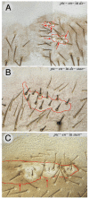
Figure 6. ptc en clones in flies lacking one or both systems
The Hh signal transduction pathway is maximally and constitutively activated in ptc en clones. Such clones reverse the polarity of hairs behind the clone both in ds flies (6A) and in stan flies (6C). However in ds stan flies, the ptc en have no discernable (consistent) effect on the surround (6B) — compare with 6A where there is a consistent effect, the hairs pointing inwards all around the clone. Clones marked with pwn. The shadowed section in 6A indicates that the image was reconstructed like Humpty Dumpty from a single bisected piece of cuticle.
Figure 7
Figure 7. A speculative model of the Ds system
The figure shows the A compartment, anterior is to the left. Ft is indicated in blue and Ds in red. The long arrows show the polarity of each cell, normal in black and reversed in red. In the wildtype (see top), there is evidence for opposing gradients of Fj protein (Fj) and of Ds (Ds) (Casal et al., 2002) as indicated by the size of the letters. Although there is no gradient of Ft protein (Ft) we envisage a gradient of Ft activity (Ft), driven by the action of Fj on Ft, as argued in the results. Active Ft can become stabilised in the membrane of one cell so that it can form heterodimers with Ds in the next cell (provided that sufficient Ds is present in that cell) . The polarity of any cell might depend on a comparison between the number of heterodimeric Ds molecules (red numbers above the cells) on the anterior and posterior faces of the cell, with the polarity of that cell pointing downwards. Note that even though the Ds gradient peaks posteriorly, there are more Ds heterodimers anteriorly. Thus the graded form of Ds and Fj expression might not be crucial, so long as one is graded it is sufficient — both uniform Fj and uniform Ds can rescue fj and ds eyes, respectively (Simon, 2004). The middle row shows the effect of a ft cell, in which all available Ds will make heterodimers with Ft on the facing (anterior) membrane of the cell on its right. Consequently, in this wildtype cell, Ds will be displaced towards the opposite (posterior) face of this wildtype cell, whose polarity will therefore become reversed. This excess of Ds molecules will bind to Ft in the nextmost cell, and again, by depleting Ds from its anterior face, will repolarise it. This effect will weaken from cell to cell. The lower row shows a UAS.ft cell that will attract more Ds to the facing membrane (posterior) of its neighbour on its left, thereby polarising that cell, the effect spreading anteriorwards.

Similar articles

Cited by

References

    1. Adler PN. Planar signaling and morphogenesis in Drosophila. Dev Cell. 2002;2:525–35. - PubMed
    1. Adler PN, Charlton J, Liu J. Mutations in the cadherin superfamily member gene dachsous cause a tissue polarity phenotype by altering frizzled signaling. Development. 1998;125:959–68. - PubMed
    1. Adler PN, Krasnow RE, Liu J. Tissue polarity points from cells that have higher Frizzled levels towards cells that have lower Frizzled levels. Curr Biol. 1997;7:940–9. - PubMed
    1. Amonlirdviman K, Khare NA, Tree DR, Chen WS, Axelrod JD, Tomlin CJ. Mathematical modeling of planar cell polarity to understand domineering nonautonomy. Science. 2005;307:423–6. - PubMed
    1. Baena-López LA, Baonza A, Garcia-Bellido A. The orientation of cell divisions determines the shape of Drosophila organs. Curr Biol. 2005;15:1640–4. - PubMed

Publication types