. 2006 Nov 7;103(45):17018-23.
doi: 10.1073/pnas.0607873103. Epub 2006 Oct 30.

Endothelial cellular senescence is inhibited by nitric oxide: implications in atherosclerosis associated with menopause and diabetes

Affiliations

Endothelial cellular senescence is inhibited by nitric oxide: implications in atherosclerosis associated with menopause and diabetes

Toshio Hayashi et al. Proc Natl Acad Sci U S A. .

Abstract

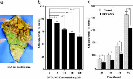
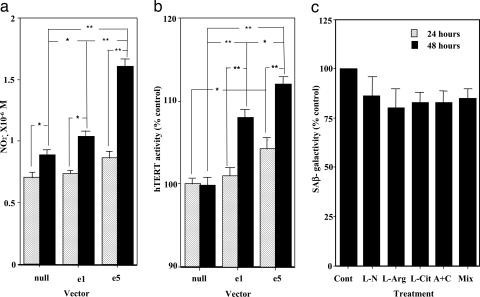
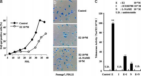
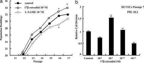
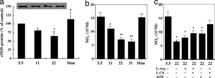
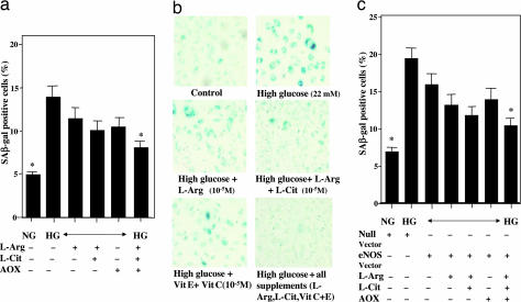
Senescence may contribute to the pathogenesis of atherosclerosis. Although the bioavailability of nitric oxide (NO) is limited in senescence, the effect of NO on senescence and its relationship to cardiovascular risk factors have not been investigated fully. We studied these factors by investigating senescence-associated beta-galactosidase (SA-beta-gal) and human telomerase activity in human umbilical venous endothelial cells (HUVECs). Treatment with NO donor (Z)-1-[2-(2-aminoethyl)-N-(2-aminoethyl)amino]diazen-1-ium-1,2-diolate (DETA-NO) and transfection with endothelial NO synthase (eNOS) into HUVECs each decreased the number of SA-beta-gal positive cells and increased telomerase activity. The NOS inhibitor N(G)-nitro-L-arginine methyl ester (L-NAME) abolished the effect of eNOS transfection. The physiological concentration of 17beta-estradiol activated hTERT, decreased SA-beta-gal-positive cells, and caused cell proliferation. However, ICI 182780, an estrogen receptor-specific antagonist, and L-NAME each inhibited these effects. Finally, we investigated the effect of NO bioavailability on high glucose-promoted cellular senescence of HUVECs. Inhibition by eNOS transfection of this cellular senescence under high glucose conditions was less pronounced. Treatment with L-arginine or L-citrulline of eNOS-transfected cells partially inhibited, and combination of L-arginine and L-citrulline with antioxidants strongly prevented, high glucose-induced cellular senescence. These data demonstrate that NO can prevent endothelial senescence, thereby contributing to the anti-senile action of estrogen. The ingestion of NO-boosting substances, including L-arginine, L-citrulline, and antioxidants, can delay endothelial senescence under high glucose. We suggest that the delay in endothelial senescence through NO and/or eNOS activation may have clinical utility in the treatment of atherosclerosis in the elderly.


Conflict of interest statement

The authors declare no conflict of interest.

Figures

Fig. 1.
Fig. 1.
SA-β-gal activity as cellular senescence. (a) SA-β-gal-positive staining was observed in atherosclerotic lesions of the intimal side of human thoracic aorta, which was obtained by autopsy. No staining was detected in the nonatherosclerotic area and advanced atherosclerotic area, including the necrotic core and ulcer complicated lesion. (b) Concentration-dependent decrease in SA-β-gal activity in HUVECs by DETA-NO. HUVECs were treated with DETA-NO for 24 h. ∗, P < 0.05; ∗∗, P < 0.01; ∗∗∗, P < 0.0001 vs. DETA-NO-untreated control. (c) Time-dependent decrease in SA-β-gal activity in HUVECs by DETA-NO. HUVECs were treated with 10 μM DETA-NO for 24–96 h. ∗, P < 0.05; ∗∗, P < 0.01 vs. the corresponding control. Control sample, which is treated for 48 h, is expressed as 100%.
Fig. 2.
Fig. 2.
Influence of eNOS modulation on cellular senescence. (a) The effect of transfection with eNOS on nitrite production by HEK 293 cells. Transfection with eNOS into cells was performed; e5 included five times the amount of eNOS vector compared with e1. The nitrite concentrations in the medium 24 and 48 h after transfection are shown. ∗, P < 0.05; ∗∗, P < 0.01. (b) The effect of transfection with eNOS on telomerase activity in HEK 293 cells. The activity of hTERT in cells 24 and 48 h after transfection are shown. ∗, P < 0.05; ∗∗, P < 0.01. (c) The effects of treatment with l-NAME (L-N, 300 μM), l-arginine (L-arg, 1 mM), and l-citrulline (L-cit, 300 μM) alone or in combination (A+C) on SA-β-gal activity in HUVECs. The treatment time was 24 h. Mix = l-arginine, l-citrulline and vitamin E plus vitamin C (each, 100 μM).
Fig. 3.
Fig. 3.
Effect of estrogen on cellular senescence. (a) The relative levels of SA-β-gal-positive staining cells in different PDL when HUVECs were untreated and treated with 10−8 M E2 for 24 h. Positive staining cells were evaluated by FACscan. (b) Representative photographs of SA-β-gal staining in control, 10−8 M E2-treated, and 10−8 M E2- and 10−4 M l-NAME-treated cells. Note that treatment with E2 decreased the number of SA-β-gal-positive cells, which was prevented by further treatment with l-NAME. Cells were used in PDL 22 at passage 7. (c) The effects of E2 (E, 10−8 M), ICI 182780 (I, 1 μM), and l-NAME (N, 1 mM) on telomerase activity in HUVECs. UD, undetectable. ∗∗, P < 0.01 vs. control.
Fig. 4.
Fig. 4.
Effects of E2 on endothelial cell proliferation. (a) The effects of E2 (10−8 M) and l-NAME (1 mM) on population doublings in each passage of HUVECs. The treatment time with E2 or l-NAME was 24 h. ∗, P < 0.05 vs. control. (b) The effects of different concentrations of E2 on relative cell division of HUVECs. Cells were used in PDL 20.2 at passage 7. ∗, P < 0.05: cell division vs. control.
Fig. 5.
Fig. 5.
Influence of high glucose on eNOS expression and nitrite production. (a) The effect of exposure to different concentrations of glucose on the level of eNOS protein expression in HUVECs. Mannitol (Man) was given as an osmolarity control. Cells were kept under different glucose conditions for 72 h. ∗, P < 0.05 vs. normal (5.5 mM) glucose. (b) The effect of exposure to different concentrations of glucose on nitrite levels in culture medium of HUVECs. ∗, P < 0.05; ∗∗, P < 0.01 vs. normal glucose. (c) The effects of l-arginine (L-arg, 1 mM), l-citrulline (L-cit, 300 μM), and antioxidants (AOX, 100 μM vitamin E plus 100 μM vitamin C) alone or in combination on nitrite levels in culture medium in HUVECs, which were reduced by 22 mM glucose. ∗, P < 0.05; ∗∗, P < 0.01 vs. normal glucose.
Fig. 6.
Fig. 6.
Influence of high glucose for 72 h on cellular senescence of HUVECs. (a) The effects of l-arginine (L-arg, 1 mM), l-citrulline (L-cit, 300 μM), and antioxidants (AOX, 100 μM vitamin E plus 100 μM vitamin C) on the increase in β-gal-positive stained cells when exposed to high (22 mM) glucose. ∗, P < 0.05 vs. high glucose without any treatment. (b) Representative photographs showing cellular senescence by staining cells with SA-β- gal. (c) Modulation by transfection with eNOS of the effects of l-arginine, l-citrulline, and antioxidants on the increase in SA-β-gal-positive-stained cells when exposed to high glucose. Null vector is control vector of eNOS Vector. ∗, P < 0.05 vs. high glucose without any treatment. NG, normal glucose; HG, high glucose (22 mM).

References

    1. Zeiher AM, Drexler H, Saurbier B, Just H. J Clin Invest. 1993;92:652–662. - PMC - PubMed
    1. Goldstein S. Science. 1990;249:1129–1133. - PubMed
    1. Hoffmann J, Haendeler J, Aicher A, Rossig L, Vasa M, Zeiher AM, Dimmeler S. Circ Res. 2001;89:709–715. - PubMed
    1. Liu JP. FASEB J. 1999;13:2091–2104. - PubMed
    1. Hsiao R, Sharma HW, Ramakrishnan S, Keith E, Narayanan R. Anticancer Res. 1997;117:827–832. - PubMed

MeSH terms

LinkOut - more resources