. 2006 Nov 7;103(45):16918-23.
doi: 10.1073/pnas.0607069103. Epub 2006 Oct 30.

Cardiac myosin binding protein C phosphorylation is cardioprotective

Affiliations

Cardiac myosin binding protein C phosphorylation is cardioprotective

Sakthivel Sadayappan et al. Proc Natl Acad Sci U S A. .

Abstract

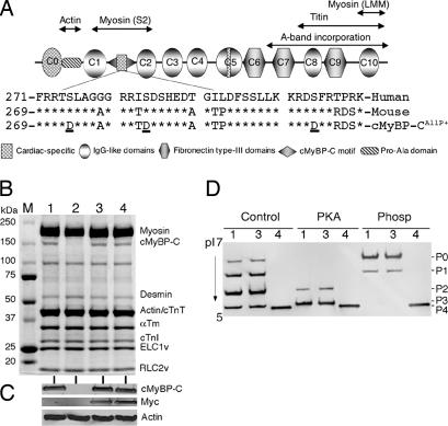
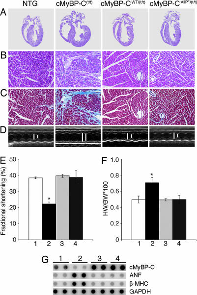
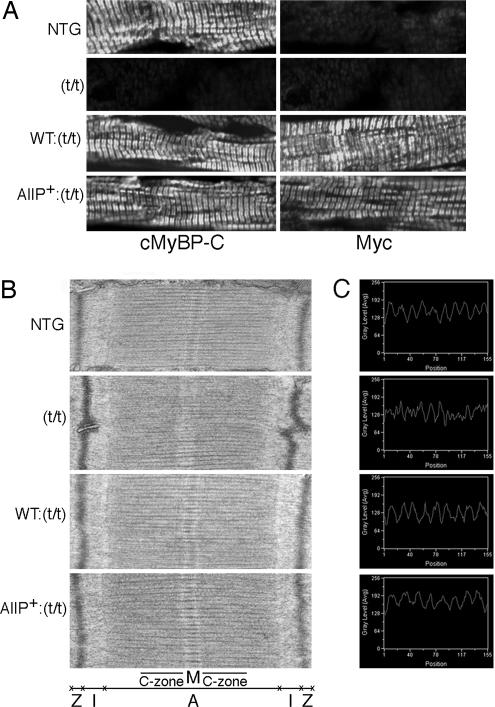
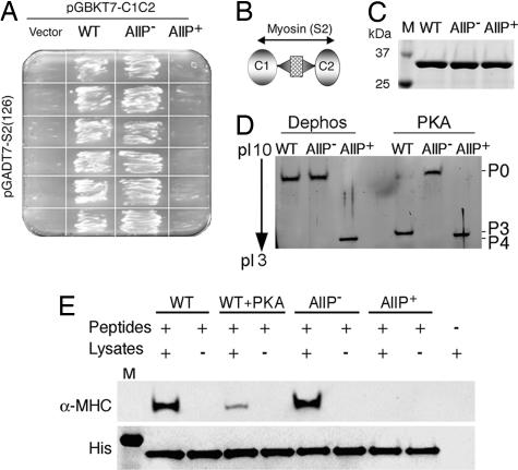
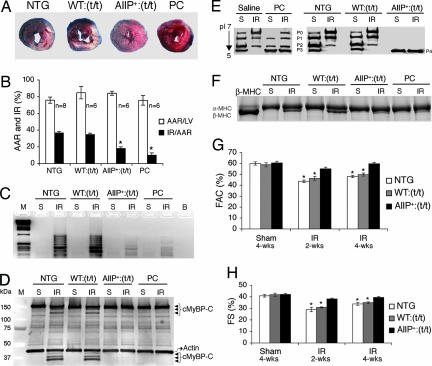
Cardiac myosin binding protein C (cMyBP-C) has three phosphorylatable serines at its N terminus (Ser-273, Ser-282, and Ser-302), and the residues' phosphorylation states may alter thick filament structure and function. To examine the effects of cMyBP-C phosphorylation, we generated transgenic mice with cardiac-specific expression of a cMyBP-C in which the three phosphorylation sites were mutated to aspartic acid, mimicking constitutive phosphorylation (cMyBP-C(AllP+)). The allele was bred into a cMyBP-C null background (cMyBP-C((t/t))) to ensure the absence of endogenous dephosphorylated cMyBP-C. cMyBP-C(AllP+) was incorporated normally into the cardiac sarcomere and restored normal cardiac function in the null background. However, subtle changes in sarcomere ultrastructure, characterized by increased distances between the thick filaments, indicated that phosphomimetic cMyBP-C affects thick-thin filament relationships, and yeast two-hybrid data and pull-down studies both showed that charged residues in these positions effectively prevented interaction with the myosin heavy chain. Confirming the physiological relevance of these data, the cMyBP-C(AllP+:(t/t)) hearts were resistant to ischemia-reperfusion injury. These data demonstrate that cMyBP-C phosphorylation functions in maintaining thick filament spacing and structure and can help protect the myocardium from ischemic injury.


Conflict of interest statement

The authors declare no conflict of interest.

Figures

Fig. 1.
Fig. 1.
cMyBP-CAllP+ transgene expression. (A) Domain organization of cMyBP-C. The cardiac-specific phosphorylation motif (sequence shown) is located between domains C1 and C2. The three known phosphorylation sites (Ser-273, Ser-282, and Ser-302, underlined) were each substituted with aspartic acid. (B) Representative Coomassie blue-stained SDS/PAGE analysis shows the lack of cMyBP-C in the homozygous cMyBP-C(t/t) hearts (lane 2), replacement of cMyBP-C to normal levels in the cMyBP-CWT:(t/t) (lane 3) and cMyBP-CAllP+:(t/t) (lane 4) crosses, and conservation of the other sarcomeric protein levels in these mice compared with a NTG sample (lane 1). (C) Western blot analysis shows the absence of endogenous cMyBP-C in the homozygous cMyBP-C(t/t) hearts and the presence of myc-tagged TG cMyBP-C in cMyBP-CWT:(t/t) and cMyBP-CAllP+:(t/t) mice. Lanes are numbered as in B. (D) 1D isoelectric focusing showing complete replacement of cMyBP-CAllP+ in the cMyBP-C null background. Samples from NTG (lane 1), cMyBP-CWT:(t/t) (lane 3), and cMyBP-CAllP+:(t/t) (lane 4) hearts were either untreated (Control) or treated with PKA or phosphatase (Phosp). Protein derived from cMyBP-C(t/t) hearts was omitted because of a lack of detectable cMyBP-C signal.
Fig. 2.
Fig. 2.
cMyBP-CAllP+ rescues the cMyBP-C(t/t) cardiac phenotype. All data and functional measurements were derived from mixed-gender, 12-week hearts. (A) Representative hearts stained with hematoxylin-eosin. (B) Hematoxylin-eosin staining. (C) Masson trichrome-stained myocardial sections to assess fibrosis. (D) M-mode echocardiographic tracings show the left ventricular chamber. B, C, and D correspond to the samples shown above in A. (E) Fractional shortening (%) measured by M-mode echocardiography (n = 6). (F) Heart (wet weight)-to-body weight ratios (n = 10). All data are presented as mean ± SE. ∗, Significant difference vs. NTG (P < 0.05). (G) RNA dot-blot analyses. In E-G, lanes 1, 2, 3, and 4 refer to NTG, cMyBP-C(t/t), cMyBP-CWT:(t/t), and cMyBP-CAllP+:(t/t) respectively. (Magnifications: A, ×4; B and C, ×20.)
Fig. 3.
Fig. 3.
Sarcomeric ultrastructural and immunohistochemical analyses. (A) Localization of cMyBP-CAllP+ into the cMyBP-C(t/t) background. Ventricular myocardial sections were immunostained for cMyBP-C by using either anti-cMyBP-C (Left) or anti-myc antibodies (Right). There is no staining of cMyBP-C in the cMyBP-C(t/t) background. (B) Transmission electron micrographs showing sarcomere ultrastructure. (C) Peaks show the distance between two thick myosin filaments, which was measured with a line scanner using Metamorph software with line length held constant at 525.83 nm (1.84501 nm per pixel). (Magnifications: A, ×60; B, ×30,000.)
Fig. 4.
Fig. 4.
cMyBP-C phosphorylation and myosin interaction. (A) Yeast two-hybrid experiments confirm that C1-C2WT and C1-C2AllP− [phosphorylation negative mimetic (14)] interact with the myosin S2, but the phosphorylation mimetic cMyBP-CAllP+ does not. (B) Schematic diagram shows the region of cMyBP-C being tested for interaction. (C) SDS/PAGE analysis of purified C1-C2WT, C1-C2AllP, and C1-C2AllP+ recombinant peptides. (D) In vitro phosphorylation of C1-C2 by PKA and stained with SYPRO Ruby (Bio-Rad). (E) C1-C2 and myosin S2 associate in vitro. Twenty micrograms of His-tagged C1-C2 (Peptides) was mixed with 200 μg of total mouse heart lysates (Lysates), the C1-C2-complex, and associated protein purified with Ni-NTA resin (see Materials and Methods), and the proteins were separated by 4–15% SDS/PAGE and blotted onto a PVDF membrane. Western blots were treated with anti-α-MHC (BA-G5, ATCC, Rockville, MD) or anti-His antibodies (Roche, Indianapolis, IN).
Fig. 5.
Fig. 5.
Phosphorylation of cMyBP-C protects the hearts from I-R injury. (A) The left anterior descending artery was ligated for 1 h to induce ischemia and it subsequently was reperfused for 24 h. Representative cross-sections were stained with triphenyl tetrazolium chloride and Evans blue to determine the extent of injury. (B) Quantification of infarct area vs. AAR after I-R injury in the indicated groups. (C and D) Assessment of DNA fragmentation by DNA ladder assay (C) and Western blot analysis (D) showing cMyBP-C and its degradation products (arrowheads). Actin was included as a loading control. M, molecular weight marker. (E and F) cMyBP-C phosphorylation levels (E) and myosin isoform (F) shift after a sham (S) procedure or I-R (IR) injury as above. (G and H) M-mode echocardiography at the indicated time points was done to determine differences in fractional area change (FAC) (G) and fractional shortening (FS) (H) after an initial 1-h ischemic injury (∗, P < 0.05 vs. sham; n = 7 from each group).

References

    1. Spirito P, Seidman CE, McKenna WJ, Maron BJ. N Engl J Med. 1997;336:775–785. - PubMed
    1. Bahler M, Moser H, Eppenberger HM, Wallimann T. Dev Biol. 1985;112:345–352. - PubMed
    1. Kawashima M, Kitani S, Tanaka T, Obinata T. J Biochem (Tokyo) 1986;99:1037–1047. - PubMed
    1. Offer G, Moos C, Starr R. J Mol Biol. 1973;74:653–676. - PubMed
    1. Moos C, Mason CM, Besterman JM, Feng IN, Dubin JH. J Mol Biol. 1978;124:571–586. - PubMed

Publication types

MeSH terms