Review
. 2006 Nov 7;103(45):16623-33.
doi: 10.1073/pnas.0606843103. Epub 2006 Oct 30.

A backbone-based theory of protein folding

Affiliations
Review

A backbone-based theory of protein folding

George D Rose et al. Proc Natl Acad Sci U S A. .

Abstract

Under physiological conditions, a protein undergoes a spontaneous disorder order transition called "folding." The protein polymer is highly flexible when unfolded but adopts its unique native, three-dimensional structure when folded. Current experimental knowledge comes primarily from thermodynamic measurements in solution or the structures of individual molecules, elucidated by either x-ray crystallography or NMR spectroscopy. From the former, we know the enthalpy, entropy, and free energy differences between the folded and unfolded forms of hundreds of proteins under a variety of solvent/cosolvent conditions. From the latter, we know the structures of approximately 35,000 proteins, which are built on scaffolds of hydrogen-bonded structural elements, alpha-helix and beta-sheet. Anfinsen showed that the amino acid sequence alone is sufficient to determine a protein's structure, but the molecular mechanism responsible for self-assembly remains an open question, probably the most fundamental open question in biochemistry. This perspective is a hybrid: partly review, partly proposal. First, we summarize key ideas regarding protein folding developed over the past half-century and culminating in the current mindset. In this view, the energetics of side-chain interactions dominate the folding process, driving the chain to self-organize under folding conditions. Next, having taken stock, we propose an alternative model that inverts the prevailing side-chain/backbone paradigm. Here, the energetics of backbone hydrogen bonds dominate the folding process, with preorganization in the unfolded state. Then, under folding conditions, the resultant fold is selected from a limited repertoire of structural possibilities, each corresponding to a distinct hydrogen-bonded arrangement of alpha-helices and/or strands of beta-sheet.


Conflict of interest statement

The authors declare no conflict of interest.

Figures
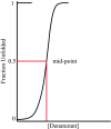
Fig. 1.
Fig. 1.
The folding transition. Many small proteins of experimental interest fold with high cooperativity so that a plot of some structure-disrupting factor, like temperature or a chemical denaturant, against the folded fraction of the population results in a sigmoidal curve. At the transition midpoint, 50% of the ensemble is folded and 50% is unfolded; the population of partially folded molecules is negligible. In this idealized plot of an actual experiment, the population is followed by a conformational probe (e.g., circular dichroism) as a function of denaturant concentration. Upon addition of sufficient denaturant, the probe signal reaches a plateau, indicating that the transition is complete. In experiments using multiple conformational probes (e.g., circular dichroism and fluorescence), all indicators trace the same sigmoidal curve after suitable normalization (6). Thus, one refers to the folding transition, not the circular dichroism folding transition, the fluorescence folding transition, etc.
Fig. 2.
Fig. 2.
The peptide unit. (A) Degrees of freedom. The peptide bond, C′-N, has partial double-bond character (23), so the six backbone atoms, -Cα-C′O-NH-Cα-, in the peptide unit (shaded rectangles) will be approximately coplanar. Consequently, there are two primary degrees of freedom in each peptide unit, the two torsion angles, φ and ψ (24). Assuming complete independence of these angles, there would be three staggered configurations per torsion, 3 × 3 = 9 conformers per peptide unit, and 9100 ≈ 1095 conformers for a 100-residue protein. (B) Residue φ,ψ distributions. Sterically allowed φ,ψ regions for the alanyl dipeptide, from model studies of Ramachandran and Sasisekharan (25), are shown in dark outline. Other regions are predicted to be unpopulated because their backbone torsion angles would cause a steric clash within the dipeptide unit. φ,ψ distributions of experimental data from the major populated regions from the coil library (88) are shown superimposed on the predicted sterically allowed regions.
Fig. 3.
Fig. 3.
A folding funnel. The funnel landscape depicts protein folding as a process that proceeds from a high entropy, disorganized state lacking in intramolecular interactions (mouth), to a low entropy, organized state with native intramolecular interactions (spout). Evolution has selected sequences that avoid frustrating traps en route from mouth to spout, smoothing what might otherwise be a rugged landscape. Under folding conditions, individual molecules can follow any route from mouth to spout, like a ball rolling down a free energy hill. One such trajectory is shown here. For a gallery of variant funnel landscapes, see ref. .
Fig. 4.
Fig. 4.
Hierarchic organization of proteins. In a hierarchy, each component is contained within the next larger component, like a series of nested boxes. Hierarchic architecture, illustrated here for ribonuclease, can be verified easily for any protein of known structure by using a simple procedure: display the structure with the first N/2 residues in magenta and the remaining N/2 residues in cyan. Then repeat this procedure, iteratively. It is apparent at a glance that at each successive level of the hierarchy, magenta and cyan regions do not intermingle. (Surface shown in gray is a place holder.) This top-down, hierarchic architecture is an experimental fact, and no hypothesis is needed to extract this result from known structures. Hierarchy suggests a bottom-up folding mechanism (40, 56, 57) in which chain segments form local structures of marginal stability, which then interact iteratively to produce intermediates of ever-increasing complexity. In this process, multiple folding routes coexist, and the stabilities of intermediates and their combinatorial associations determine the dominant pathways (49). Picture was rendered by using Pymol (127).
Fig. 5.
Fig. 5.
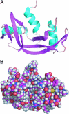
Ribonuclease. (A) The fold. The molecule is depicted as a color-coded ribbon diagram: α-helices are shown in cyan, β-strands are shown in magenta. (B) The all-atom structure. Atoms are depicted as space-filling spheres with radii proportional to their van der Waals radii. Color-code is carbon, magenta; nitrogen, blue; oxygen, red; hydrogen, white; sulfur, yellow. Picture was rendered by using Pymol (127).

References

    1. Haber E, Anfinsen CB. J Biol Chem. 1961;236:422–424. - PubMed
    1. Anfinsen CB. Science. 1973;181:223–230. - PubMed
    1. Wu H. Chin J Physiol. 1931;5:321–344.
    1. Mirsky AE, Pauling L. Proc Natl Acad Sci USA. 1936;22:439–447. - PMC - PubMed
    1. Myers JK, Pace CN, Scholtz JM. Protein Sci. 1995;4:2138–2148. - PMC - PubMed

Publication types