. 2006 Nov 7;103(45):16846-51.
doi: 10.1073/pnas.0607771103. Epub 2006 Oct 31.

Quantum dot/peptide-MHC biosensors reveal strong CD8-dependent cooperation between self and viral antigens that augment the T cell response

Affiliations

Quantum dot/peptide-MHC biosensors reveal strong CD8-dependent cooperation between self and viral antigens that augment the T cell response

Nadia Anikeeva et al. Proc Natl Acad Sci U S A. .

Abstract

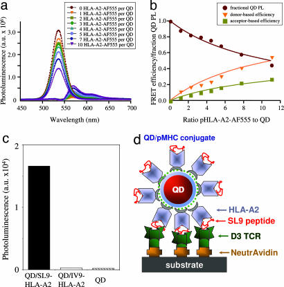
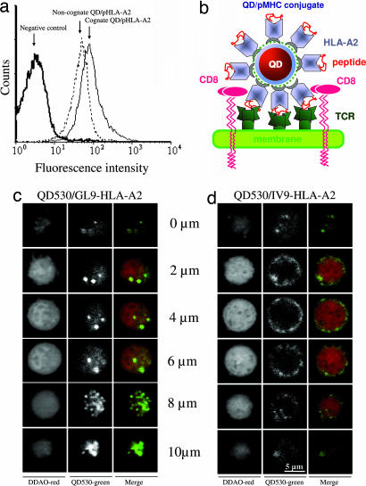
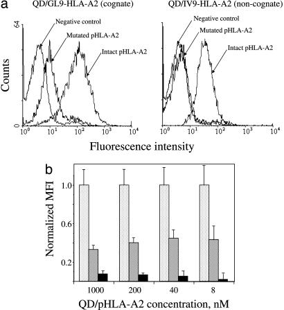
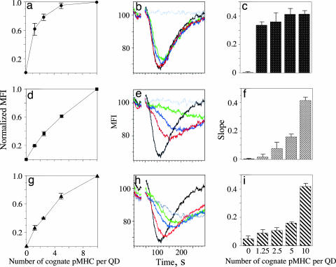
Cytotoxic T lymphocytes (CTL) can respond to a few viral peptide-MHC-I (pMHC-I) complexes among a myriad of virus-unrelated endogenous self pMHC-I complexes displayed on virus-infected cells. To elucidate the molecular recognition events on live CTL, we have utilized a self-assembled biosensor composed of semiconductor nanocrystals, quantum dots, carrying a controlled number of virus-derived (cognate) and other (noncognate) pMHC-I complexes and examined their recognition by antigen-specific T cell receptor (TCR) on anti-virus CD8(+) T cells. The unique architecture of nanoscale quantum dot/pMHC-I conjugates revealed that unexpectedly strong multivalent CD8-MHC-I interactions underlie the cooperative contribution of noncognate pMHC-I to the recognition of cognate pMHC-I by TCR to augment T cell responses. The cooperative, CD8-dependent spread of signal from a few productively engaged TCR to many other TCR can explain the remarkable ability of CTL to respond to virus-infected cells that present few cognate pMHC-I complexes.


Conflict of interest statement

The authors declare no conflict of interest.

Figures

Fig. 1.
Fig. 1.
Formation of functional QD/pHLA-A2 conjugates. (a) Deconvoluted photoluminescence (PL) spectra of QD and Alexa Fluor 555 (AF555)-labeled pHLA-A2 for different dye-to-protein ratios are shown. a.u., atomic units. (b) Changes of the fluorescence intensity versus the dye-to-QD ratio show that up to 12 pHLA-A2 complexes can be attached to each QD. FRET, Förster resonance energy transfer. (c) Immobilized D3 TCR specifically recognize cognate (QD/SL9-HLA-A2) but not noncognate (QD/IV9-HLA-A2) conjugates at 150 nM. Data shown are representative of four independent experiments. (d) Schematic drawing of QD/SL9-HLA-A2 conjugate bound to TCR immobilized on a plastic surface.
Fig. 2.
Fig. 2.
Binding of QD/pHLA-A2 conjugates to the surface of live CTL. (a) Although cognate QD/GL9-HLA-A2 conjugate binds specifically to CER43 CTL, noncognate QD/IV9-HLA-A2 conjugate shows high TCR-independent binding to the CTL. QD concentration was 1 μM; the binding was performed for 30 min at 4°C. (b) Schematic drawing of QD/HLA-A2 and peptide complexed with a TCR bound to the membrane of a CD8+ cell. (c and d) Binding of cognate (QD/GL9-HLA-A2) (c) but not noncognate (QD/IV9-HLA-A2) (d) conjugates to CER43 CTL leads to internalization of the QD conjugates. Images of the distribution of QD conjugates (green) in various z sections of CTL are shown.
Fig. 3.
Fig. 3.
Binding of noncognate QD/pHLA-A2 conjugates to live CTL is CD8-dependent. (a) A single mutation in HLA-A2 that disrupts HLA-A2–CD8 interactions abolishes binding of noncognate QD conjugates (Right) and significantly reduced binding of cognate QD conjugates (Left) to live CTL. (b) The dependence of normalized mean fluorescence intensity (MFI) of CER43 CTL incubated with cognate QD/GL9-HLA-A2 (stippled bars), noncognate QD/IV9-HLA-A2 (hatched bars), and cognate QD/GL9-HLA-A2 with A245V mutation (solid bars) upon concentration of the QD/pHLA-A2 conjugates is shown. MFIs measured with either cognate QD/pHLA-A2 or noncognate QD/pHLA-A2 or cognate QD/mutated pHLA-A2 were divided by MFI measured with cognate QD/pHLA-A2 for every given concentration to determine normalized MFI values. Data represent mean ± SD.
Fig. 4.
Fig. 4.
CD8-dependent cooperation of noncognate and cognate pHLA-A2 recognized by CTL. QDs were loaded with a mixture of intact cognate and noncognate (ac), intact cognate and mutated (A245V) noncognate (df), and intact and mutated (A245V) cognate (gi) pHLA-A2 complexes. The total number of pHLA-A2 molecules (10 per QD) was fixed, whereas the number of intact cognate pHLA-A2 molecules per QD was 0 (gray), 1.25 (green), 2.5 (blue), 5 (red), and 10 (black). Effectiveness of the binding of QD/pHLA-A2 to live CTL (a, d, and g) was characterized by normalized MFI. Slope (c, f, and i) of the Ca2+ flux was determined from the initial decay of the kinetic curve in each experiment (b, e, and h). Results of representative experiments are shown.

References

    1. Rudolph MG, Wilson IA. Curr Opin Immunol. 2002;14:52–65. - PubMed
    1. Gao GF, Rao Z, Bell JI. Trends Immunol. 2002;23:408–413. - PubMed
    1. Hugues S, Malherbe L, Filippi C, Glaichenhaus N. J Immunol Methods. 2002;268:83–92. - PubMed
    1. Gestwicki JE, Cairo CW, Strong LE, Oetjen KA, Kiessling LL. J Am Chem Soc. 2002;124:14922–14933. - PubMed
    1. Mattoussi H, Mauro JM, Goldman ER, Anderson GP, Sundar VC, Mikulec FV, Bawendi MG. J Am Chem Soc. 2000;122:12142–12150.

Publication types