. 2006 Nov 7;103(45):16806-11.
doi: 10.1073/pnas.0603916103. Epub 2006 Oct 31.

Wnt and TGF-beta signaling are required for the induction of an in vitro model of primitive streak formation using embryonic stem cells

Affiliations

Wnt and TGF-beta signaling are required for the induction of an in vitro model of primitive streak formation using embryonic stem cells

Paul Gadue et al. Proc Natl Acad Sci U S A. .

Abstract

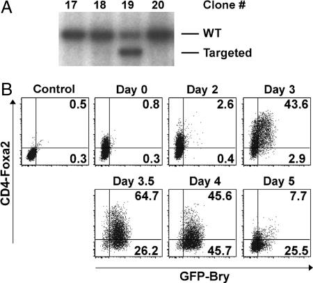
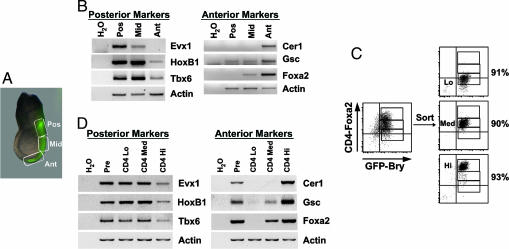
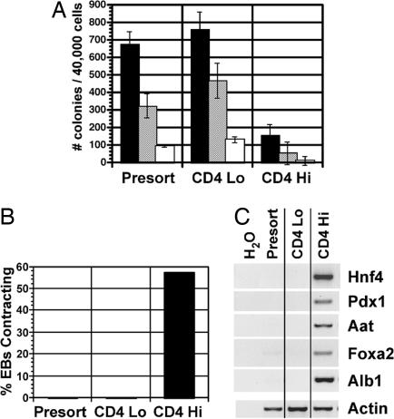
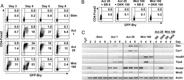
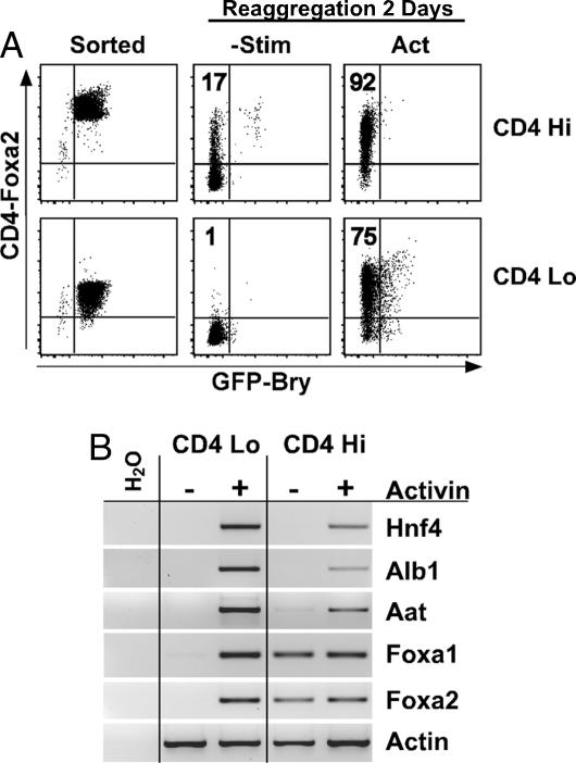
The establishment of the primitive streak and its derivative germ layers, mesoderm and endoderm, are prerequisite steps in the formation of many tissues. To model these developmental stages in vitro, an ES cell line was established that expresses CD4 from the foxa2 locus in addition to GFP from the brachyury locus. A GFP-Bry(+) population expressing variable levels of CD4-Foxa2 developed upon differentiation of this ES cell line. Analysis of gene-expression patterns and developmental potential revealed that the CD4-Foxa2(hi)GFP-Bry(+) population displays characteristics of the anterior primitive streak, whereas the CD4-Foxa2(lo)GFP-Bry(+) cells resemble the posterior streak. Using this model, we were able to demonstrate that Wnt and TGF-beta/nodal/activin signaling simultaneously were required for the generation of the CD4-Foxa2(+)GFP-Bry(+) population. Wnt or low levels of activin-induced a posterior primitive streak population, whereas high levels of activin resulted in an anterior streak fate. Finally, sustained activin signaling was found to stimulate endoderm commitment from the CD4-Foxa2(+)GFP-Bry(+) ES cell population. These findings demonstrate that the early developmental events involved in germ-layer induction in the embryo are recapitulated in the ES cell model and uncover insights into the signaling pathways involved in the establishment of mesoderm and endoderm.


Conflict of interest statement

The authors declare no conflict of interest.

Figures

Fig. 1.
Fig. 1.
Targeting human CD4 to the Foxa2 locus. (A) Southern blot analysis depicting a targeted clone (#19). (B) Flow cytometric analysis of CD4-Foxa2 and GFP-Bry expression in EBs differentiated for varying periods of time. Day 0 is undifferentiated ES cells. Control represents unstained ES cells.
Fig. 2.
Fig. 2.
Gene-expression analysis of regions of the PS and CD4-Foxa2+GFP-Bry+ EB subpopulations. (A) An embryonic day 7.25 mouse GFP-Bry embryo expressing GFP in the PS. Boxes indicate the posterior (Pos), middle (Mid), and anterior (Ant) PS regions. (B) RT-PCR gene-expression analysis of the different regions of the PS. (C) The presort (Pre), CD4loGFP+ (CD4 Lo), CD4medGFP+ (CD4 Med), and CD4hiGFP+ (CD4 Hi) populations were isolated by cell sorting from day 3.25 serum-stimulated EBs. (D) RT-PCR gene-expression analysis of the CD4 Lo, CD4 Med, and CD4 Hi populations.
Fig. 3.
Fig. 3.
The functional potential of the CD4loGFP+ and CD4hiGFP+ populations. CD4loGFP+ and CD4hiGFP+ cells were isolated from serum-induced EBs as indicated in Fig. 2 and analyzed as described below. (A) Isolated populations were reaggregated for 24 h in serum-free media and then assayed for hematopoietic potential by colony assay. The number of primitive erythroid (filled bars), macrophage (hatched bars), and macrophage/erythroid (open bars) colonies were counted after 5–7 days of culture. Data are presented as mean number of colonies from three cultures. Bars represent SEM. (B) Isolated populations were reaggregated in serum-free media for 24 h, and the aggregates were allowed to adhere onto matrigel-coated dishes in serum-free media. The proportion of aggregates with contracting cells was scored 1–2 days later. (C) Aggregates were maintained for 7 days on matrigel-coated dishes in serum-free media, and gene expression was analyzed by RT-PCR.
Fig. 4.
Fig. 4.
Activin and Wnt can induce the development of PS cells in the absence of serum. ES cells were differentiated in serum-free media alone for 2 days. The EBs were dispersed and reaggregated in serum-free media with activin A (Act; in ng/ml), Wnt3a (Wnt; in ng/ml), SB-431542 (SB; in μM), and DKK1 (DKK; in ng/ml) as indicated. The percentages of CD4hiGFP+ and CD4lo/medGFP+ are indicated on the plots. (A) Kinetics of CD4 and GFP expression in cultures induced with activin or Wnt. (B) Expression of CD4 and GFP in day 4 EB cultures after the indicated treatments. (C) Cells were harvested at the indicated times, and gene-expression analysis was performed by RT-PCR.
Fig. 5.
Fig. 5.
Activin induces endoderm formation from CD4+GFP+ cells. ES cells were differentiated in serum-free cultures in the presence of activin A (2 ng/ml) and Wnt3a (10 ng/ml). At day 4, the CD4loGFP+ and CD4hiGFP+ cells were isolated and reaggregated in serum-free media alone or with activin (10 ng/ml) as indicated. (A) Expression of GFP and CD4 are shown for sorted cells and 2 days after aggregation. (B) After 2 days of aggregation, aggregates were plated onto dishes. After 5–7 days of culture, gene expression was analyzed by RT-PCR.

References

    1. Tam PP, Behringer RR. Mech Dev. 1997;68:3–25. - PubMed
    1. Lawson KA, Meneses JJ, Pedersen RA. Development (Cambridge, UK) 1991;113:891–911. - PubMed
    1. Lawson KA, Pedersen RA. In: Ciba Found Symp. 1992. pp. 3–21. discussion 21–26. - PubMed
    1. Tam PP, Beddington RS. In: Ciba Found Symp. 1992. pp. 27–41. discussion 42–49. - PubMed
    1. Kinder SJ, Tsang TE, Wakamiya M, Sasaki H, Behringer RR, Nagy A, Tam PP. Development (Cambridge, UK) 2001;128:3623–3634. - PubMed

MeSH terms