. 2006 Nov 7;103(45):16995-7000.
doi: 10.1073/pnas.0603386103. Epub 2006 Oct 31.

MyoD expression restores defective myogenic differentiation of human mesoangioblasts from inclusion-body myositis muscle

Affiliations

MyoD expression restores defective myogenic differentiation of human mesoangioblasts from inclusion-body myositis muscle

Roberta Morosetti et al. Proc Natl Acad Sci U S A. .

Abstract

Inflammatory myopathies (IM) are acquired diseases of skeletal muscle comprising dermatomyositis (DM), polymyositis (PM), and inclusion-body myositis (IBM). Immunosuppressive therapies, usually beneficial for DM and PM, are poorly effective in IBM. We report the isolation and characterization of mesoangioblasts, vessel-associated stem cells, from diagnostic muscle biopsies of IM. The number of cells isolated, proliferation rate and lifespan, markers expression, and ability to differentiate into smooth muscle do not differ among normal and IM mesoangioblasts. At variance with normal, DM and PM mesoangioblasts, cells isolated from IBM, fail to differentiate into skeletal myotubes. These data correlate with lack in connective tissue of IBM muscle of alkaline phosphatase (ALP)-positive cells, conversely dramatically increased in PM and DM. A myogenic inhibitory basic helix-loop-helix factor B3 is highly expressed in IBM mesoangioblasts. Indeed, silencing this gene or overexpressing MyoD rescues the myogenic defect of IBM mesoangioblasts, opening novel cell-based therapeutic strategies for this crippling disorder.


Conflict of interest statement

The authors declare no conflict of interest.

Figures

Fig. 1.
Fig. 1.
Cell morphology, growth curve, and cell cycle. (A) From the organ culture on, refractive triangular, adherent, and round loosely adherent/floating cells were observed. (Scale bar: 40 μm.) (B) Cell growth was assessed after 24 h, 48 h, and 72 h. Viable cells were judged by trypan blue exclusion. Results are expressed as absolute counts. Bars represent mean ± SD of triplicate samples of one representative experiment of three. (C) Cell cycle distribution of proliferating normal human SDMC and mesoangioblasts from three controls, three DM, three PM, and six IBM (run in duplicate) after 24 h of culture was assessed by propidium iodide and FACS. For each sample, percentage of cells in G0/G1, S, or G2/M phases of cell cycle is indicated. One representative experiment of three is shown.
Fig. 2.
Fig. 2.
FACS, immunophenotyping, ALP histochemistry, and gene expression profiling of IM mesoangioblasts. (A) More than 90% of cells from all samples were strongly positive for CD44 and CD13 with high percentage of cells CD49b-positive. None of the markers positive in murine mesoangioblasts were significantly expressed. Bars represent the mean ± SD of 36 samples from the 12 patients with IM (3 DM, 3 PM, and 6 IBM) (each performed in triplicate). (B) IM mesoangioblasts in vitro are all ALP-positive. After simultaneous staining in the same culture conditions, more intensely labeled cells can be observed in PM and to a lesser extent in DM, whereas IBM mesoangioblasts are only weakly positive. (Scale bar: 20 μm.) (C) Clustering results show two main classes: mesoangioblasts from normal controls (lanes 5–8), mesoangioblasts from DM (lanes 1 and 2), and IBM (lanes 3 and 4). Clustering procedure pairs together DM and IBM replicates.
Fig. 3.
Fig. 3.
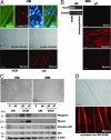
Skeletal muscle differentiation. (A) (Top) Immunofluorescence for myosin and human lamin A/C. Mesoangioblasts from DM efficiently fused with C2C12 murine myoblasts into mature myosin-positive myotubes, as indicated by the presence of nuclei expressing human lamin A/C. (Scale bar: 10 μm.) (Middle and Bottom) DM mesoangioblasts exposed 4 days to SDMC-conditioned growth medium (CM) and subsequently cultured for 7 days in differentiation medium (Dif.M) spontaneously fuse into differentiated myotubes. (Scale bar: 40 μm.) Each experiment was performed in duplicate at least three times. (B) IBM mesoangioblasts do not constitutively express MyoD (RT-PCR) and when exposed 4 days to CM and 7 days to Dif.M are negative for MyoD and myosin, but display a strong desmin immunoreactivity. (Scale bar: 20 μm.) (C) Mesoangioblasts from DM, but not from IBM, differentiate into multinucleated myotubes (phase contrast). (Scale bar: 40 μm.) For Western blot analysis, cells were harvested at day 0, after 4 days in CM and after 3 and 7 days in Dif.M. A marked up-regulation of myogenin and myosin is observed already at day 3 of differentiation for DM mesoangioblasts; no up-regulation of myosin and myogenin is visible in IBM cells. For both DM and IBM mesoangioblasts, activation of p38 (phospho-p38) is observed. One representative experiment of six is shown. (D) SDMC from primary muscle cultures from all six IBM biopsies normally differentiate in multinucleated myotubes (phase contrast, Upper) and show strong myosin-immunoreactivity (Lower). (Scale bar: 20 μm.) Two representative cultures are shown. NCM, nonconditioned medium.
Fig. 4.
Fig. 4.
ALP histochemistry in IM muscle biopsies. (A) In normal muscle only small arteries show ALP-positive staining. Intense ALP staining is present in PM and DM biopsies in perymisial and endomysial connective tissue, particularly strong in round cells associated to vessels' walls. IM biopsies contain also a variable number of small-sized regenerating muscle fibers with slight punctuate ALP positivity. IBM muscle shows no ALP connective tissue staining. (Scale bar: 40 μm.) (B) Immunohistochemistry for MyoD or Pax7 with peroxidase-antiperoxidase (PAP) on the same unfixed frozen sections of biopsies from PM, DM, IBM, and normal muscle from which mesoangioblasts were isolated. Representative fields of a PM biopsy showing a round vessel-associated cell (open arrowhead) strongly ALP-positive containing a MyoD-positive nucleus. Several PAX7-positive cells (filled arrowheads), identified as activated satellite cells, are associated with muscle fibers showing regenerative aspects with slight ALP positivity, whereas the roundish ALP-positive cells in the interstitium are PAX7-negative (arrow). CTRL, control. (Scale bar: 10 μm.)
Fig. 5.
Fig. 5.
MyoD transduction and induction of differentiation in IBM mesoangioblasts. (A) As shown by immunofluorescence and Western blot, AdenoMyoD-transduced IBM mesoangioblasts after 7 days in differentiation medium, fused into multinucleated myosin-positive myotubes. Approximately 90% of cells were MyoD-positive. (Scale bar: 10 μm.) Efficiency of transduction was evaluated 48 h after the infection by Western blot showing a marked up-regulation of MyoD. β-actin was used as loading control. A representative experiment of three, each performed in duplicate, is shown. (B) Double immunohistochemistry for human dystrophin and lamin A/C. In vivo myogenicity of IM mesoangioblasts was tested by intramuscular transplantion in scid/mdx mice. After two consecutive transplants, the TA of mice injected with IBM mesoangioblasts showed the presence of occasional human nuclei (blue with AMCA-labeled human lamin A/C, arrowheads) within muscle fibers and in the interstitium, but dystrophin expression was undetectable (Left). On the contrary, after IBM adenoMyoD-infected mesoangioblasts transplantation (Middle), large areas of injected muscle were reconstituted with fibers containing human lamin A/C-positive myonuclei (blue, arrowheads) and expressing dystrophin along the sarcolemma (red with Texas red-labeled specific antibody). When DM mesoangioblasts were used (Right), the majority of muscle fibers in the injected areas expressed dystrophin and contained human nuclei (positive for lamin A/C, arrowheads). (Scale bars: 10 μm.) SYTOX green staining on the same sections was used to identify both human and murine nuclei.
Fig. 6.
Fig. 6.
Induction of myogenic differentiation of IBM mesoangioblasts by BHLHB3 siRNA. (A) BHLHB3 siRNA (Hs 1) was transfected into IBM mesoangioblasts. Twenty-four hours after transfection, cells were harvested for RNA extraction and RT-PCR analysis. (B) MAPK siRNA was used as positive control; 24 h after transfection, cells were harvested for Western blot. (C) BHLHB3 siRNA-transfected cells were shifted to differentiation medium after 24 h. After 7-day culture, cells were stained with an anti-myosin antibody or harvested for Western analysis. (Scale bar: 40 μm.) A representative culture and immunoblot of one of three independent experiments (each one run in duplicate) are shown.

References

    1. Dalakas MC, Hohlfeld R. Lancet. 2003;362:971–982. - PubMed
    1. Mastaglia FL, Garlepp MJ, Phillips BA, Zilko PJ. Muscle Nerve. 2003;27:407–425. - PubMed
    1. Askanas V, Engel WK. Am J Pathol. 1998;153:1673–1677. - PMC - PubMed
    1. Askanas V, Engel WK. J Child Neurol. 2003;18:185–190. - PubMed
    1. Broccolini A, Ricci E, Pescatori M, Papacci M, Gliubizzi C, D'Amico A, Servidei S, Tonali P, Mirabella M. J Neuropathol Exp Neurol. 2004;63:650–659. - PubMed

Publication types

MeSH terms

Substances

LinkOut - more resources