. 2006 Dec;12(12):2160-70.
doi: 10.1261/rna.201406. Epub 2006 Oct 31.

The canonical UPF1-dependent nonsense-mediated mRNA decay is inhibited in transcripts carrying a short open reading frame independent of sequence context

Affiliations

The canonical UPF1-dependent nonsense-mediated mRNA decay is inhibited in transcripts carrying a short open reading frame independent of sequence context

Ana Luísa Silva et al. RNA. 2006 Dec.

Abstract

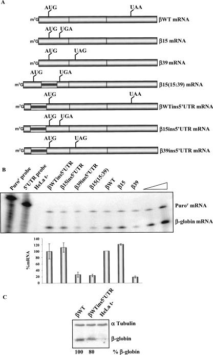
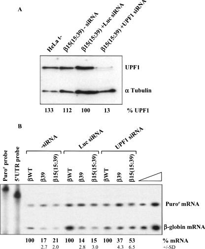
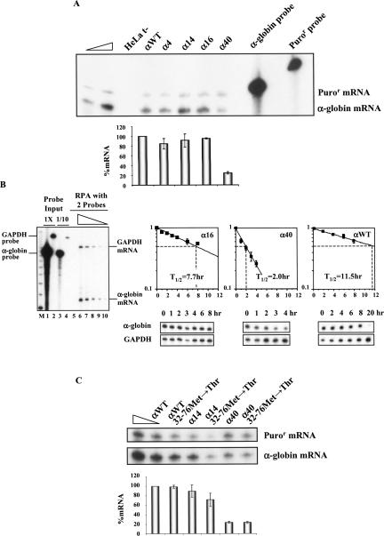
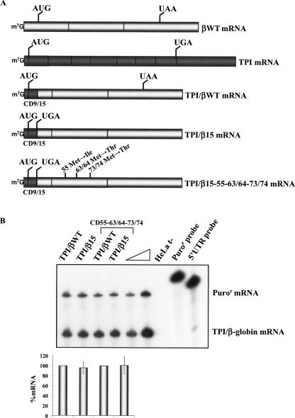
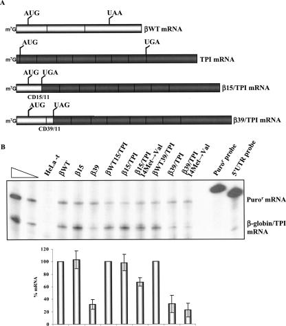
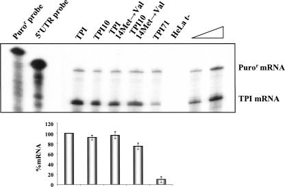
Nonsense-mediated mRNA decay (NMD) is a surveillance mechanism that degrades mRNAs carrying premature translation termination codons. Generally, NMD is elicited if translation terminates >50-54 nucleotides (nt) upstream of an exon-exon junction. We have previously reported that human beta-globin mRNAs carrying 5'-proximal nonsense mutations (e.g., beta15) accumulate to normal levels, suggesting an exception to the "50-54-nt boundary rule." In the present report, we demonstrate that the strength of the UPF1-dependent NMD of mutant beta-globin mRNAs is specifically determined by the proximity of the nonsense codon to the initiation AUG. This conclusion is supported by a parallel effect of the short ORF size on NMD of nonsense-containing alpha-globin mRNAs. To determine whether the short-ORF effect on NMD response is conserved in heterologous transcripts, we assessed its effects on a set of beta-globin/triosephosphate isomerase (TPI) hybrid mRNAs and on the TPI mRNA. Our data support the conclusion that nonsense mutations resulting in a short ORF are able to circumvent the full activity of the canonical UPF1-dependent NMD pathway.


Figures

FIGURE 1.
FIGURE 1.
The proximity of the nonsense codon to the AUG, rather than the distance to a putative 5′-UTR determinant, inhibits mRNA decay. (A) Schematic representation of the studied human β-globin mRNAs. The position of initiation and termination (native or premature) codons is represented. The inverted-shadowed box represents the spacer insertion. The name of each transcript is indicated on the right. (B) Representative RPA of RNA isolated from HeLa cells untransfected (t−) or transfected with constructs specified above each lane. Increasing amounts of RNA (indicated by a triangle) from HeLa cells transfected with the βWT gene were also analyzed to demonstrate that the experimental RPA was carried out in probe excess; the amount of each of the input riboprobes used per analysis is also shown. The positions of the human β-globin or puromycin-resistance (Puror) mRNAs are indicated on the right of the autoradiograph. Levels of human β-globin mRNA were normalized relatively to Puror and then compared to wild type (WT). Average values from five independent experiments and standard deviations are plotted below the autoradiograph. (C) Western blot analysis of the HeLa cells extracts untransfected (t−) or transfected with the constructs specified above each lane. Immunoblotting was performed using a human β-globin-specific antibody and a α-tubulin-specific antibody to control for variations in protein loading.
FIGURE 2.
FIGURE 2.
Down-regulating human UPF1 protein results in an up-regulation of the β15(15:39) transcripts. HeLa cells were transiently cotransfected with synthetic small-interfering RNA (siRNA) duplexes directed to human UPF1 or to a non-endogenous target (Luciferase; Luc) used as control, and plasmids with the β-globin gene variants [normal (βWT), β39, and β15(15:39); plasmid also contains the puromycin resistance (Puror) gene]; (t−) untransfected cells. About 24 h after siRNA treatment, cells were transfected with the β-globin constructs specified above each lane. Two days after transfection, protein and RNA were isolated. (A) Western blot analysis of the HeLa cell extracts transfected with human UPF1 siRNA. Immunoblotting was performed using a human UPF1-specific antibody and an α-tubulin-specific antibody to control for variations in protein loading. (B) Representative RPA to quantify the level of human β-globin transcript, normalized to the level of puromycin-resistance (Puror) mRNA. Increasing amounts of RNA (indicated by a triangle) from HeLa cells transfected with βWT gene were also analyzed to demonstrate that the experimental RPA was carried out in probe excess; the amount of each of the input riboprobes used per analysis is also shown. Levels of β39 and β15(15:39) transcripts were compared to βWT mRNA levels (defined as 100%) in the absence (−siRNA) or in the presence of each siRNA. The mRNA percentage average values and standard deviations from three independent experiments are indicated at the bottom.
FIGURE 3.
FIGURE 3.
The AUG-proximity effect on NMD is observed in human α-globin transcripts. (A) HeLa cells were transfected with the α-globin constructs specified above each lane [the plasmid also contains the puromycin-resistance (Puror) gene]. Total RNA from either transfected or untransfected (t−) HeLa cells was isolated and analyzed by RPA. Levels of human α-globin mRNA were normalized relatively to the expression level of the Puror mRNA and compared with expression of the normal (αWT) gene. Increasing amounts of RNA (indicated by a triangle) from HeLa cells transfected with an αWT gene were also analyzed to demonstrate that the experimental RPA was carried out in probe excess; the amount of each of the input riboprobes used per analysis is also shown. The percentage mRNA values were plotted for each construct, and standard deviations from four independent experiments are shown. (B) MEL cells stably expressing the tet transactivator (MEL/tTA cells) were transiently transfected with αWT, α16, or α40 genes under the transcriptional control of a tTA-regulated promoter. Cells were pulsed with the respective α-globin mRNAs for 4 h by transfer to Tet(−) medium and then transferred back to Tet(+) medium for analysis of decay rates. Total mRNA was isolated at various time intervals during the transcriptional chase period and analyzed by RPA. On the left, a representative autoradiograph of a titration RPA is presented to show that experimental RPAs were carried out in probe excess and in the linear range of detection. The triangle above lanes 610 represents decreasing amounts (2×, 1×, 1/2, 1/4, and 1/8) of αWT RNA. Lanes 1 and 2 show the amount of each input α-globin or murine GAPDH probe used per RNA sample. Lanes 3 and 4 show 1/10 of each input probe. (M) Molecular weight marker. On the right, representative autoradiographs are presented to show the decay rate of each α-globin variant. RNase protection bands corresponding to human α-globin mRNA and to the constitutively expressed mouse GAPDH mRNA (loading control) are indicated. The intensities of the α-globin mRNA bands were quantified and normalized relatively to the GAPDH mRNA band. The data were recorded in the graphs presented. To calculate the α-globin mRNA half-lives, each data time point was expressed as a ratio of α-globin:GAPDH mRNA and normalized to the average value of all time points from a single transfection. The ratios were then renormalized to the average initial time point from all transfections (time 0 = 1). Each point represents the mean ± standard deviation from four independent experiments. Linear regression analysis was performed by standard techniques. The half-lives (T 1/2) of the mRNAs are indicated. (C) The two potential reinitiating AUGs at codons 32 and 76 were converted to ACG triplets in each of the index mRNAs. A representative RPA of RNA isolated from HeLa cells transiently transfected with αWT, α14, α40, αWT–32–76Met→Thr, α14–32–76Met→Thr, or α40–32–76Met→Thr genes is shown. Identification of the α-globin and Puror protected fragments is indicated to the right of the autoradiograph. Levels of α-globin mRNA were quantified relatively to the Puror mRNA, and these values are plotted beneath each respective lane (average and standard deviations) normalized to the expression level of the αWT gene. Increasing amounts of RNA (indicated by a triangle) from HeLa cells transfected with an αWT gene were also analyzed to demonstrate that the experimental RPA was carried out in probe excess. For each case, three independent experiments were performed.
FIGURE 4.
FIGURE 4.
Human β15 globin mRNA carrying the first nine-codon TPI ORF is resistant to NMD. (A) Physical maps of the hybrid TPI/β-globin constructs. The bright rectangles represent β-globin exons, and dark rectangles represent TPI exons. CD9/15 represents the position of the junction between the TPI and β-globin genes. The position of initiation and termination (premature or native) codons is indicated. The name of each transcript is indicated to the right. (B) HeLa cells were transfected with the constructs specified above each lane [the plasmid also contains the puromycin-resistance (Puror) gene]. Total RNA was isolated and analyzed by RPA. The levels of each nonsense-mutated TPI/β-globin mRNA were quantified relatively to the Puror mRNA and normalized to the expression level of the corresponding nonsense-free gene. Increasing amounts of RNA (indicated by a triangle) from HeLa cells transfected with a TPI/βWT gene were also analyzed to demonstrate that the experimental RPA was carried out in probe excess; the amount of each of the input riboprobes used per analysis is also shown. The percentage mRNA values were plotted for each construct, and standard deviations from three independent experiments are shown.
FIGURE 5.
FIGURE 5.
Human TPI mRNA carrying the first 15 codons of the β15 globin ORF escapes NMD. (A) Physical maps of the hybrid β-globin/TPI mRNAs. The bright rectangles depict β-globin exons, and dark rectangles depict TPI exons. CD15/11 and CD39/11 indicate the junction position between the β-globin and TPI genes. The vertical lines represent the initiation and termination (native or premature) codons. The identification of each mRNA is indicated to the right. (B) HeLa cells were transfected with the constructs specified above each lane [the plasmid also contains the puromycin-resistance (Puror) gene]. Total RNA was isolated and analyzed by RPA. Levels of each nonsense-mutated β-globin/TPI mRNA were quantified relative to the Puror mRNA and normalized to the expression level of the corresponding nonsense-free gene. Increasing amounts of RNA (indicated by a triangle) from HeLa cells transfected with a βWT gene were also analyzed to demonstrate that the experimental RPA was carried out in probe excess; the amount of each of the input riboprobes used per analysis is also shown. The percentage mRNA values were plotted for each construct, and standard deviations from three independent experiments are shown.
FIGURE 6.
FIGURE 6.
TPI transcripts bearing a short ORF escape NMD. HeLa cells were transfected with the constructs specified above each lane [the plasmid also contains the puromycin-resistance (Puror) gene]. Total RNA was isolated and analyzed by RPA. Levels of each TPI mRNA were quantified relative to the Puror mRNA and normalized to the expression level of the wild-type TPI mRNA. Increasing amounts of RNA (indicated by a triangle) from HeLa cells transfected with a normal TPI gene were also analyzed to demonstrate that the experimental RPA was carried out in probe excess; the amount of each of the input riboprobes used per analysis is also shown. The percentage mRNA values relative to normal were plotted for each construct, and standard deviations from three independent experiments are shown.

Similar articles

Cited by

References

    1. Fribourg, S., Gatfield, D., Izaurralde, E., Conti, E. A novel mode of RBD-protein recognition in the Y14-Mago complex. Nat. Struct. Biol. 2003;10:433–439. - PubMed
    1. Gaba, A., Jacobson, A., Sachs, M.S. Ribosome occupancy of the yeast CPA1 upstream open reading frame termination codon modulates nonsense-mediated mRNA decay. Mol. Cell. 2005;20:449–460. - PubMed
    1. Gehring, N.H., Neu-Yilik, G., Schell, T., Hentze, M.W., Kulozik, A.E. Y14 and hUpf3b form an NMD-activating complex. Mol. Cell. 2003;11:939–949. - PubMed
    1. Ghilardi, N., Skoda, R.C. A single-base deletion in the thrombopoietin (TPO) gene causes familial essential thrombocythemia through a mechanism of more efficient translation of TPO mRNA. Blood. 1999;94:1480–1482. - PubMed
    1. Inácio, A., Silva, A.L., Pinto, J., Ji, X., Morgado, A., Almeida, F., Faustino, P., Lavinha, J., Liebhaber, S.A., Romão, L. Nonsense mutations in close proximity to the initiation codon fail to trigger full nonsense-mediated mRNA decay. J. Biol. Chem. 2004;279:32170–32180. - PubMed

Publication types