. 2006 Dec;16(12):1548-56.
doi: 10.1101/gr.5565706. Epub 2006 Oct 31.

Identification of an infectious progenitor for the multiple-copy HERV-K human endogenous retroelements

Affiliations

Identification of an infectious progenitor for the multiple-copy HERV-K human endogenous retroelements

Marie Dewannieux et al. Genome Res. 2006 Dec.

Abstract

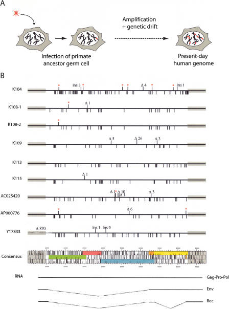
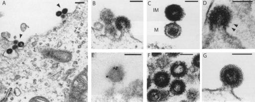
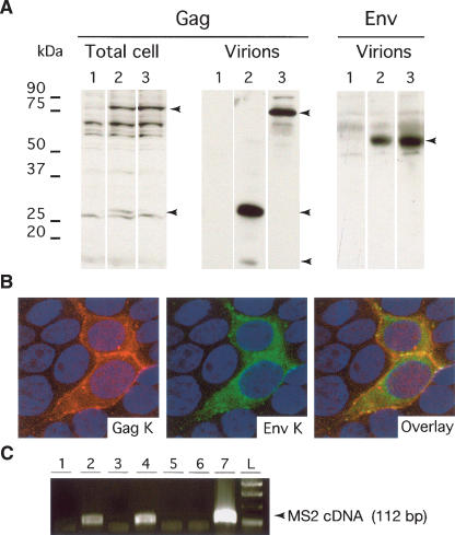
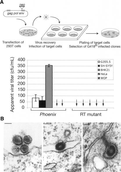
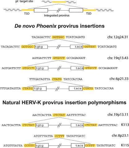
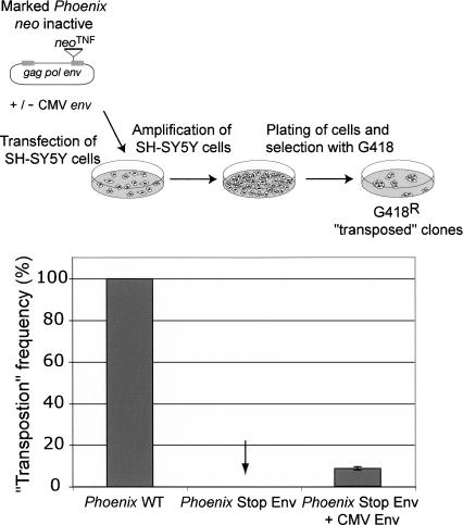
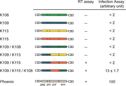
Human Endogenous Retroviruses are expected to be the remnants of ancestral infections of primates by active retroviruses that have thereafter been transmitted in a Mendelian fashion. Here, we derived in silico the sequence of the putative ancestral "progenitor" element of one of the most recently amplified family - the HERV-K family - and constructed it. This element, Phoenix, produces viral particles that disclose all of the structural and functional properties of a bona-fide retrovirus, can infect mammalian, including human, cells, and integrate with the exact signature of the presently found endogenous HERV-K progeny. We also show that this element amplifies via an extracellular pathway involving reinfection, at variance with the non-LTR-retrotransposons (LINEs, SINEs) or LTR-retrotransposons, thus recapitulating ex vivo the molecular events responsible for its dissemination in the host genomes. We also show that in vitro recombinations among present-day human HERV-K (also known as ERVK) loci can similarly generate functional HERV-K elements, indicating that human cells still have the potential to produce infectious retroviruses.


Figures

Figure 1.
Figure 1.
HERV-K(HML2) “endogenization” and present-day human proviruses. (A) Evolutionary scheme for HERV-K(HML2) entry into and invasion of the genome of primates. (B) Map of the full-length 9.4-kb long human-specific HERV-K(HML2) proviruses and comparison with the in silico-engineered consensus sequence. Each provirus is represented by a solid dark line, with the amino acid substitutions in Gag, Pro, Pol, and Env as compared with the consensus element indicated below the line, and the insertions/deletions (ins/Δ) and premature Stop codons (red stars) indicated above the line. The ORF map of the consensus provirus is shown, with gag in green, pro in pink, pol in blue, env in orange and yellow, the bipartite rec in orange, and the two LTRs as gray boxes. (Note that the first coding exon of rec belongs to the env ORF). The transcripts responsible for the expression of the viral proteins, with the corresponding spliced out domains (dotted lines), are schematized below the ORF map.
Figure 2.
Figure 2.
Electron microscopy of the viral-like particles generated by the Phoenix provirus. Human 293T cells were transfected with an expression vector for Phoenix (A–E), or mutants (F,G), and observed 48 h post-transfection. (A) Low magnification of particles assembled at the cell membrane. (B) Representative image of particles budding from the plasma membrane. (C) High magnification of two particles, one of which (bottom) discloses a mature (M) morphology with a condensed core, while the other appears to be still immature (IM) with two dark peripheral rings surrounding an electron-lucent core. (D) High magnification of a particle with prominent spikes, corresponding to the Env protein. (E) Image of a particle after labeling with an antibody specific for the HERV-K envelope protein and a secondary antibody linked to gold beads, obtained by immuno-electron microscopy. Quantification of the labeling on 11 independent fields demonstrates association of the gold beads with the viral particles: 307 ± 121 gold beads/μm2 for the viral particles, versus 4.9 ± 3.2 and 1.1 ± 1.5 gold beads/μm2 for the cytoplasm and particle-free extracellular space, respectively (P < 0.001 between viral particles and any of the two other compartments, Student’s t-test). (F) Image of representative particles obtained after transfection with an expression vector for the Phoenix pro mutant. All of them disclosed an immature morphology (41 of 41 identified “free” particles, i.e., no more in the budding process, for the pro mutant, vs. 15 of 37 for Phoenix WT). (G) High magnification of a particle obtained after transfection with an expression vector for the Phoenix env mutant. The membrane surrounding the particle is clearly detectable, without any spike. Scale bars: (A): 200 nm, (B–G):100 nm.
Figure 3.
Figure 3.
Characterization of Phoenix. (A) Immunoblot analysis of the Phoenix products present in the lysates of transiently transfected 293T cells, or in the concentrated supernatant. Cells were transfected with (1) a control plasmid (pCMV-β), (2) an expression vector for Phoenix, or (3) an expression vector for a pro mutant. The membranes were hybridized with an antibody directed against the Gag protein (left) or the Env protein (right). (B) Immunofluorescence study of 293T cells transfected with Phoenix. Gag is marked in red, Env in green, and the nuclei are stained in blue. Note the colocalization of the two proteins, especially at the membrane that separates the two adjacent marked cells. (C) Detection of RT activity in the concentrated supernatant of 293T cells transfected with Phoenix and a series of proviral and control vectors. Concentrated virions were used as a source of RT activity to reverse transcribe synthetic MS2 RNA. Presence of cDNA was revealed via a PCR amplification using appropriate primers. (1) pCMV-β; (2) Phoenix; (3) Phoenix mutated in RT; (4) chimera K109-K115; (5) chimera K109-K113; (6) chimera K109-K108; (7) MLV core proteins).
Figure 4.
Figure 4.
Infectious properties of Phoenix encoded particles. (A) Infectivity of the HERV-K retroviral particles. The apparent viral titer observed with the neo-marked HERV-K(HML2) provirus is indicated for a panel of target cell lines (the quantitative results for the 293 cell line is not shown since their restricted adherence impaired the proper quantification of individual G418R clones), together with that obtained with a mutant for the RT domain tested in the same conditions (no clone observed); data are the results from three to eight independent experiments, with the standard deviation indicated. The other mutants (gag, pro, and env) gave the same, negative, results when assayed, with no clone observed in three independent experiments (difference measured between Phoenix WT and mutants significant with P <0.001 for G355.5 and BHK21 cells and P <0.01 for SH-SY5Y cells, Student’s t-test). The rationale of the assay is schematized at top. (B) Infection of cells by Phoenix particles. On the left, two mature particles appear to interact with the cell membrane of the closest cell, with a thickening of the membrane at the exact point of interaction with the particle. In the middle and on the right, two images suggestive of a cell-to-cell infection are presented, with a particle still budding from its progenitor cell already in contact with the neighboring cell.
Figure 5.
Figure 5.
Structure of three de novo integration sites of Phoenix provirus, and comparison with the structure of natural HERV-K provirus insertions. The complete characterization of inserted Phoenix elements and insertion sites was performed using individual clones from human SH-SY5Y cells after infection and G418 selection. A provirus insertion and the corresponding empty site are schematized at the top. The sequences of three characterized Phoenix de novo insertions are shown below, with the flanking DNA in uppercase and the proviral sequences in lowercase; target-site duplications of 5/6 bp (TSD, yellow) are found in all cases, associated with full-length LTRs. For comparison, the corresponding structures of two resident HERV-K proviruses with a polymorphic insertion in humans (Turner et al. 2001) are presented in the lower part, one with a 5-bp TSD and one with a 6-bp TSD.
Figure 6.
Figure 6.
Assay for Phoenix “retrotransposition.” (Top) Rationale of the assay. Phoenix was marked with the neoTNF indicator gene for retrotransposition (Esnault et al. 2002), in which neo becomes active only after a complete retrotransposition cycle (i.e., transcription, reverse transcription, and integration), and the marked element was introduced by transfection into SH-SY5Y human cells permissive for Phoenix infection. The cells were then amplified and subjected to G418 selection for 2 wk; the number of G418R clones (several of which were assayed by PCR to demonstrate splicing out of the intron within the neo indicator gene) yielded the frequency of retrotransposition of the marked elements. (Bottom) “Retrotransposition” is dependent on the presence of a functional env gene, either present within the native Phoenix provirus (Phoenix WT) or added in trans to an env-mutated Phoenix provirus (Phoenix Stop Env) by cotransfection of the cells with an env expression vector (CMV Env). The results presented here correspond to three independent experiments, performed with 5–10 × 106 cells, and are given as relative transposition frequencies as compared to neoTNF-marked Phoenix (whose transposition frequency is 2 × 10⁻⁵ clone/seeded cell under these conditions).
Figure 7.
Figure 7.
An infectious HERV-K(HML2) retrovirus can be generated by in vitro recombination from cloned HERV-K loci. Structure of the cloned HERV-K(HML2) proviruses and of the in vitro constructed chimera (see Methods), with the RT activities in the supernatant of 293T cells transfected with the corresponding plasmids measured as in Figure 3C, and the associated infection efficiencies measured by quantitative PCR on the target cell genomic DNA (BHK21 cells, see Methods).

References

    1. Bannert N., Kurth R., Kurth R. Retroelements and the human genome: New perspectives on an old relation. Proc. Natl. Acad. Sci. 2004;101:14572–14579. - PMC - PubMed
    1. Barbulescu M., Turner G., Seaman M.I., Deinard A.S., Kidd K.K., Lenz J., Turner G., Seaman M.I., Deinard A.S., Kidd K.K., Lenz J., Seaman M.I., Deinard A.S., Kidd K.K., Lenz J., Deinard A.S., Kidd K.K., Lenz J., Kidd K.K., Lenz J., Lenz J. Many human endogenous retrovirus K (HERV-K) proviruses are unique to humans. Curr. Biol. 1999;9:861–868. - PubMed
    1. Belshaw R., Pereira V., Katzourakis A., Talbot G., Paces J., Burt A., Tristem M., Pereira V., Katzourakis A., Talbot G., Paces J., Burt A., Tristem M., Katzourakis A., Talbot G., Paces J., Burt A., Tristem M., Talbot G., Paces J., Burt A., Tristem M., Paces J., Burt A., Tristem M., Burt A., Tristem M., Tristem M. Long-term reinfection of the human genome by endogenous retroviruses. Proc. Natl. Acad. Sci. 2004;25:25. - PMC - PubMed
    1. Belshaw R., Dawson A.L., Woolven-Allen J., Redding J., Burt A., Tristem M., Dawson A.L., Woolven-Allen J., Redding J., Burt A., Tristem M., Woolven-Allen J., Redding J., Burt A., Tristem M., Redding J., Burt A., Tristem M., Burt A., Tristem M., Tristem M. Genomewide screening reveals high levels of insertional polymorphism in the human endogenous retrovirus family HERV-K(HML2): Implications for present-day activity. J. Virol. 2005;79:12507–12514. - PMC - PubMed
    1. Benit L., Dessen P., Heidmann T., Dessen P., Heidmann T., Heidmann T. Identification, phylogeny, and evolution of retroviral elements based on their envelope genes. J. Virol. 2001;75:11709–11719. - PMC - PubMed

Publication types

LinkOut - more resources