

Structures of PmSOD1 and PmSOD2, two superoxide dismutases from the protozoan parasite Perkinsus marinus
- PMID: 17077482
- PMCID: PMC2225229
- DOI: 10.1107/S1744309106040425
Structures of PmSOD1 and PmSOD2, two superoxide dismutases from the protozoan parasite Perkinsus marinus
Abstract
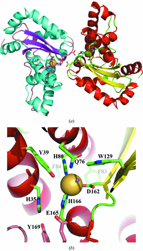
Perkinsus marinus, a facultative intracellular parasite of the eastern oyster Crassostrea virginica, is responsible for mass mortalities of oyster populations. P. marinus trophozoites survive and proliferate within oyster hemocytes, invading most tissues and fluids, thus causing a systemic infection that eventually kills the host. The phagocytosis of P. marinus trophozoites lacks a respiratory burst, suggesting that the parasite has mechanisms that actively abrogate the host's oxidative defense responses. One mechanism and the first line of defense against oxidative damage is the dismutation of superoxide radical to molecular oxygen and hydrogen peroxide by superoxide dismutases (SODs). P. marinus possesses two iron-cofactored SODs, PmSOD1 and PmSOD2. Here, the crystallization and X-ray structures of both PmSOD1 and PmSOD2 are presented.
Figures
References
-
- Ahmed, H., Schott, E. J., Gauthier, J. D. & Vasta, G. R. (2003). Anal. Biochem.318, 132–141. - PubMed
-
- Anderson, R. S., Oliver, L. M. & Brubacher, L. L. (1992). J. Invert. Pathol.59, 303–307.
-
- Anderson, R. S., Patel, K. M. & Roesijadi, G. (1999). Dev. Comput. Immunol.23, 443–449. - PubMed
-
- Brünger, A. T. (1992a). X-PLOR. A System for X-ray Crystallography & NMR, v.3.1. New Haven, CT, USA: Yale University Press.
Publication types
MeSH terms
Substances
LinkOut - more resources
Full Text Sources
