Purification and crystallization of the human EF-hand tumour suppressor protein S100A2
- PMID: 17077493
- PMCID: PMC2225223
- DOI: 10.1107/S1744309106039881
Purification and crystallization of the human EF-hand tumour suppressor protein S100A2
Abstract
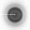
S100A2 is a Ca(2+)-binding EF-hand protein that is mainly localized in the nucleus. There, it acts as a tumour suppressor by binding and activating p53. Wild-type S100A2 and a S100A2 variant lacking cysteines have been purified. CD spectroscopy showed that there are no changes in secondary-structure composition. The S100A2 mutant was crystallized in a calcium-free form. The crystals, with dimensions 30 x 30 x 70 microm, diffract to 1.7 A and belong to space group P2(1)2(1)2(1), with unit-cell parameters a = 43.5, b = 57.8, c = 59.8 A, alpha = beta = gamma = 90 degrees. Preliminary analysis of the X-ray data indicates that there are two subunits per asymmetric unit.
Figures


Similar articles
-
Crystallization and calcium/sulfur SAD phasing of the human EF-hand protein S100A2.Acta Crystallogr Sect F Struct Biol Cryst Commun. 2010 Sep 1;66(Pt 9):1032-6. doi: 10.1107/S1744309110030691. Epub 2010 Aug 26. Acta Crystallogr Sect F Struct Biol Cryst Commun. 2010. PMID: 20823519 Free PMC article.
-
Purification, crystallization and preliminary X-ray diffraction studies on human Ca2+-binding protein S100B.Acta Crystallogr Sect F Struct Biol Cryst Commun. 2005 Jul 1;61(Pt 7):673-5. doi: 10.1107/S1744309105018014. Epub 2005 Jun 15. Acta Crystallogr Sect F Struct Biol Cryst Commun. 2005. PMID: 16511125 Free PMC article.
-
Metal ions modulate the folding and stability of the tumor suppressor protein S100A2.FEBS J. 2009 Mar;276(6):1776-86. doi: 10.1111/j.1742-4658.2009.06912.x. FEBS J. 2009. PMID: 19267779
-
S100A2 in cancerogenesis: a friend or a foe?Amino Acids. 2011 Oct;41(4):849-61. doi: 10.1007/s00726-010-0623-2. Epub 2010 Jun 3. Amino Acids. 2011. PMID: 20521072 Review.
-
S100A2 protein and non-small cell lung cancer. The dual role concept.Tumour Biol. 2014 Aug;35(8):7327-33. doi: 10.1007/s13277-014-2117-4. Epub 2014 May 27. Tumour Biol. 2014. PMID: 24863947 Review.
Cited by
-
Crystallization and calcium/sulfur SAD phasing of the human EF-hand protein S100A2.Acta Crystallogr Sect F Struct Biol Cryst Commun. 2010 Sep 1;66(Pt 9):1032-6. doi: 10.1107/S1744309110030691. Epub 2010 Aug 26. Acta Crystallogr Sect F Struct Biol Cryst Commun. 2010. PMID: 20823519 Free PMC article.
References
-
- Böhm, G., Muhr, R. & Jaenicke, R. (1992). Protein Eng.5, 191–195. - PubMed
-
- Fritz, G., Mittl, P. R., Vasak, M., Grütter, M. G. & Heizmann, C. W. (2002). J. Biol. Chem.277, 33092–33098. - PubMed
-
- Guex, N. & Peitsch, M. C. (1997). Electrophoresis, 18, 2714–2723. - PubMed
-
- Heizmann, C. W., Fritz, G. & Schäfer, B. W. (2002). Front. Biosci.7, d1356–d1368. - PubMed
-
- Kabsch, W. (1993). J. Appl. Cryst.26, 795–800.
Publication types
MeSH terms
Substances
Associated data
- Actions
LinkOut - more resources
Full Text Sources
Research Materials
Miscellaneous

