

doi: 10.1107/S1744309106041959.
Epub 2006 Oct 20.
Expression, purification and crystallization of a PCI domain from the COP9 signalosome subunit 7 (CSN7)
Affiliations
- PMID: 17077498
- PMCID: PMC2225213
- DOI: 10.1107/S1744309106041959
Item in Clipboard
Expression, purification and crystallization of a PCI domain from the COP9 signalosome subunit 7 (CSN7)
Acta Crystallogr Sect F Struct Biol Cryst Commun.
.
Abstract
A core fragment of Arabidopsis thaliana COP9 signalosome (CSN) subunit 7 was expressed in Escherichia coli. The protein was purified to homogeneity and screened for crystallization. Crystallization conditions were refined using the sitting-drop vapour-diffusion method. Crystals were obtained using polyethylene glycol 8000 as a precipitant and have a thick rod-like morphology. Their crystallographic symmetry is orthorhombic, space group C222(1), with unit-cell parameters a = 57.2, b = 86.2, c = 72.6 A and a diffraction limit of 2.06 A.
Figures
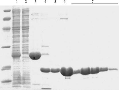
SDS–PAGE of the AtCSN7 core purification steps. Lane 1, material loaded onto Ni2+-chelate column; lane 2, sample of unbound material; lane 3, elution from the Ni2+ -chelate column obtained with 50 mM imidazole; lane 4, protein after overnight TEV protease digestion; lane 5, sample of unbound material from second Ni2+-chelate column; lane 6, sample of material loaded onto Superdex 200 gel-filtration column; lane 7, relevant gel-filtration fractions. MW markers (extreme left) are from top to bottom: 97, 66, 45, 31, 21, 14 kDa. The gel was Coomassie-stained.
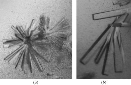
(a) A cluster of rod-shaped crystals of the AtCSN7 core as obtained in the initial screen. The cystals are approximately 0.05–0.1 mm in length. (b) Rod-shaped crystals of the AtCSN7 core obtained after lowering the magnesium acetate concentration. The crystals are approximately 0.15–0.3 mm in the longest dimension. The crystallization conditions for these crystals were a protein concentration of 10 mg ml−1 with a reservoir comprising 23%(w/v) PEG 8000, 0.1 M sodium cacodylate pH 7.2 and 0.08 M magnesium acetate.
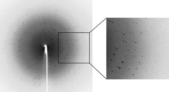
Oscillation image (1°) of AtCSN7 core protein crystals. The crystal-to-detector distance was 180 mm. Diffraction data were observed to 2.06 Å. Data were collected and processed to a d
min of 2.1 Å. Crystallization conditions were the same as for Fig. 2 ▶(b).
References
-
- Chamovitz, D. A. & Glickman, M. (2002). Curr. Biol.12, R232. - PubMed
-
- Freilich, S., Oron, E., Kapp, Y., Nevo-Caspi, Y., Orgad, S., Segal, D. & Chamovitz, D. A. (1999). Curr. Biol.9, 1187–1190. - PubMed
-
- Hofmann, K. & Bucher, P. (1998). Trends Biochem. Sci.23, 204–205. - PubMed
-
- Kapelari, B., Bech-Otschir, D., Hegerl, R., Schade, R., Dumdey, R. & Dubiel, W. (2000). J. Mol. Biol.300, 1169–1178. - PubMed
Publication types
MeSH terms
Substances
LinkOut - more resources
Full Text Sources
Molecular Biology Databases
Miscellaneous
