. 2006;8(6):R64.
doi: 10.1186/bcr1617.

GATA3 protein as a MUC1 transcriptional regulator in breast cancer cells

Affiliations

GATA3 protein as a MUC1 transcriptional regulator in breast cancer cells

Martín C Abba et al. Breast Cancer Res. 2006.

Abstract

Introduction: Recent studies have demonstrated that members of the GATA-binding protein (GATA) family (GATA4 and GATA5) might have pivotal roles in the transcriptional upregulation of mucin genes (MUC2, MUC3 and MUC4) in gastrointestinal epithelium. The zinc-finger GATA3 transcription factor has been reported to be involved in the growth control and differentiation of breast epithelial cells. In SAGE (serial analysis of gene expression) studies we observed an intriguing significant correlation between GATA3 and MUC1 mRNA expression in breast carcinomas. We therefore designed the present study to elucidate whether MUC1 expression is regulated by GATA3 in breast cancer cells.

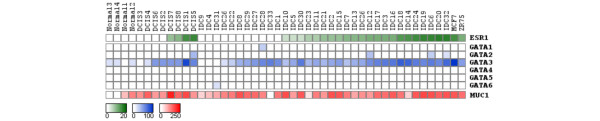
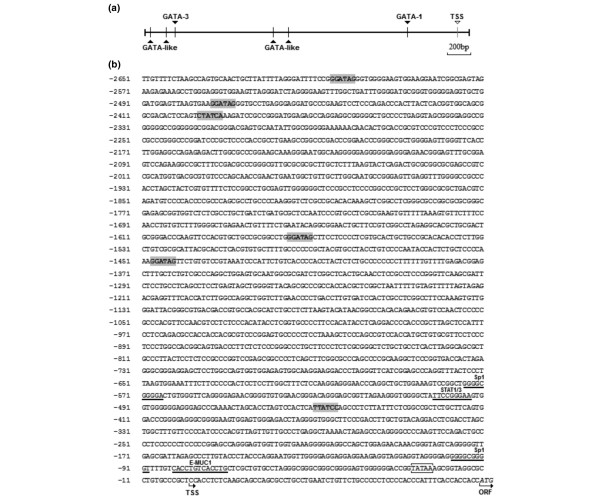
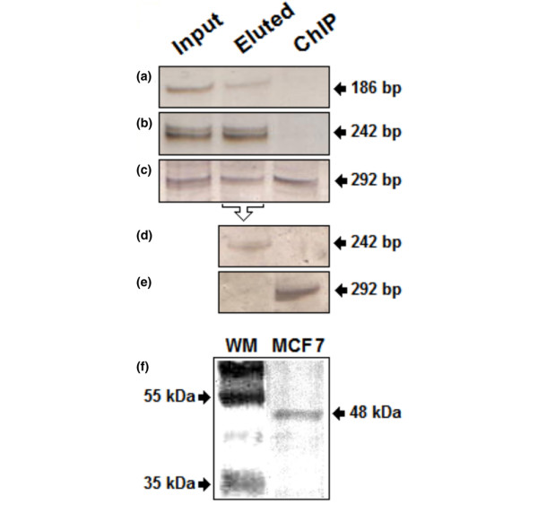
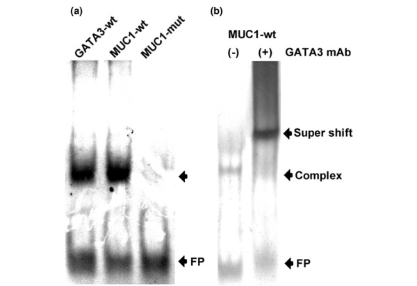
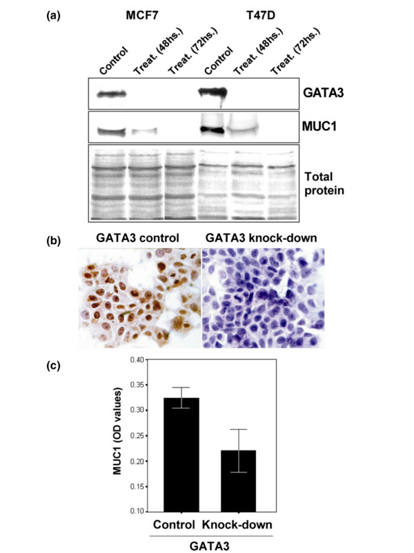
Methods: Promoter sequence analysis of the MUC1 gene identified six GATA cis consensus elements in the 5' flanking region (GATA1, GATA3 and four GATA-like sequences). Chromatin immunoprecipitation and electrophoretic mobility-shift assays were employed to study the presence of a functional GATA3-binding site. GATA3 and MUC1 expression was analyzed in vitro with a GATA3 knockdown assay. Furthermore, expression of GATA3 and MUC1 genes was analyzed by real-time RT-PCR and immunohistochemistry on breast cancer-specific tissue microarrays.

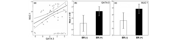
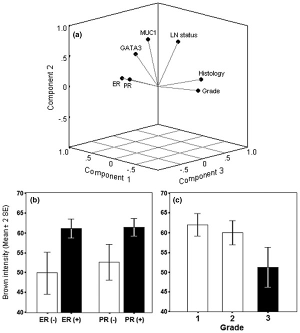
Results: We confirmed the presence of a functional GATA3-binding site on the MUC1 promoter region in the MCF7 cell line. We determined that GATA3 knockdown assays led to a decrease in MUC1 protein expression in MCF7 and T47D cells. In addition, we detected a statistically significant correlation in expression between GATA3 and MUC1 genes at the mRNA and protein levels both in normal breast epithelium and in breast carcinomas (p = 0.01). GATA3 expression was also highly associated with estrogen receptor and progesterone receptor status (p = 0.0001) and tumor grade (p = 0.004) in breast carcinomas.

Conclusion: Our study provides evidence indicating that GATA3 is probably a mediator for the transcriptional upregulation of MUC1 expression in some breast cancers.


Figures

Figure 1
Figure 1
Digital northern analysis based on SAGE data of GATA family members, ESR1 and MUC1 genes in 47 breast SAGE libraries. The color scale at the bottom represents expression level based on numbers of transcripts (tags) per library. SAGE, serial analysis of gene expression.
Figure 2
Figure 2
GATA3 and MUC1 real-time RT-PCR analysis in 36 invasive breast carcinomas. (a) Linear regression analysis of GATA3 and MUC1 mRNA expression levels in invasive ductal carcinoma (IDC) with 95% mean prediction interval (r = 0.6; p < 0.0001). (b,c) GATA3 (p = 0.008) (b) and MUC1 (p = 0.009) (c) mRNA expression of IDC in association with ERα status. Results are real-time RT-PCR values of the assayed gene relative to 18S rRNA used as normalizing control and are shown as means ± 2 SEM based on a log2 transformation.
Figure 3
Figure 3
Tissue microarray analysis of 263 breast normal and cancer tissue samples. (a) Component plot in rotated space showing factors positively correlated, namely estrogen receptor/progesterone receptor (ER/PR; p = 0.0001), ER–PR/GATA3 (p = 0.001), and MUC1/lymph node status (p = 0.006), GATA3/MUC1 (p = 0.01); and factors negatively correlated, namely ER–PR/tumor grade (p = 0.0001) and GATA3/tumor grade (p = 0.002). (b,c) Univariate analysis showing highly statistical association between GATA3 immunostaining and ER and PR status (p = 0.0001) (b) and statistical decrease trend in GATA3 immunostaining and the tumor grade of invasive component (p = 0.004) (c). Results are based on the brown intensity of immunostaining as described in the Materials and methods section, and are shown as means ± 2 SEM.
Figure 4
Figure 4
MUC1 promoter sequence. (a) GATA cis-elements map identified in MUC1 promoter region. (b) DNA sequence showing the TATA box (white box), the transcriptional start site +1 (TSS arrow) and the open reading frame (ORF arrow). Gray boxes indicate putative binding sites for GATA transcription factors showing the following: a GATA1 sequence (-448), a GATA3 sequence (-2395) and four GATA-like sequences (-1446, -1574, -2472, and -2604). Known regulatory elements are underlined (Sp1, STAT1/3, E-MUC1).
Figure 5
Figure 5
Chromatin immunoprecipitation assay of GATA3 in MCF7 breast cancer cells. Chromatin complexes were crosslinked in vivo with formaldehyde (see the Materials and methods section). The GATA3-associated DNA fragments were immunoprecipitated (IP) with mouse monoclonal antibody against GATA3 (HG3-31). DNA samples were isolated before IP (lane labeled 'input'), after elution the unbound proteins from the Protein A–Sepharose batch (lane labeled 'eluted') and after specific IP (lane labeled 'ChIP', for chromatin immunoprecipitation). (a–c) PCR was performed with (a) flanking primers amplifying a GATA1 binding site on the MUC1 promoter (186 bp; see Figure 2), (b) flanking primers for amplification of exonic sequences of the HLA-DQA1 gene (242 bp; negative control), and (c) flanking primers for amplification of the GATA3-binding site on the MUC1 promoter (292 bp) as shown in Figure 2. (d–e) Second ChIP (reChIP) assay using the first eluted DNA–protein complex as input sample. (f) Western blot analysis of GATA3 protein expression: a sample of MCF7 protein extract was run on a 10% SDS-PAGE gel and assayed for GATA3 expression with the monoclonal antibody used in ChIP assays (HG3-31). The expected wild-type band of 48 kDa was immunodetected. Lane WM shows an SDS-7B prestained SDS-molecular mass standard (Sigma-Aldrich).
Figure 6
Figure 6
Electrophoretic mobility-shift assay employing cell nuclear extract from MCF7. (a) Gel shift analyses were performed with double-stranded oligonucleotides (27 bp) as indicated: positive control probe containing the GATA3-binding element (GATA3-wt), a mimetic wild-type MUC1 probe containing the predictive GATA3-binding site (MUC1-wt), and a probe sequence with predictive GATA3-binding site mutations (MUC1-mut). (b) The presence of GATA3 was confirmed by supershift assay. Nuclear extract prepared from MCF7 cells was preincubated with GATA3 antibody, and then incubated with the mimetic wild-type MUC1 probe containing the GATA3-binding site (MUC1-wt). Arrows to the right indicate the position of the free probe (FP), the probe–nuclear-protein complex (Complex), and the specifically retarded protein–GATA3-antibody–probe complex (Supershift).
Figure 7
Figure 7
GATA3 knock-down analysis in MCF7 and T47D breast cancer cell lines. (a) Western blot analysis of GATA3 and MUC1 proteins in non-treated (control) and GATA3 anti-sense treated cells (48 and 72 hours) in both breast cancer cell lines. SDS-PAGE silver protein staining showed equal amounts of total protein loaded. (b) MCF7 immunocytochemistry of GATA3 anti-sense transfected cells showing no immunoreactivity to the anti-GATA3 antibody, and matching control cells show GATA3-positive nuclear localization. (c) Quantification of MUC1 protein expression by MCF7 culture ELISA assay. GATA3 anti-sense treated cells (48 hours) showed a statistically significant decrease in MUC1 expression compared with matched control cells (p = 0.001). Each experiment was performed in triplicate.

References

    1. Merika M, Orkin SH. DNA-binding specificity of GATA family transcription factors. Mol Cell Biol. 1993;13:3999–4010. - PMC - PubMed
    1. Patient RK, McGhee JD. The GATA family (vertebrates and invertebrates) Curr Opin Genet Dev. 2002;12:416–422. doi: 10.1016/S0959-437X(02)00319-2. - DOI - PubMed
    1. Abba MC, Hu Y, Sun H, Drake JA, Gaddis S, Baggerly K, Sahin A, Aldaz CM. Gene expression signature of estrogen receptor α status in breast cancer. BMC Genomics. 2005;6:37. doi: 10.1186/1471-2164-6-37. - DOI - PMC - PubMed
    1. Hoch RV, Thompson DA, Baker RJ, Weigel RJ. GATA-3 is expressed in association with estrogen receptor in breast cancer. Int J Cancer. 1999;84:122–128. doi: 10.1002/(SICI)1097-0215(19990420)84:2<122::AID-IJC5>3.0.CO;2-S. - DOI - PubMed
    1. Perou CM, Sorlie T, Eisen MB, van de Rijn M, Jeffrey SS, Rees CA, Pollack JR, Ross DT, Johnsen H, Akslen LA, et al. Molecular portraits of human breast tumors. Nature. 2000;406:747–752. doi: 10.1038/35021093. - DOI - PubMed

Publication types