. 2006;8(6):R166.
doi: 10.1186/ar2074.

Interactions between IL-32 and tumor necrosis factor alpha contribute to the exacerbation of immune-inflammatory diseases

Affiliations

Interactions between IL-32 and tumor necrosis factor alpha contribute to the exacerbation of immune-inflammatory diseases

Hirofumi Shoda et al. Arthritis Res Ther. 2006.

Abstract

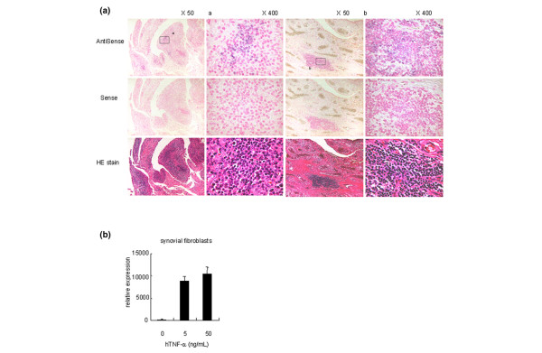
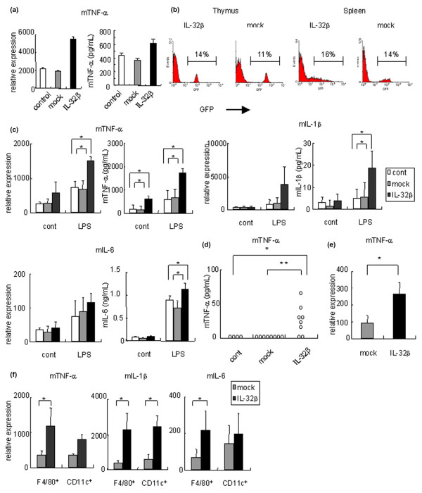
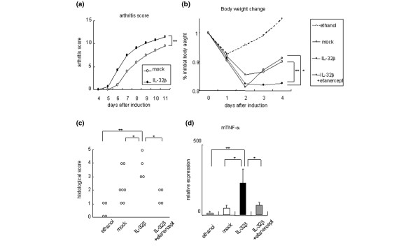
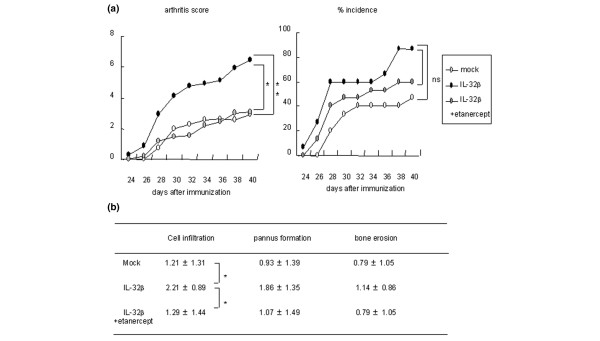
IL-32 is a newly described cytokine in the human found to be an in vitro inducer of tumor necrosis factor alpha (TNFalpha). We examined the in vivo relationship between IL-32 and TNFalpha, and the pathologic role of IL-32 in the TNFalpha-related diseases - arthritis and colitis. We demonstrated by quantitative PCR assay that IL-32 mRNA was expressed in the lymphoid tissues, and in stimulated peripheral T cells, monocytes, and B cells. Activated T cells were important for IL-32 mRNA expression in monocytes and B cells. Interestingly, TNFalpha reciprocally induced IL-32 mRNA expression in T cells, monocyte-derived dendritic cells, and synovial fibroblasts. Moreover, IL-32 mRNA expression was prominent in the synovial tissues of rheumatoid arthritis patients, especially in synovial-infiltrated lymphocytes by in situ hybridization. To examine the in vivo relationship of IL-32 and TNFalpha, we prepared an overexpression model mouse of human IL-32beta (BM-hIL-32) by bone marrow transplantation. Splenocytes of BM-hIL-32 mice showed increased expression and secretion of TNFalpha, IL-1beta, and IL-6 especially in response to lipopolysaccharide stimulation. Moreover, serum TNFalpha concentration showed a clear increase in BM-hIL-32 mice. Cell-sorting analysis of splenocytes showed that the expression of TNFalpha was increased in resting F4/80+ macrophages, and the expression of TNFalpha, IL-1beta and IL-6 was increased in lipopolysaccharide-stimulated F4/80+ macrophages and CD11c+ dendritic cells. In fact, BM-hIL-32 mice showed exacerbation of collagen-antibody-induced arthritis and trinitrobenzen sulfonic acid-induced colitis. In addition, the transfer of hIL-32beta-producing CD4+ T cells significantly exacerbated collagen-induced arthritis, and a TNFalpha blockade cancelled the exacerbating effects of hIL-32beta. We therefore conclude that IL-32 is closely associated with TNFalpha, and contributes to the exacerbation of TNFalpha-related inflammatory arthritis and colitis.


Figures

Figure 1
Figure 1
Examination of tissue and cell expression of IL-32 by quantitative real-time PCR. (a) Tissue expression of IL-32. WBC, white blood cells. (b) Human peripheral blood mononuclear cells (PBMCs) expressed IL-32. PBMCs were cultured with or without concanavalin A. PBMCs were also stimulated by immobilized anti-human CD3 and anti-human CD28 antibodies. Cont, control. (c) IL-32 expression of monocytes and B cells after the depletion of CD3+ cells. (d) Peripheral CD4+ T cells were cultured with the indicated inflammatory cytokines for 24 hours. (e) Human monocyte-derived dendritic cells (MoDCs) were cultured with lipopolysaccharide (LPS) or tumor necrosis factor alpha (TNFα) for 24 hours to induce maturation. The data are representative of at least three independent studies.
Figure 2
Figure 2
IL-32 was abundantly expressed in the synovial tissues of rheumatoid arthritis patients. (a) In situ hybridization of the synovial tissues from rheumatoid arthritis (RA) patients. IL-32β was expressed in the synovial-infiltrated lymphocytes of RA patients. HE stain, hematoxylin and eosin stain. We examined the tissue samples from four RA patients, and show representative examples. (b) IL-32 expression of the synovial fibroblasts derived from four RA patients in response to human tumor necrosis factor alpha (hTNFα).
Figure 3
Figure 3
Inflammatory cytokines were induced by human IL-32β in mice. (a) Raw 267.4 was cultured with the supernatant of human IL-32β (h IL-32β) or mock-transfected mammalian cells (293T) for 24 hours. Left, relative expression of mouse tumor necrosis factor alpha (mTNFα), compared with β-actin; right, secreted TNFα protein level measured by ELISA. (b) We generated hIL-32β overexpressed mice by transplantation of hIL-32β-transduced bone marrow cells. The expression of green fluorescent protein, was analyzed by flow cytometry 6–9 weeks after transplantation. (c) Expression of mTNFα, mIL-1β and mIL-6 in the cultured splenocytes of the control group (white bars; n = 3), or bone-marrow chimeric mice of the mock group (BM-Mock mice) (gray bars; n = 4), or hIL-32β (BM-hIL-32) (black bars; n = 4) with or without 1 μg/ml lipopolysaccharide (LPS). Concentrations of indicated cytokines of the cultured supernatants are shown in the right-hand figures. (d) Serum concentration of mTNFα determined in control mice (n = 4), in BM-Mock mice (n = 8), and in BM-hIL-32 mice (n = 8). (e) Expression of mTNFα in splenic F4/80+ CD11c- macrophages of BM-Mock mice (gray bars; n = 4) and in BM-hIL-32 mice (black bars; n = 4). (f) Expression of mTNFα, mIL-1β, and mIL-6 in LPS-stimulated splenic F4/80+ CD11c- macrophages and CD11c+, CD3-, and CD19- dendritic cells in BM-Mock mice (gray bars; n = 4), and in BM-hIL-32 mice (black bars; n = 4). Data are representative of at least three independent studies. *P < 0.05, **P < 0.01, BM-hIL-32 mice versus BM-Mock mice or control mice.
Figure 4
Figure 4
Exacerbation of murine models of tumor necrosis factor alpha-related inflammatory diseases in BM-hIL-32 mice. (a) Collagen-antibody-induced arthritis was induced in bone-marrow chimeric human IL-32β mice (BM-hIL-32) (n = 6) and bone-marrow chimeric mice of the mock group (BM-Mock) (n = 4). Mean arthritis scores are shown. (b) Body weight change after induction of trinitrobenzen sulfonic acid (TNBS)-induced colitis in BM-Mock mice (n = 7), in BM-hIL-32 mice (n = 4), and in BM-hIL-32 mice + 200 μg/day intraperitoneal administration of etanercept (n = 4). Control mice (n = 5) were administered only 50% ethanol with PBS. Percentage of initial body weight is shown. (c) Histological scores of TNBS-induced colitis. (d) Relative expression of mouse tumor necrosis factor alpha (mTNFα) in the colon of TNBS-induced colitis mice. *P < 0.05, **P < 0.01, BM-hIL-32 mice versus BM-Mock mice or BM-hIL-32 mice + etanercept.
Figure 5
Figure 5
Transfer of human IL-32β-transduced CD4+ T cells exacerbated collagen-induced arthritis. Human IL-32β-transduced CD4+ T cells were transferred to collagen-immunized mice before the onset of arthritis (day 23). In one group (IL-32β + etanercept group), 50 μg/day etanercept was administered intraperitoneally for 14 days after transfer of CD4+ T cells. Each group consisted of 14 mice. (a) Arthritis scores and the percentage incidence of arthritis. (b) Cell infiltration, pannus formation, and bone erosion in CIA mice are quantified. Histological scores are shown as the mean ± standard deviation. *P < 0.05, **P < 0.01, IL-32β group versus mock group or IL-32β + etanercept group. ns, not significant.

References

    1. Keffer J, Probert L, Cazlaris H, Georgopoulos S, Kaslaris E, Kioussis D, Kollias G. Transgenic mice expressing human tumour necrosis factor: a predictive genetic model of arthritis. Embo J. 1991;10:4025–4031. - PMC - PubMed
    1. Feldmann M, Brennan FM, Maini RN. Role of cytokines in rheumatoid arthritis. Annu Rev Immunol. 1996;14:397–440. doi: 10.1146/annurev.immunol.14.1.397. - DOI - PubMed
    1. Lipsky PE, van der Heijde DM, St Clair EW, Furst DE, Breedveld FC, Kalden JR, Smolen JS, Weisman M, Emery P, Feldmann M, et al. Infliximab and methotrexate in the treatment of rheumatoid arthritis. Anti-Tumor Necrosis Factor Trial in Rheumatoid Arthritis with Concomitant Therapy Study Group. N Engl J Med. 2000;343:1594–1602. doi: 10.1056/NEJM200011303432202. - DOI - PubMed
    1. Hata H, Sakaguchi N, Yoshitomi H, Iwakura Y, Sekikawa K, Azuma Y, Kanai C, Moriizumi E, Nomura T, Nakamura T, et al. Distinct contribution of IL-6, TNF-alpha, IL-1, and IL-10 to T cell-mediated spontaneous autoimmune arthritis in mice. J Clin Invest. 2004;114:582–588. doi: 10.1172/JCI200421795. - DOI - PMC - PubMed
    1. Elliott MJ, Maini RN, Feldmann M, Long-Fox A, Charles P, Bijl H, Woody JN. Repeated therapy with monoclonal antibody to tumour necrosis factor alpha (cA2) in patients with rheumatoid arthritis. Lancet. 1994;344:1125–1127. doi: 10.1016/S0140-6736(94)90632-7. - DOI - PubMed

Publication types

MeSH terms