. 2007 Jan 1;313(1):109-20.
doi: 10.1016/j.yexcr.2006.09.020. Epub 2006 Sep 28.

Discrete functional elements required for initiation activity of the Chinese hamster dihydrofolate reductase origin beta at ectopic chromosomal sites

Affiliations

Discrete functional elements required for initiation activity of the Chinese hamster dihydrofolate reductase origin beta at ectopic chromosomal sites

Steven J Gray et al. Exp Cell Res. .

Abstract

The Chinese hamster dihydrofolate reductase (DHFR) DNA replication initiation region, the 5.8 kb ori-beta, can function as a DNA replicator at random ectopic chromosomal sites in hamster cells. We report a detailed genetic analysis of the DiNucleotide Repeat (DNR) element, one of several sequence elements necessary for ectopic ori-beta activity. Deletions within ori-beta identified a 132 bp core region within the DNR element, consisting mainly of dinucleotide repeats, and a downstream region that are required for ori-beta initiation activity at non-specific ectopic sites in hamster cells. Replacement of the DNR element with Xenopus or mouse transcriptional elements from rDNA genes restored full levels of initiation activity, but replacement with a nucleosome positioning element or a viral intron sequence did not. The requirement for the DNR element and three other ori-beta sequence elements was conserved when ori-beta activity was tested at either random sites or at a single specific ectopic chromosomal site in human cells. These results confirm the importance of specific cis-acting elements in directing the initiation of DNA replication in mammalian cells, and provide new evidence that transcriptional elements can functionally substitute for one of these elements in ori-beta.


Figures

Figure 1
Figure 1
The DHFR origin beta at endogenous and ectopic locations. (A) Diagram of the endogenous DHFR ori-beta IR in hamster cell. Preferred start sites of DNA replication (ori-β , ori-β ’, and ori γ) within a 55-kb initiation zone between the genes DHFR and 2BE2121 are indicated. The 5.8 kb DNA fragment containing ori-beta extends from the BamHI (nucleotide position 1) to the KpnI restriction enzyme site. Previously identified [8, 9] functional elements of ori-beta are indicated: IR, initiation region; RIP60, 60 kDa replication initiation protein binding sites; BEND, sequence-induced stable bend in the DNA; AT, (AT)n repeats and AT-rich sequences; DNR, GA+CA dinucleotide repeat element. (B) 5.8 kb ori-beta sequence cloned into pUC19 (pMCD) and integrated at non-specific ectopic locations in DR12 and Hela cells. Locations of pp8, pp2, pp6 and pp3 PCR primer sites are indicated by gray diamonds. (C) 5.8 kb ori-beta sequence integrated at the FRT site in Hela 406 cells [10]. The locations of the FRT sites, hygromycin-neomycin resistance fusion gene (HygR NeoR) with transcription start site (bent arrow), poly-adenylation and transcription termination site (pA), and thymidine kinase (TK) gene are indicated. The 5.6 kb EcoRI fragment used for Southern blot analysis, and diagnostic PCR primers 1, 2, and 3 for specific site integration are shown. The target sites for ppHyg and pp2b are indicated with gray diamonds.
Figure 2
Figure 2
Initiation activity of ori-beta constructs at multiple ectopic chromosomal sites in pooled stably transfected DR12 cells. (A) Integration of full-length ori-beta into DR12 cells was assayed by Southern blot analysis of BamHI/KpnI-digested genomic DNA hybridized to ori-beta probes 1 and 2, or probe GNAI3 as a loading control. “DR12” refers to untransfected cells, “ori-beta CHOK1” refers to the endogenous ori-beta, “ori-beta DR12” refers to stably transfected ori-beta sequences in DR12 cells, and “ori-beta plasmid” refers to 30 pg of pMCD. Densitometry quantitation of ectopic ori-beta was first calculated in each lane as [(intensity of 5.8 kb band)/(intensity of 3.5 kb band)], then expressed as a percentage (shown below the blot) of the same densitometry quantitation done in the CHOK1 lane corresponding to the same μ g quantity of DNA. (B) Summary of the methodology used to prepare and analyze nascent DNA samples. (C) Diagram of ori-beta constructs, with primer sets pp2 and pp3 shown (gray diamonds). (C, rows I, ii, and vii) Cartoon representation of the DHFR ori-beta, with deleted regions shown as dashed lines. (C, rows iii-vi) DNR replacement and partial deletion constructs, in the context of the entire ori-beta fragment, as described in the Materials and Methods. The DNR region and replacement elements are drawn to scale +/− 5 bp. All construct diagrams in (C) correspond to the adjacent name and bar graph in (D). In panel D, “initiation activity” is calculated as the abundance of target sequences detected with primer set pp2 in nascent-enriched DNA fractions, divided by those detected with primer set pp3. For comparison, all initiation activities are expressed as a percentage of the initiation activity of pMCD (unmodified 5.8 kb ectopic ori-beta, row i). Each bar is the average initiation activity measured from at least 3 independent transfections and nascent DNA preparations. Brackets indicate SEM. *, significantly lower than the pMCD construct; +, significantly greater than the ΔDNR construct (student’s t-test, p value<0.05).
Figure 3
Figure 3
Loss of initiation activity in DNR-deleted ori-beta can be restored by transcriptional elements. (A) Summary of the methodology used to prepare and analyze nascent DNA samples. (B) Diagram of ori-beta constructs, with primer sets pp2 and pp3 shown (gray diamonds). The DNR region and replacement elements are drawn to scale +/− 5 bp. (B, row i) Wild type ectopic ori-beta in DR12 cells. (B, row ii) Arrows above and below the SB2 element indicate the polar impediment of replication forks moving toward ori-beta (X) mediated by TTF-1 at its binding site (thick line). (B, row iii) A gray arrow indicates the transcription direction of 5S RNA gene, with the nucleosome positioning element (NPE, thick line) and TFIIIA binding site (black box) highlighted. (B, row iv-v) Partial 5S RNA gene replacements containing the NPE (black bar) without the TFIIIA binding site, in either orientation. Dashed lines indicate deleted regions. All construct diagrams in (B) correspond to the adjacent name and bar graph in (C). In panel C, “initiation activity” and notations are as described in the legend of Figure 2.
Figure 4
Figure 4
Initiation activity of mutant DNR ori-beta constructs at the specific chromosomal site in Hela cells. (A) Following transfection with ori-beta constructs and drug selection, genomic DNA from clonal cell lines was tested for site-specific integration by PCR. A PCR product using primers 1+2 indicates no integration at the FRT site, but a product using primers 1+3 indicates integration in the correct orientation. (B) Southern blots EcoRI-digested genomic DNA from clonal lines was hybridized to either the Hyg probe or Neo probe as described in the Materials and Methods. The Hyg probe hybridizes to a 2.3 kb fragment in the acceptor cell line and to a 5.6 kb fragment when ori-beta is integrated. The Neo probe hybridizes to a 5.6 kb fragment only in the integrated ori-beta. (C) Summary of the methodology used to prepare and analyze nascent DNA samples as previously described [10]. (D) Analysis of the initiation activity of ori-beta constructs at the specific integration site in Hela. 406, Hela acceptor cell line; WT, full-length ori-beta fragment; ΔDNR, DNR deletion construct; DNRrev, construct in which the DNR orientation was reversed; DNRspcr, replacement of DNR with a 235 bp SV40 intron fragment. “Initiation activity” is defined as the abundance of target sequences in nascent DNA-enriched fractions detected at primer sets pp2b (black bars) or ppHyg (white bars), divided by that at primer set ppGlobin. Brackets indicate SEM for WT.
Figure 5
Figure 5
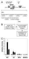
Initiation activity of ori-beta constructs at multiple ectopic chromosomal sites in pooled stably transfected Hela cells. (A) Diagram of WT ori-beta and mutated residues. The sequences necessary for the bent DNA region (underlined) and downstream RIP60 binding site (bold) are shown. Nucleotide substitutions for the BENDx and RIP60 mutants are in parentheses and brackets, respectively. (B) Summary of the methodology used to prepare and analyze nascent DNA samples as previously described for hamster cells [9]. (C) “Initiation activity” was calculated by dividing the quantity of target sequences in nascent-enriched DNA fractions detected at primer sets pp8, pp2, or pp6 by that detected at primer set pp3. Primer set locations are given in Figure 1B. Construction of these ori-beta plasmids is described in [8, 9]. Bars are the average quantitation of target sequences in at least 2 independent transfections and nascent DNA isolations. Brackets indicate SEM.
Figure 6
Figure 6
Initiation activity of ori-beta constructs at the specific chromosomal site in Hela cells. (A) PCR-based analysis of clonal cell lines containing ori-beta constructs, as described in the Materials and Methods section and Figure 4A. (B) Southern blot analysis of EcoRI-digested genomic DNA from clonal cell lines, as described in the Materials and Methods section and Figure 4B. (C) Summary of the methodology used to prepare and analyze nascent DNA samples as previously described [10]. (D) Analysis of the initiation activity of ori-beta constructs at the specific integration site in Hela. 406, Hela acceptor cell line; WT, full-length ori-beta fragment. Mutant constructs are described in the Materials and Methods and Figure 5. “Initiation activity” is defined as the abundance of target sequences in nascent-enriched DNA fractions detected at primer set pp2b (black bars) or ppHyg (white bars), divided by that at primer set ppGlobin. Brackets indicate SEM for WT.

Similar articles

Cited by

References

    1. Jacob F, Brenner S. [On the regulation of DNA synthesis in bacteria: the hypothesis of the replicon.] C R Hebd Seances Acad Sci. 1963;256:298–300. - PubMed
    1. Gilbert DM. Making sense of eukaryotic DNA replication origins. Science. 2001;294:96–100. - PMC - PubMed
    1. Gilbert DM. In search of the holy replicator. Nat Rev Mol Cell Biol. 2004;5:848–55. - PMC - PubMed
    1. Bell SP, Dutta A. DNA replication in eukaryotic cells. Annu Rev Biochem. 2002;71:333–74. - PubMed
    1. Kobayashi T, Rein T, DePamphilis ML. Identification of primary initiation sites for DNA replication in the hamster dihydrofolate reductase gene initiation zone. Mol Cell Biol. 1998;18:3266–77. - PMC - PubMed

Publication types