. 2007 Feb;81(3):1297-304.
doi: 10.1128/JVI.01336-06. Epub 2006 Nov 1.

Hsp70 negatively controls rotavirus protein bioavailability in caco-2 cells infected by the rotavirus RF strain

Affiliations

Hsp70 negatively controls rotavirus protein bioavailability in caco-2 cells infected by the rotavirus RF strain

Alexis H Broquet et al. J Virol. 2007 Feb.

Abstract

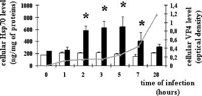
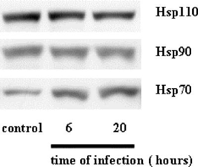
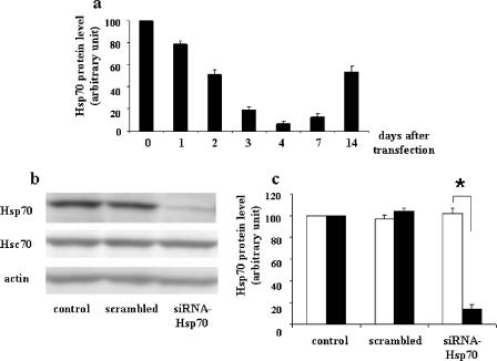
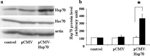
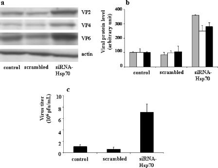
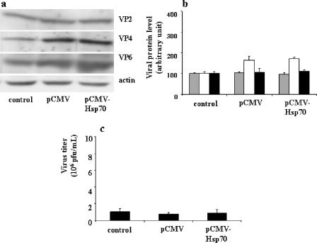
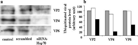
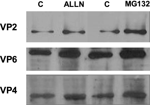
Previous studies demonstrated that the induction of the heat shock protein Hsp70 in response to viral infection is highly specific and differs from one cell to another and for a given virus type. However, no clear consensus exists so far to explain the likely reasons for Hsp70 induction within host cells during viral infection. We show here that upon rotavirus infection of intestinal cells, Hsp70 is indeed rapidly, specifically, and transiently induced. Using small interfering RNA-Hsp70-transfected Caco-2 cells, we observed that Hsp70 silencing was associated with an increased virus protein level and enhanced progeny virus production. Upon Hsp70 silencing, we observed that the ubiquitination of the main rotavirus structural proteins was strongly reduced. In addition, the use of proteasome inhibitors in infected Caco-2 cells was shown to induce an accumulation of structural viral proteins. Together, these results are consistent with a role of Hsp70 in the control of the bioavailability of viral proteins within cells for virus morphogenesis.


Figures

FIG. 1.
FIG. 1.
Rotavirus infection increases Hsp70 level in a time-dependent manner. Hsp70 (white and black histograms) and VP4 (gray curve) levels were analyzed by ELISA, as detailed in Materials and Methods. Caco-2 cell homogenates were harvested at selected times during the course of infection (1 h to 20 h) (10 PFU/cell) from infected cells (black histograms) or noninfected cells (white histograms). Data are expressed as means ± SEM (n = 4). *, P < 0.05. Note that the increased level of Hsp70 is only transient and that VP4 starts to be detectable as soon as 2 h p.i.
FIG. 2.
FIG. 2.
Rotavirus infection specifically increases Hsp70 level. Caco-2 cells were infected with rotavirus strain RF (10 PFU/cell) for 6 h or 20 h. Cells were harvested, and cell lysates were analyzed by immunoblotting using monoclonal anti-Hsp70 (SPA-810), monoclonal anti-Hsc70 (SPA-815), monoclonal anti-Hsp90 (SPA-835), and monoclonal anti-Hsp110 (SPA-1103) as described in Materials and Methods. A representative experiment of four total experiments is shown. Note that only the Hsp70 level increases, whereas the Hsc70, Hsp90, and Hsp110 levels remain unaffected.
FIG. 3.
FIG. 3.
Transient and specific inhibition of Hsp70 in Caco-2 cells. (a) Kinetics of inhibition of the Hsp70 protein level after specific siRNA targeting (0 to 14 days). A Caco-2 cell suspension of 5 × 10⁵ cells was supplied with 2 μg of siRNA-Hsp70 and then electroporated. Cells were plated and cultured at the indicated times after electroporation. Cells were harvested, and lysates were analyzed by immunoblotting as described in Materials and Methods. Hsp70 levels were quantified by immunoblot scanning. Results of this quantification are means ± SEM (n = 4). Note that the maximal inhibition of the Hsp70 level was observed 4 days after siRNA electroporation. (b) Specificity of siRNA-Hsp70. Caco-2 cells were plated and cultured for 96 h in the presence of 100 pmol scrambled siRNA in 100 μl of T solution from Amaxa, and 5 × 10⁵ cells were subjected to electroporation and 2 μg of siRNA-Hsp70. Cell lysates were harvested and analyzed by immunoblotting as described in Materials and Methods, using monoclonal anti-Hsp70 or anti-Hsc70 antibodies. A representative experiment of four total experiments is shown. Only the Hsp70 level was affected by siRNA treatment, whereas the Hsc70 level remained unaffected. (c) Quantitative analysis of Hsp70 levels in siRNA-Hsp70-transfected cells. Caco-2 cells were treated as described for panel b, and the resulting immunoblots (four independent experiments) were quantified using immunoblot scanning. Hsc70 (white histograms) and Hsp70 (black histograms) were measured. *, P < 0.05. Note that at this time (4 days after electroporation) only the Hsp70 level decreased, whereas the Hsc70 level remained unaffected.
FIG. 4.
FIG. 4.
Transient, specific overexpression of Hsp70 in Caco-2 cells. (a) A suspension of 5 × 10⁵ Caco-2 cells was supplied with 5 μg of pCMV-Neo (control) or 5 μg of pCMV-Hsp70 and then electroporated. Cells were plated and cultured for 96 h. Cells were harvested, and lysates were subjected to immunoblotting as described in the legend to Fig. 3. A representative experiment of four total experiments is shown. (b) Quantitative analysis of Hsp70 levels in pCMV-Hsp70-transfected cells. Caco-2 cells were treated as described for panel a, and the resulting immunoblots (four independent experiments) were quantified using immunoblot scanning. Hsc70 (white histograms) and Hsp70 (black histograms) were measured. *, P < 0.05. Note that the Hsp70 level increased 96 h after pCMV-Hsp70 electroporation, whereas the Hsc70 level was not affected.
FIG. 5.
FIG. 5.
Effects of siRNA-Hsp70 transfection on rotavirus protein level (a and b) and on progeny virus production (c) in infected Caco-2 cells. (a) A cell suspension of 5 × 10⁵ Caco-2 cells was supplied with 2 μg of siRNA-Hsp70 and then electroporated. Transfected cells were infected with rotavirus strain RF (1 PFU/cell) for 6 h as described in Materials and Methods. Cells were harvested, and lysates were subjected to immunoblotting using monoclonal anti-VP4 (clone 7.7) and anti-VP6 (clone RV133) as described in Materials and Methods. A representative experiment of four total experiments is shown. (b) Quantitative analysis of VP2, VP4, and VP6 levels in siRNA-transfected cells. Caco-2 cells were treated as described for panel a and subjected to immunoblot analysis (a representative experiment of four total experiments is displayed). VP2 (gray histograms), VP4 (white histograms), and VP6 (black histograms) were measured. (c) Quantification of virus production by siRNA-transfected and infected Caco-2 cells. Caco-2 cells were treated as described for panel a (1 PFU/cell, 18-h infection). Supernatants were titrated by a standard plaque assay with MA104 cells, as described in Materials and Methods (22). Results are expressed as PFU/ml and represent means ± SEM for six experiments. Note that in siRNA-transfected Caco-2 cells, the levels of virus structural proteins (VP2, VP4, and VP6) were significantly increased and associated with an increase of virus production.
FIG. 6.
FIG. 6.
Effects of pCMV-Hsp70 transfection on rotavirus protein level (a and b) and on progeny virus production (c) in infected Caco-2 cells. (a) A suspension of 5 × 10⁵ Caco-2 cells was supplied with 5 μg of pCMV-Neo (control) or 5 μg of pCMV-Hsp70 and then electroporated. Cells were plated and cultured for 90 h and then infected with rotavirus strain RF (1 PFU/cell for 6 h), as described in Materials and Methods. Cells were harvested, and lysates were subjected to immunoblotting as described in the legend to Fig. 5b. A representative experiment of four total experiments is shown. (b) Quantitative analysis of VP2, VP4, and VP6 levels in pCMV-Hsp70-transfected cells. Caco-2 cells were treated as described for panel a and subjected to immunoblot analysis (a representative experiment of four total experiments is displayed). VP2 (gray histograms), VP4 (white histograms), and VP6 (black histograms) levels were measured. (c) Quantification of virus production by pCMV-Hsp70-transfected and infected Caco-2 cells. Caco-2 cells were treated as described for panel a (1 PFU/cell, 18 h). Supernatants were titrated by a standard plaque assay with MA104 cells as described in Materials and Methods (22). Results are expressed as PFU/ml and represent means ± SEM for three experiments. Note that the overexpression of Hsp70 in Caco-2 cells was not followed by any change in viral protein level or virus production.
FIG. 7.
FIG. 7.
siRNA-Hsp70 effect on virus protein ubiquitination in infected Caco-2 cells. (a) A cell suspension of 5 × 10⁵ Caco-2 cells was supplied with 2 μg of siRNA-Hsp70 and then electroporated. Transfected cells were infected with rotavirus strain RF (1 PFU/cell) for 18 h as described in Materials and Methods. Infected lysates were subjected to immunoprecipitation using anti-ubiquitin antibodies, as described in Materials and Methods. Immunopurified ubiquitinated proteins were subjected to immunoblot analysis using anti-VP2, -VP4, and -VP6 antibodies as described in Materials and Methods. (b) Quantitative analysis of ubiquitin-labeled VP2, VP4, and VP6 levels in siRNA-transfected and rotavirus-infected Caco-2 cells. Caco-2 cells were treated as described for panel a, and the amounts of ubiquitinated VP2, VP4, and VP6 were quantified using immunoblot scanning. Control (gray histograms), scrambled siRNA-treated (white histograms), and siRNA-Hsp70-treated (black histograms) cells were studied. Note that the decrease in expression level of Hsp70 in Caco-2 cells results in a significant decrease of the ubiquitination of virus structural proteins.
FIG. 8.
FIG. 8.
Effects of proteasome inhibitors on viral structural protein levels. Caco-2 cells infected with rotavirus strain RF (10 PFU/cell) for 12 h were subjected to proteasome inhibitor treatment with either ALLN (10 μM), MG132 (10 μM), or vehicle (lanes C) for the last 2 h of infection. Cells were harvested, and cell lysates were analyzed by immunoblotting using monoclonal anti-VP2, -VP6, and -VP4 as described in Materials and Methods. Note that treatment with proteasome inhibitors resulted in significant increases of viral structural protein levels, as follows: for VP2, 2.1-fold with ALLN and 2.8-fold with MG132; for VP6, 2.2-fold with ALLN and 2.4-fold with MG132; and for VP4, 2.3-fold with ALLN and 3-fold with MG132.

References

    1. Beere, H. M., B. B. Wolf, K. Cain, D. D. Mosser, A. Mahboubi, T. Kuwana, P. Tailor, R. I. Morimoto, G. M. Cohen, and D. R. Green. 2000. Heat-shock protein 70 inhibits apoptosis by preventing recruitment of procaspase-9 to the Apaf-1 apoptosome. Nat. Cell Biol. 2:469-475. - PubMed
    1. Brenner, B. G., and M. A. Wainberg. 1999. Heat shock protein-based therapeutic strategies against human immunodeficiency virus type 1 infection. Infect. Dis. Obstet. Gynecol. 7:80-90. - PMC - PubMed
    1. Broquet, A. H., G. Thomas, J. Masliah, G. Trugnan, and M. Bachelet. 2003. Expression of the molecular chaperone Hsp70 in detergent-resistant microdomains correlates with its membrane delivery and release. J. Biol. Chem. 278:21601-21606. - PubMed
    1. Bukau, B., and A. L. Horwich. 1998. The Hsp70 and Hsp60 chaperone machines. Cell 92:351-366. - PubMed
    1. Burns, J. W., H. B. Greenberg, R. D. Shaw, and M. K. Estes. 1988. Functional and topographical analyses of epitopes on the hemagglutinin (VP4) of the simian rotavirus SA11. J. Virol. 62:2164-2172. - PMC - PubMed

Publication types

MeSH terms

LinkOut - more resources