. 2007 Jan;81(2):945-53.
doi: 10.1128/JVI.01354-06. Epub 2006 Nov 1.

Analysis of CD127 and KLRG1 expression on hepatitis C virus-specific CD8+ T cells reveals the existence of different memory T-cell subsets in the peripheral blood and liver

Affiliations

Analysis of CD127 and KLRG1 expression on hepatitis C virus-specific CD8+ T cells reveals the existence of different memory T-cell subsets in the peripheral blood and liver

Bertram Bengsch et al. J Virol. 2007 Jan.

Abstract

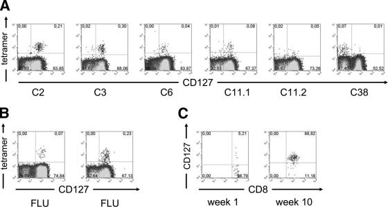
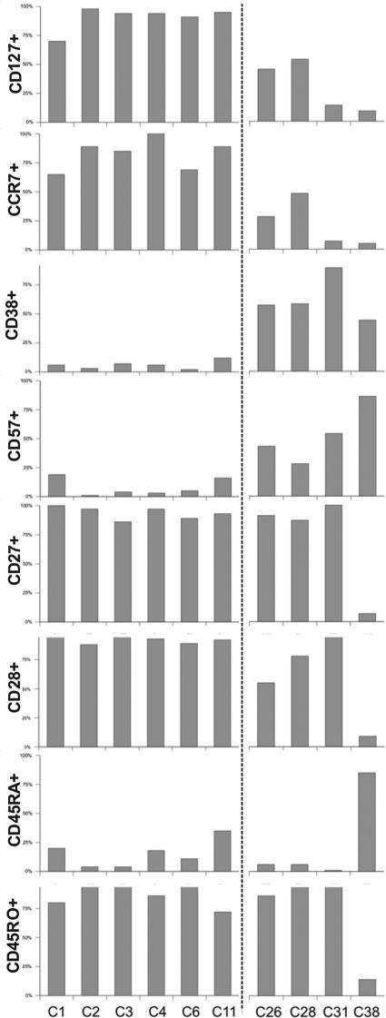
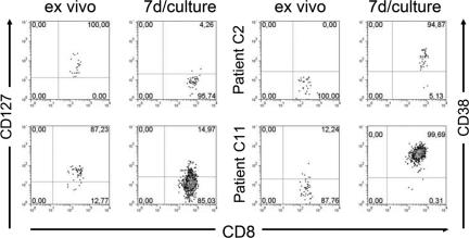
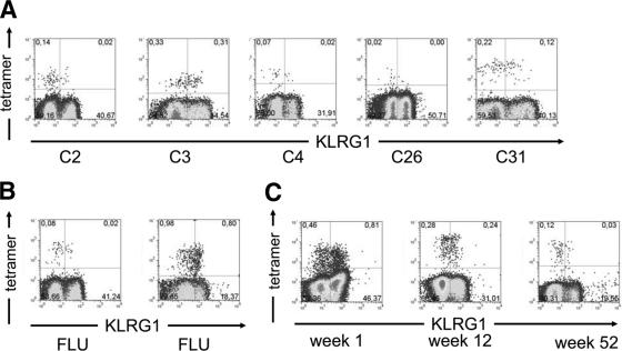
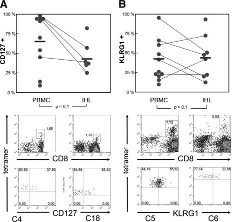
The differentiation and functional status of virus-specific CD8+ T cells is significantly influenced by specific and ongoing antigen recognition. Importantly, the expression profiles of the interleukin-7 receptor alpha chain (CD127) and the killer cell lectin-like receptor G1 (KLRG1) have been shown to be differentially influenced by repetitive T-cell receptor interactions. Indeed, antigen-specific CD8+ T cells targeting persistent viruses (e.g., human immunodeficiency virus and Epstein-Barr virus) have been shown to have low CD127 and high KLRG1 expressions, while CD8+ T cells targeting resolved viral antigens (e.g., FLU) typically display high CD127 and low KLRG1 expressions. Here, we analyzed the surface phenotype and function of hepatitis C virus (HCV)-specific CD8+ T cells. Surprisingly, despite viral persistence, we found that a large fraction of peripheral HCV-specific CD8+ T cells were CD127+ and KLRG1- and had good proliferative capacities, thus resembling memory cells that usually develop following acute resolving infection. Intrahepatic virus-specific CD8+ T cells displayed significantly reduced levels of CD127 expression but similar levels of KLRG1 expression compared to the peripheral blood. These results extend previous studies that demonstrated central memory (CCR7+) and early-differentiated phenotypes of HCV-specific CD8+ T cells and suggest that insufficient stimulation of virus-specific CD8+ T cells by viral antigen may be responsible for this alteration in HCV-specific CD8+ T-cell differentiation during chronic HCV infection.


Figures

FIG. 1.
FIG. 1.
(A and B) CD127 expression on CD8+ T cells. (A) Representative plots of five patients are shown. HCV-specific CD8+ T cells were detected by positive tetramer binding. Patient C11 was analyzed using NS5 2594 (C11.1) and NS3 1406 (C11.2) tetramers. (B) In addition, two representative controls tested for CD127 expression on FLU-specific CD8+ T cells are shown. Gates were set on total CD8+ T cells. (C) Emergence of CD127 expression in acute infection. Patient A1 was analyzed for CD127 expression on HCV-specific CD8+ T cells during the acute phase of HCV infection. Upon clinical presentation, the majority of HCV-specific CD8+ tetramer-positive cells lack CD127 expression. After 10 weeks, the phenotype is reversed, with almost all HCV-specific CD8+ T cells expressing CD127. Gates were set on total tetramer-positive CD8+ T cells.
FIG. 2.
FIG. 2.
Correlation of CD127 expression with other phenotypical markers. Correlation of CD127 expression on HCV-specific CD8+ T cells with other phenotypical markers of 10 patients with chronic HCV infection is shown. Two subgroups with a differential expression profile based on CD127 and CD38 expression are distinguishable.
FIG. 3.
FIG. 3.
Effect of activation on HCV-specific CD8+ T cells. PBMCs were stimulated with epitope-specific peptide and cultured for 7 days. Analyses of CD127 and CD38 expression on HCV-specific CD8+ T cells are shown for two patients. In both, a CD127+ CD38 phenotype prior to stimulation was switched to a CD127 CD38+ phenotype after stimulation. Gates were set on the HCV-specific CD8+ tetramer-positive population.
FIG. 4.
FIG. 4.
(A and B) KLRG1 expression on CD8+ T cells. (A) Representative plots of five patients are shown. HCV-specific CD8+ T cells were detected by positive tetramer binding. (B) In addition, two representative controls tested for KLRG1 expression on FLU-specific CD8+ T cells are shown. Gates were set on total CD8+ T cells. (C) KLRG1 expression in acute infection. Patient A2 was analyzed for KLRG1 expression on HCV-specific CD8+ T cells during the acute phase of HCV infection. Upon clinical presentation, the majority of HCV-specific CD8+ tetramer-positive cells expressed KLRG1. After 12 weeks, KLRG1 expression was observed on 45% of HCV-specific CD8+ T cells and 52 weeks after presentation, only 18% of tetramer-positive CD8+ cells showed KLRG1 expression. Gates were set on total CD8+ T cells.
FIG. 5.
FIG. 5.
Expression of CD127 and KLRG1 on HCV-specific CD8+ T cells in blood and liver. (A) CD127 expression and (B) KLRG1 expression on HCV-specific CD8+ T cells derived from blood (PBMCs) or liver tissue (IHL) from chronically infected patients. Each circle represents one patient. Corresponding PBMC and IHL data from the same patient are connected by lines. Mean values are indicated by bars. The P value was calculated using a standard Student t test. Individual plots of patients C4 and C18 (CD127) as well as C5 and C6 (KLRG1) are shown below.
FIG. 6.
FIG. 6.
Proliferation of HCV-specific CD8+ T cells. (A) PBMCs from several patients were preincubated with CFSE, stimulated with epitope-specific peptide, and incubated for 7 days. HCV-specific CD8+ T cells from patients with an original CD127+ phenotype proliferated vigorously (C2, C3, C6, and C11), but HCV-specific CD8+ T cells with a CD127 background failed to proliferate (C38), as measured by the decrease in CFSE fluorescence per cell. (B) IFN-γ production was tested after epitope-specific stimulation of HCV-specific CD8+ T cells. In contrast to ex vivo assays, significant IFN-γ production could be detected after in vitro culture in several patients. Gates were set on total CD8+ T cells.

References

    1. Accapezzato, D., V. Francavilla, M. Paroli, M. Casciaro, L. V. Chircu, A. Cividini, S. Abrignani, M. U. Mondelli, and V. Barnaba. 2004. Hepatic expansion of a virus-specific regulatory CD8+ T cell population in chronic hepatitis C virus infection. J. Clin. Investig. 113:963-972. - PMC - PubMed
    1. Accapezzato, D., V. Francavilla, P. Rawson, A. Cerino, A. Cividini, M. U. Mondelli, and V. Barnaba. 2004. Subversion of effector CD8+ T cell differentiation in acute hepatitis C virus infection: the role of the virus. Eur. J. Immunol. 34:438-446. - PubMed
    1. Appay, V., P. R. Dunbar, M. Callan, P. Klenerman, G. M. Gillespie, L. Papagno, G. S. Ogg, A. King, F. Lechner, C. A. Spina, S. Little, D. V. Havlir, D. D. Richman, N. Gruener, G. Pape, A. Waters, P. Easterbrook, M. Salio, V. Cerundolo, A. J. McMichael, and S. L. Rowland-Jones. 2002. Memory CD8+ T cells vary in differentiation phenotype in different persistent virus infections. Nat. Med. 8:379-385. - PubMed
    1. Appay, V., and S. L. Rowland-Jones. 2004. Lessons from the study of T-cell differentiation in persistent human virus infection. Semin. Immunol. 16:205-212. - PubMed
    1. Bachmann, M. F., R. R. Beerli, P. Agnellini, P. Wolint, K. Schwarz, and A. Oxenius. 2006. Long-lived memory CD8+ T cells are programmed by prolonged antigen exposure and low levels of cellular activation. Eur. J. Immunol. 36:842-854. - PubMed

Publication types

MeSH terms