. 2007 Jan;81(2):514-24.
doi: 10.1128/JVI.01265-06. Epub 2006 Nov 1.

Inhibition of retinoic acid-inducible gene I-mediated induction of beta interferon by the NS1 protein of influenza A virus

Affiliations

Inhibition of retinoic acid-inducible gene I-mediated induction of beta interferon by the NS1 protein of influenza A virus

Masaki Mibayashi et al. J Virol. 2007 Jan.

Abstract

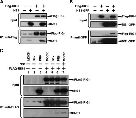
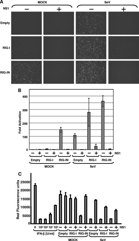
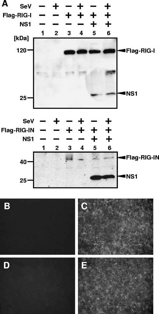
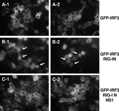
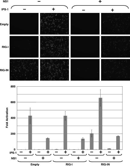
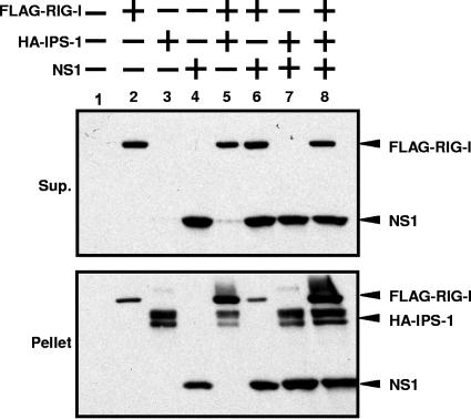
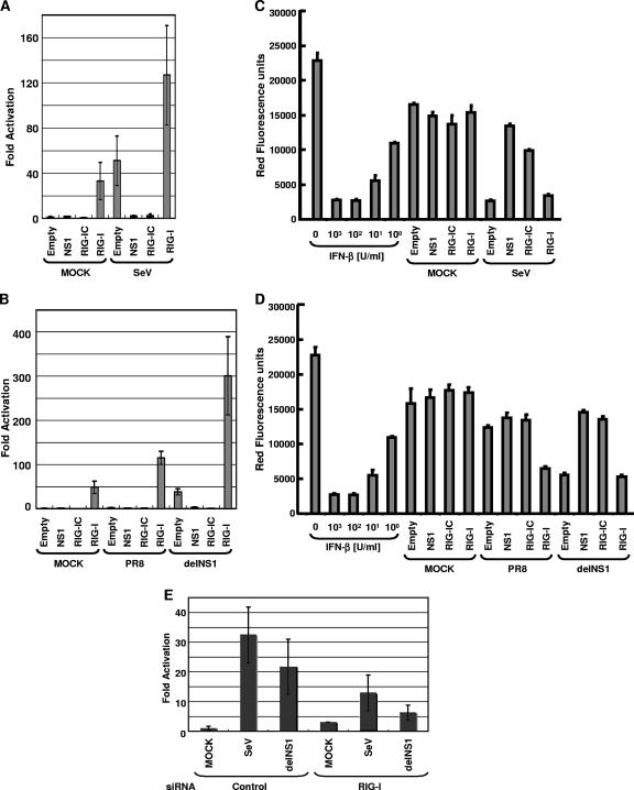
The retinoic acid-inducible gene I product (RIG-I) has been identified as a cellular sensor of RNA virus infection resulting in beta interferon (IFN-beta) induction. However, many viruses are known to encode viral products that inhibit IFN-beta production. In the case of influenza A virus, the viral nonstructural protein 1 (NS1) prevents the induction of the IFN-beta promoter by inhibiting the activation of transcription factors, including IRF-3, involved in IFN-beta transcriptional activation. The inhibitory properties of NS1 appear to be due at least in part to its binding to double-stranded RNA (dsRNA), resulting in the sequestration of this viral mediator of RIG-I activation. However, the precise effects of NS1 on the RIG-I-mediated induction of IFN-beta have not been characterized. We now report that the NS1 of influenza A virus interacts with RIG-I and inhibits the RIG-I-mediated induction of IFN-beta. This inhibition was apparent even when a mutant RIG-I that is constitutively activated (in the absence of dsRNA) was used to trigger IFN-beta production. Coexpression of RIG-I, its downstream signaling partner, IPS-1, and NS1 resulted in increased levels of RIG-I and NS1 within an IPS-1-rich, solubilization-resistant fraction after cell lysis. These results suggest that RIG-I, IPS-1, and NS1 become part of the same complex. Consistent with this idea, NS1 was also found to inhibit IFN-beta promoter activation by IPS-1 overexpression. Our results indicate that, in addition to sequestering dsRNA, the NS1 of influenza A virus binds to RIG-I and inhibits downstream activation of IRF-3, preventing the transcriptional induction of IFN-beta.


Figures

FIG. 1.
FIG. 1.
RIG-I and NS1 interact in coimmunoprecipitation experiments. IP, immunoprecipitation. (A) 293T cells were transfected with 3.5 μg of pCAGGS-Flag-RIG-I (third and fourth lanes) and/or 3.5 μg of pCAGGS-NS1 (second and fourth lanes) by use of Lipofectamine 2000. The total DNA amount was normalized with empty pCAGGS plasmid. At 48 h posttransfection, cells were subjected to reversible cross-linking immunoprecipitation assays using anti-Flag antibodies. RIG-I and NS1 were detected by Western blotting using monoclonal anti-Flag and polyclonal anti-NS1 antibodies. (B) 293T cells were transfected with 3.5 μg of pCAGGS-Flag-RIG-I (second and fourth lanes) and/or 3.5 μg of pCAGGS-NS1-GFP (third and fourth lanes) by use of Lipofectamine 2000. The total DNA amount was normalized with empty pCAGGS plasmid. At 48 h posttransfection, cells were subjected to the reversible cross-linking immunoprecipitation assays using monoclonal anti-GFP antibodies. RIG-I and NS1-GFP were detected by Western blotting using monoclonal anti-Flag and monoclonal anti-GFP antibodies. (C) 293T cells were transfected with 3.5 μg of pCAGGS-Flag-RIG-I (lanes 4 to 7) with (lane 7) or without 3.5 μg of pCAGGS-NS1 by use of Lipofectamine 2000. The total DNA amount was normalized with empty pCAGGS plasmid. At 48 h posttransfection, cells were mock infected (lanes 1, 4, and 7) or infected with SeV (lanes 2 and 5) or PR8 (lanes 3 and 6). Cells were collected at 9 h postinfection and subjected to the reversible cross-linking immunoprecipitation assays using anti-Flag antibodies. RIG-I and NS1 were detected by Western blotting using monoclonal anti-Flag and polyclonal anti-NS1 antibodies. Weak bands in the first and second lanes and lanes 1, 2, and 3, of the third blots in panels A and C, respectively, most likely correspond to anti-Flag antibody used in the IP and appearing at low levels after peptide elution of immunocomplexes from the beads.
FIG. 2.
FIG. 2.
Inhibition of RIG-I-mediated IFN-β production by NS1. (A) Fluorescent reporter assays. 293T cells were transfected with 0.5 μg of pIFN-β-GFP/CAT, 0.5 μg of pCAGGS-Luc, 250 ng of pCAGGS-Flag-RIG-I or pCAGGS-Flag-RIG-IN (as indicated at the left of each panel), and 2.5 μg of pCAGGS-NS1 (as indicated at the top of each panel) by using CaPO4. The total DNA amount was normalized with empty pCAGGS plasmid. At 24 h posttransfection, cells were mock infected or infected with SeV at an MOI of 10 (as indicated at the top). Twenty-four hours postinfection, the cells were observed by use of a fluorescence microscope. (B) Enzymatic reporter assays. After the fluorescence was observed, cells were collected and lysed in reporter lysis buffer (Promega). The lysates were cleared by centrifugation, and supernatants were subjected to CAT and luciferase assays. Relative CAT activity was normalized to the values obtained for luciferase activity. The y axis indicates activation (n-fold) of the IFN-β promoter by setting the reporter activity in the absence of RIG-I, RIG-IN, and NS1 at 1. The reporter assays were conducted in triplicate. Error bars indicate standard deviations. (C) Bioassays. Before the collection of cells, culture media were harvested and subjected to a bioassay for determination of IFN levels/production based on RFP expression in NDV-mRFP-infected Vero cells. The y axis indicates the red fluorescence produced by NDV-mRFP.
FIG. 3.
FIG. 3.
Expression levels of RIG-I and SeV infection in cells expressing NS1. (A) Expression level of RIG-I in cells expressing NS1. 293T cells were transfected with 0.5 μg of the pHISG54-GFP/CAT reporter plasmid (lanes 1 to 6), 0.25 μg of the pCAGGS-RIG-I (top panel, lanes 3 to 6) or pCAGGS-RIG-IN (bottom panel, lanes 3 to 6) plasmid, and 2 μg of empty plasmid (lanes 1 to 4) or NS1 expression plasmid (lanes 5 and 6). At 24 h posttransfection, cells were mock infected (lanes 1, 3, and 5) or infected with SeV (lanes 2, 4, and 6). Cells were collected and lysed in SDS-PAGE gel loading buffer at 24 h postinfection. Proteins were detected by Western blotting by using monoclonal anti-Flag M2 and monoclonal NS1 antibodies. An unspecific weak band can be seen in the top panel between Flag-RIG-I and NS1 signals. (B through E) SeV infection levels in cells expressing NS1. Cells transfected with empty plasmid (B and C) or NS1 expression plasmid (D and E) were mock infected (B and D) or infected with SeV (C and E). At 24 h postinfection, cells were fixed and viral proteins were detected by indirect immunofluorescence assays with SeV-specific antibodies.
FIG. 4.
FIG. 4.
Inhibition of RIG-IN-induced nuclear translocation of GFP-IRF-3 by NS1. 293T cells were transfected with 250 ng of the pEGFP-IRF-3 expression plasmid in the absence (A) or presence of 250 ng of RIG-IN expression plasmid alone (B) or with 2 μg of NS1 expression plasmid (C) as indicated. Nuclear translocation of GFP-IRF-3 was monitored at 48 h posttransfection under a fluorescence microscope. Representative cells with nuclear translocation of GFP-IRF-3 are indicated with arrows. Two representative fields per transfection are shown.
FIG. 5.
FIG. 5.
Inhibition of IPS-1 by NS1. (Upper panel) Fluorescent reporter assays. 293T cells were transfected with 0.5 μg of pIFN-β-GFP/CAT and 0.5 μg of pCAGGS-Luc, 250 ng of pCAGGS-Flag-RIG-I or pCAGGS-Flag-RIG-IN (as indicated at the left), 0.25 μg of pCAAGGS-HA-IPS-1 (as indicated in the top), and 2.5 μg of pCAGGS-NS1 (as indicated in the top) by using CaPO4. The total DNA amount was normalized with empty pCAGGS plasmid. Expression of GFP driven by the IFN-β promoter was observed by fluorescence microscopy. (Lower panel) Enzymatic reporter assays. After the fluorescence was observed, cells were collected and lysed in reporter lysis buffer (Promega). The lysates were cleared by centrifugation and supernatants were subjected to CAT and luciferase assays. Relative CAT activity was normalized to the values obtained for luciferase activity. The y axis indicates activation (n-fold) of the IFN-β promoter by setting the reporter activity in the absence of IPS-1, RIG-I, RIG-IN, and NS1 at 1. The reporter assays were conducted in triplicate. Error bars indicate standard deviations.
FIG. 6.
FIG. 6.
Copurification of IPS-1, RIG-I, and NS1 in detergent insoluble complexes. A total of 2 × 10⁶ 293T cells were transfected with 2.5 μg of pCAGGS-FLAG-RIG-I (lanes 2, 5, 6, and 8), 2.5 μg of pCAGGS-HA-IPS-1 (lanes 3, 5, 7, and 8), and/or 2.5 μg of pCAGGS-NS1 (lanes 4, 6, 7, and 8). Amounts of transfected DNA were adjusted with empty pCAGGS plasmid. At 48 h posttransfection, cells were subjected to reversible cross-linking reaction and then lysed in 500 μl of RIPA buffer. After centrifugation at 10,000 × g for 5 min, cell supernatants (Sup.) and pellets were separated. The supernatant was mixed with equal volume of 2 × SDS gel loading buffer. The pellet was resuspended in 50 μl of SDS gel loading buffer. The samples were boiled for 5 min and then subjected to 12% SDS-PAGE. Expression levels of Flag-RIG-I, HA-IPS-1, and NS1 were detected by Western blotting using anti-Flag, anti-HA, and anti-NS1 antibodies, respectively.
FIG. 7.
FIG. 7.
Involvement of RIG-I in SeV- and influenza A virus-induced IFN-β production. (A and B) RIG-IC inhibits activation of IFN-β promoter by viral infection. 293T cells were transfected with 0.5 μg of pIFNβ-CAT, 0.5 μg of pCAGGS-Luc, and 0.5 μg of pCAGGS, pCAGGS-NS1, pCAGGS-Flag-RIG-IC, or pCAGGS-Flag-RIG-I, as indicated, using CaPO4. The reporter assays were conducted in triplicate. At 24 h posttransfection, cells were mock infected or infected with SeV, influenza A/PR/8/34 virus, or delNS1 virus at an MOI of 1. Cells were collected at 24 h postinfection and subjected to CAT assays. Relative CAT activity was normalized against relative luciferase activity. The y axis indicates activation (n-fold) of the IFN-β promoter by setting the reporter activities of empty pCAGGS-transfected and mock-infected cells at 1. (C and D) Suppression of virus-induced IFN-β production by RIG-IC and NS1. Before cells were collected, culture media were harvested and subjected to a bioassay for determination of IFN levels/production based on RFP expression in NDV-mRFP-infected Vero cells. The y axis indicates the red fluorescence produced by NDV-mRFP. (E) siRNA for RIG-I inhibits activation of IFN-β promoter by viral infection. To knock down the expression of RIG-I, RNA interference was performed with siRNA (see Materials and Methods). Expression of RIG-I was downregulated in Stealth_1199-transfected cells but not in Stealth_control_1199-transfected cells (data not shown), and that of β-actin was not affected by the transfection of both Stealth_1199 and Stealth_control_1199 (data not shown). After incubation, cells were mock infected or infected with SeV or delNS1. After the infection, cells were incubated for 24 h and then cells were collected and subjected to CAT assay. The y axis indicates activation (n-fold) of IFN-β promoter by setting the reporter activity of Stealth_control_1199 and the reporter plasmid-transfected and mock-infected cells at 1.

References

    1. Akira, S., and K. Takeda. 2004. Toll-like receptor signalling. Nat. Rev. Immunol. 4:499-511. - PubMed
    1. Alexopoulou, L., A. C. Holt, R. Medzhitov, and R. A. Flavell. 2001. Recognition of double-stranded RNA and activation of NF-kappaB by Toll-like receptor 3. Nature 413:732-738. - PubMed
    1. Andrejeva, J., K. S. Childs, D. F. Young, T. S. Carlos, N. Stock, S. Goodbourn, and R. E. Randall. 2004. The V proteins of paramyxoviruses bind the IFN-inducible RNA helicase, mda-5, and inhibit its activation of the IFN-beta promoter. Proc. Natl. Acad. Sci. USA 101:17264-17269. - PMC - PubMed
    1. Basler, C. F., A. Mikulasova, L. Martinez-Sobrido, J. Paragas, E. Muhlberger, M. Bray, H. D. Klenk, P. Palese, and A. García-Sastre. 2003. The Ebola virus VP35 protein inhibits activation of interferon regulatory factor 3. J. Virol. 77:7945-7956. - PMC - PubMed
    1. Bergmann, M., A. García-Sastre, E. Carnero, H. Pehamberger, K. Wolff, P. Palese, and T. Muster. 2000. Influenza virus NS1 protein counteracts PKR-mediated inhibition of replication. J. Virol. 74:6203-6206. - PMC - PubMed

Publication types

MeSH terms

LinkOut - more resources