. 2007 Jan;81(2):1000-12.
doi: 10.1128/JVI.01629-06. Epub 2006 Nov 1.

Inefficient human immunodeficiency virus replication in mobile lymphocytes

Affiliations

Inefficient human immunodeficiency virus replication in mobile lymphocytes

Marion Sourisseau et al. J Virol. 2007 Jan.

Abstract

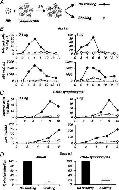
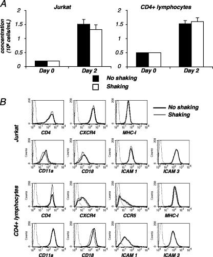
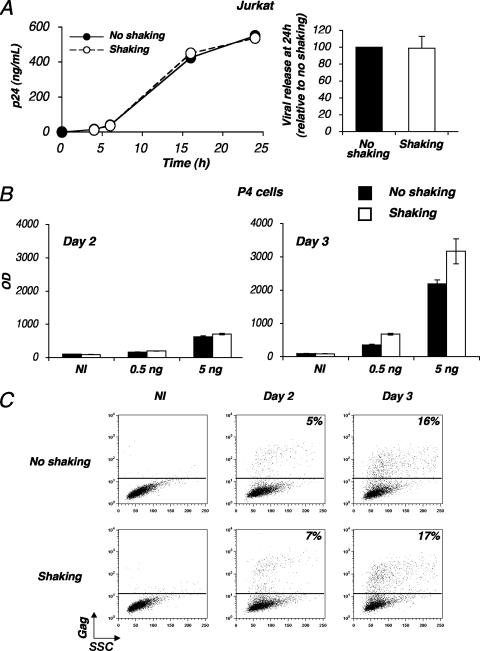
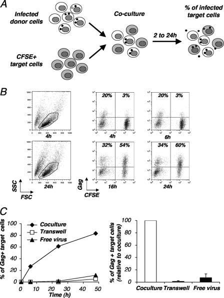
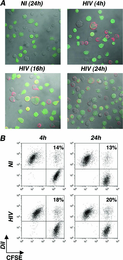
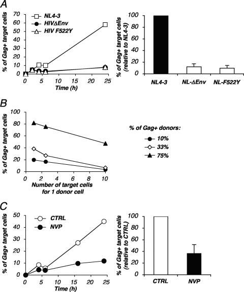
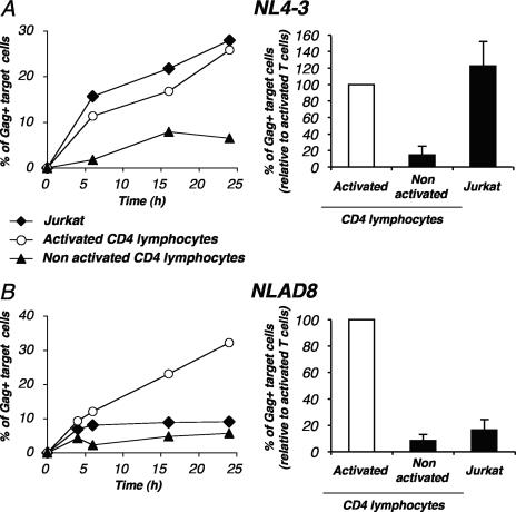
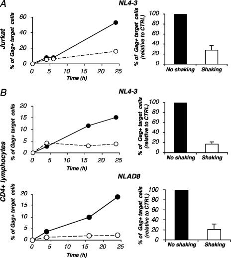
Cell-to-cell viral transfer facilitates the spread of lymphotropic retroviruses such as human immunodeficiency virus (HIV) and human T-cell leukemia virus (HTLV), likely through the formation of "virological synapses" between donor and target cells. Regarding HIV replication, the importance of cell contacts has been demonstrated, but this phenomenon remains only partly characterized. In order to alter cell-to-cell HIV transmission, we have maintained cultures under continuous gentle shaking and followed viral replication in this experimental system. In lymphoid cell lines, as well as in primary lymphocytes, viral replication was dramatically reduced in shaken cultures. To document this phenomenon, we have developed an assay to assess the relative contributions of free and cell-associated virions in HIV propagation. Acutely infected donor cells were mixed with carboxyfluorescein diacetate succinimidyl ester-labeled lymphocytes as targets, and viral production was followed by measuring HIV Gag expression at different time points by flow cytometry. We report that cellular contacts drastically enhance productive viral transfer compared to what is seen with infection with free virus. Productive cell-to-cell viral transmission required fusogenic viral envelope glycoproteins on donor cells and adequate receptors on targets. Only a few syncytia were observed in this coculture system. Virus release from donor cells was unaffected when cultures were gently shaken, whereas virus transfer to recipient cells was severely impaired. Altogether, these results indicate that cell-to-cell transfer is the predominant mode of HIV spread and help to explain why this virus replicates so efficiently in lymphoid organs.


Figures

FIG. 1.
FIG. 1.
Inefficient HIV-1 replication in shaken cultures of lymphocytes. (A) Experimental protocol. Jurkat or primary CD4+ T cells were exposed to HIV (NL4-3 strain) for 2 h, unbound virus was removed, and cells were cultivated in static conditions (no shaking) or placed on a rocker and continuously and gently shaken (40 movements/min). Viral replication was then measured at different days p.i. (B to D) HIV-1 replication in static or shaken Jurkat (B) and primary CD4+ (C) T cells. Cells were exposed to the indicated HIV inocula (0.1 and 1 ng p24/106 cells). Viral replication is depicted as the percentage of Gag+ cells as measured by flow cytometry (upper panels) and as Gag p24 production in supernatants as measured by ELISA (lower panels). Data are representative of three independent experiments. (D) Means ± standard deviations (SD) of three independent experiments are depicted, with 100% corresponding to supernatant Gag p24 values obtained at the peak of infection (day 6 or day 8 p.i.).
FIG. 2.
FIG. 2.
Characteristics of shaken T cells. (A) Growth kinetics of shaken cells. Noninfected Jurkat cells (left panel) or primary CD4+ lymphocytes (right panel), at 2 × 10⁵ cells/ml, were grown with or without rocking. At the indicated time points, concentrations of living cells were measured in cultures. Data are means ± SD of triplicates and are representative of four independent experiments. (B) Surface expression of various receptors in shaken lymphocytes. Noninfected Jurkat cells (upper panels) or primary CD4+ lymphocytes (lower panels) were kept static or shaken for 24 or 48 h, respectively. Cells were then stained with antibodies against the indicated surface receptors and analyzed by flow cytometry. An isotypic mAb was used as a negative control (dotted line). Data are representative of three independent experiments. MHC-I, major histocompatibility complex I.
FIG. 3.
FIG. 3.
Effects of shaking on HIV replication. (A) Shaking does not affect viral release from lymphocytes. Jurkat cells were productively infected with HIV (NL4-3 strain). After a few days of culturing, when about 25% of the cells expressed Gag antigens, cells were treated with NVP to prevent further viral spread. (Left panel) Viral release from infected cells was followed by measuring Gag p24 content in supernatants at the indicated time points. Data are representative of three independent experiments. (Right panel) Relative efficiency of each experimental condition. A mean ± SD of three independent experiments (24-h time point) is depicted, with 100% corresponding to values obtained without shaking. (B and C) HIV infection of static or shaken HeLa CD4 cells. HeLa CD4 cells (P4 clone) were infected with HIV (NL4-3 strain) at the indicated viral doses (0.5 or 5 ng p24/0.5 × 106 cells for panel B; 100 ng p24/1 × 106 cells for panel C). Cells were then kept static or were gently shaken. (B) Infection was assessed at days 2 and 3 p.i. by measuring β-galactosidase activity in cell extracts. Data are means ± SD of triplicates and are representative of three independent experiments. (C) Infection was assessed by measuring Gag p24 expression by flow cytometry. Data are representative of three independent experiments. NI, noninfected cells; OD, optical density; SSC, side scatter.
FIG. 4.
FIG. 4.
Analysis of cell-to-cell HIV transfer by flow cytometry. (A) Principle of the flow cytometry assay. Lymphocytes were productively infected with HIV. After a few days of culturing, when about 10 to 75% of the cells expressed Gag antigens, cells were cocultivated with recipient cells stained with CFSE. Gag expression was then measured for target (CFSE+) cells at various time points by flow cytometry. (B) A representative experiment. Productively infected Jurkat cells (20% of Gag+ cells at the beginning of the assay) were cocultivated with target CFSE+ Jurkat cells at a 1/1 ratio. The percentages of Gag+ cells among donors and targets are indicated at 4, 6, 16, and 24 h postcoculture. Cell viability was not significantly affected by the coculture, as visualized by side (SSC) and forward (FSC) scatter plots at 4 h and 24 h. Data are representative of at least 10 independent experiments. (C) Efficient HIV spread requires contact between infected and target lymphocytes. Productively infected Jurkat cells were cocultivated with target CFSE+ Jurkat cells at a 1/1 ratio either directly or in a Transwell chamber in which donors and recipient cells were separated by a virus-permeable membrane. Targets were also directly exposed to a high concentration of free virus (200 ng/ml). (Left panel) Results are presented as the percentages of Gag+ cells within CFSE+ Jurkat targets. (Right panel) Relative efficiency of each experimental condition. Means ± SD of three independent experiments (24-h time point) are depicted, with 100% corresponding to values obtained for target cells by direct coculturing.
FIG. 5.
FIG. 5.
Analysis of syncytium formation in Jurkat cells. (A) Confocal microscopy analysis. Jurkat cells were productively infected with HIV. After a few days of culturing, when about 70% expressed Gag antigens, cells were cocultivated at a 1/1.5 ratio with recipient cells stained with CFSE. Gag (red) and CFSE stainings are depicted at various time points of the coculture. One out of three representative experiments is shown. (B) Flow cytometry analysis. Productively infected Jurkat cells (20% of Gag+ cells) were stained with the fluorescent probe DiI (red) and cocultivated with target CFSE+ Jurkat cells at a 1/1 ratio. The percentages of double-fluorescent cells (DiI+ CFSE+ cells among total CFSE+ cells), which correspond to cell-cell clustering or fusion, are depicted at the indicated time points. Data are representative of three independent experiments. NI, noninfected cells.
FIG. 6.
FIG. 6.
Roles of viral envelope glycoproteins and reverse transcriptase in HIV cell-to-cell transfer. (A) HIV cell-to-cell transfer requires fusogenic envelope glycoproteins. Jurkat cells infected with wild-type (NL4-3) or envelope-deleted (HIVΔenv) viruses or with a mutant virus (HIV F522Y mutant) carrying a nonfusogenic gp120 were used as donors in the cell-to-cell transfer assay. Infection of donors was performed with VSV-G-pseudotyped viruses in order to obtain about 30 to 45% Gag+ cells. These cells were then cocultivated with target CFSE+ Jurkat cells at a 1/2.5 ratio for the indicated time points. (Left panel) Results are presented as the percentages of Gag-positive cells within CFSE+ Jurkat targets. (Right panel) Relative efficiency of each experimental condition. Means ± SD of four independent experiments (24-h time point) are depicted, with 100% corresponding to values obtained for target cells with the wild-type virus. (B) Quantitative analysis of HIV cell-to-cell transfer. Productively HIV-infected Jurkat cells (10%, 33%, and 75% Gag+ cells, respectively, at the beginning of the assay) were cocultivated with target CFSE+ Jurkat cells at the indicated number of targets for one donor cell. The percentages of Gag+ cells among CFSE+ targets at 24 h postcoculture are depicted. Data are representative of five independent experiments. (C) HIV cell-to-cell transfer is inhibited by nevirapine. Productively infected Jurkat cells were cocultivated with target CFSE+ Jurkat cells with or without NVP, a reverse transcriptase inhibitor. (Left panel) Results are presented as the percentages of Gag-positive cells within CFSE+ Jurkat targets. (Right panel) Relative efficiency of each experimental condition. A mean ± SD of four independent experiments (24-h time point) is depicted, with 100% corresponding to values obtained for target cells without NVP. CTRL, control.
FIG. 7.
FIG. 7.
HIV cell-to-cell transfer in primary lymphocytes. Primary CD4+ T cells were activated with PHA and maintained in IL-2 and then infected with the X4 strain NL4-3 (A) or with the R5 strain NLAD8 (B) in order to obtain about 30% Gag+ cells. These cells were then used as donors in the flow cytometry-based assay of viral transfer. Infected cells were cocultivated with the indicated target CFSE+ cells, i.e., Jurkat cells, or autologous nonactivated or activated CD4 lymphocytes, at a 1/1 ratio at the indicated time points. (Left panels) Results are presented as the percentages of Gag-positive cells within CFSE+ targets. One out of four independent experiments is shown. (Right panels) Relative efficiency of each experimental condition. Means ± SD of four independent experiments (24-h time point) are depicted, with 100% corresponding to values obtained with activated CD4+ lymphocytes as targets.
FIG. 8.
FIG. 8.
Inefficient HIV cell-to-cell transfer in shaken lymphocytes. Productively infected Jurkat cells (A) or primary CD4+ cells (B) (20 to 25% Gag+ cells) were cocultivated with target CFSE+ Jurkat cells or primary CD4+ cells, respectively, with or without shaking. (Left panels) Results are presented as the percentages of Gag+ cells within CFSE+ targets. (Right panels) Relative efficiency of each experimental condition. Means ± SD of three independent experiments (24-h time point) are depicted, with 100% corresponding to values obtained for target cells without shaking. CTRL, control.

Similar articles

Cited by

References

    1. Aiken, C., and D. Trono. 1995. Nef stimulates human immunodeficiency virus type 1 proviral DNA synthesis. J. Virol. 69:5048-5056. - PMC - PubMed
    1. Alfsen, A., H. Yu, A. Magerus-Chatinet, A. Schmitt, and M. Bomsel. 2005. HIV-1-infected blood mononuclear cells form an integrin- and agrin-dependent viral synapse to induce efficient HIV-1 transcytosis across epithelial cell monolayer. Mol. Biol. Cell 16:4267-4279. - PMC - PubMed
    1. Arrighi, J. F., M. Pion, E. Garcia, J. M. Escola, Y. van Kooyk, T. B. Geijtenbeek, and V. Piguet. 2004. DC-SIGN-mediated infectious synapse formation enhances X4 HIV-1 transmission from dendritic cells to T cells. J. Exp. Med. 200:1279-1288. - PMC - PubMed
    1. Bar, S., and M. Alizon. 2004. Role of the ectodomain of the gp41 transmembrane envelope protein of human immunodeficiency virus type 1 in late steps of the membrane fusion process. J. Virol. 78:811-820. - PMC - PubMed
    1. Barnard, A. L., T. Igakura, Y. Tanaka, G. P. Taylor, and C. R. Bangham. 2005. Engagement of specific T-cell surface molecules regulates cytoskeletal polarization in HTLV-1-infected lymphocytes. Blood 106:988-995. - PubMed

Publication types

LinkOut - more resources