

Sheep-passaged bovine spongiform encephalopathy agent exhibits altered pathobiological properties in bovine-PrP transgenic mice
- PMID: 17079295
- PMCID: PMC1797487
- DOI: 10.1128/JVI.01356-06
Sheep-passaged bovine spongiform encephalopathy agent exhibits altered pathobiological properties in bovine-PrP transgenic mice
Abstract
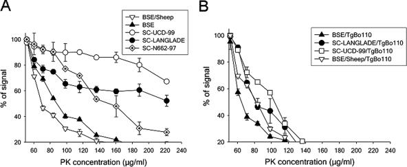
Sheep can be experimentally infected with bovine spongiform encephalopathy (BSE), and the ensuing disease is similar to scrapie in terms of pathogenesis and clinical signs. BSE infection in sheep is an animal and human health concern. In this study, the transmission in BoPrP-Tg110 mice of prions from BSE-infected sheep was examined and compared to the transmission of original cattle BSE in cattle and sheep scrapie prions. Our results indicate no transmission barrier for sheep BSE prions to infect BoPrP-Tg110 mice, but the course of the disease is accelerated compared to the effects of the original BSE isolate. The shortened incubation period of sheep BSE in the model was conserved in subsequent passage in BoPrP-Tg110 mice, indicating that it is not related to infectious titer differences. Biochemical signature, lesion profile, and PrP(Sc) deposition pattern of both cattle and sheep BSE were similar. In contrast, all three sheep scrapie isolates tested showed an evident transmission barrier and further adaptation in subsequent passage. Taken together, those data indicate that BSE agent can be altered by crossing a species barrier, raising concerns about the virulence of this new prion towards other species, including humans. The BoPrP-Tg110 mouse bioassay should be considered as a valuable tool for discriminating scrapie and BSE in sheep.
Figures

Similar articles
-
Transmission barriers for bovine, ovine, and human prions in transgenic mice.J Virol. 2005 May;79(9):5259-71. doi: 10.1128/JVI.79.9.5259-5271.2005. J Virol. 2005. PMID: 15827140 Free PMC article.
-
Early detection of PrPres in BSE-infected bovine PrP transgenic mice.Arch Virol. 2003 Apr;148(4):677-91. doi: 10.1007/s00705-002-0958-4. Arch Virol. 2003. PMID: 12664293
-
Compelling transgenetic evidence for transmission of bovine spongiform encephalopathy prions to humans.Proc Natl Acad Sci U S A. 1999 Dec 21;96(26):15137-42. doi: 10.1073/pnas.96.26.15137. Proc Natl Acad Sci U S A. 1999. PMID: 10611351 Free PMC article.
-
Transmission of bovine spongiform encephalopathy and scrapie to mice: strain variation and the species barrier.Philos Trans R Soc Lond B Biol Sci. 1994 Mar 29;343(1306):405-11. doi: 10.1098/rstb.1994.0036. Philos Trans R Soc Lond B Biol Sci. 1994. PMID: 7913758 Review.
-
Transgenic models of prion disease.Arch Virol Suppl. 2000;(16):113-24. doi: 10.1007/978-3-7091-6308-5_10. Arch Virol Suppl. 2000. PMID: 11214913 Review.
Cited by
-
Prion Diseases in Animals and Zoonotic Potential.Food Saf (Tokyo). 2016 Dec 7;4(4):105-109. doi: 10.14252/foodsafetyfscj.2016021. eCollection 2016 Dec. Food Saf (Tokyo). 2016. PMID: 32231913 Free PMC article. Review.
-
Enhanced virulence of sheep-passaged bovine spongiform encephalopathy agent is revealed by decreased polymorphism barriers in prion protein conversion studies.J Virol. 2014 Mar;88(5):2903-12. doi: 10.1128/JVI.02446-13. Epub 2013 Dec 26. J Virol. 2014. PMID: 24371051 Free PMC article.
-
Transgenic Mouse Bioassay: Evidence That Rabbits Are Susceptible to a Variety of Prion Isolates.PLoS Pathog. 2015 Aug 6;11(8):e1004977. doi: 10.1371/journal.ppat.1004977. eCollection 2015 Aug. PLoS Pathog. 2015. PMID: 26247589 Free PMC article.
-
Transgenic mice expressing porcine prion protein resistant to classical scrapie but susceptible to sheep bovine spongiform encephalopathy and atypical scrapie.Emerg Infect Dis. 2009 Aug;15(8):1214-21. doi: 10.3201/eid1508.081218. Emerg Infect Dis. 2009. PMID: 19751582 Free PMC article.
-
Molecular characterisation of atypical BSE prions by mass spectrometry and changes following transmission to sheep and transgenic mouse models.PLoS One. 2018 Nov 8;13(11):e0206505. doi: 10.1371/journal.pone.0206505. eCollection 2018. PLoS One. 2018. PMID: 30408075 Free PMC article.
References
-
- Andreoletti, O., C. Lacroux, A. Chabert, L. Monnereau, G. Tabouret, F. Lantier, P. Berthon, F. Eychenne, S. Lafond-Benestad, J. M. Elsen, and F. Schelcher. 2002. PrP(Sc) accumulation in placentas of ewes exposed to natural scrapie: influence of foetal PrP genotype and effect on ewe-to-lamb transmission. J. Gen. Virol. 83:2607-2616. - PubMed
-
- Bartz, J. C., R. F. Marsh, D. I. McKenzie, and J. M. Aiken. 1998. The host range of chronic wasting disease is altered on passage in ferrets. Virology 251:297-301. - PubMed
-
- Bellworthy, S. J., G. Dexter, M. Stack, M. Chaplin, S. A. Hawkins, M. M. Simmons, M. Jeffrey, S. Martin, L. Gonzalez, and P. Hill. 2005. Natural transmission of BSE between sheep within an experimental flock. Vet. Rec. 157:206. - PubMed
-
- Bruce, M. E., R. G. Will, J. W. Ironside, I. McConnell, D. Drummond, A. Suttie, L. McCardle, A. Chree, J. Hope, C. Birkett, S. Cousens, H. Fraser, and C. J. Bostock. 1997. Transmissions to mice indicate that ‘new variant’ CJD is caused by the BSE agent. Nature 389:498-501. - PubMed
-
- Brun, A., J. Castilla, M. A. Ramirez, K. Prager, B. Parra, F. J. Salguero, D. Shiveral, C. Sanchez, J. M. Sanchez-Vizcaino, A. Douglas, and J. M. Torres. 2004. Proteinase K enhanced immunoreactivity of the prion protein-specific monoclonal antibody 2A11. Neurosci. Res. 48:75-83. - PubMed
Publication types
MeSH terms
Substances
LinkOut - more resources
Full Text Sources
Molecular Biology Databases
Research Materials
