. 2007 Feb;81(3):1461-71.
doi: 10.1128/JVI.01880-06. Epub 2006 Nov 1.

White spot syndrome virus annexes a shrimp STAT to enhance expression of the immediate-early gene ie1

Affiliations

White spot syndrome virus annexes a shrimp STAT to enhance expression of the immediate-early gene ie1

Wang-Jing Liu et al. J Virol. 2007 Feb.

Abstract

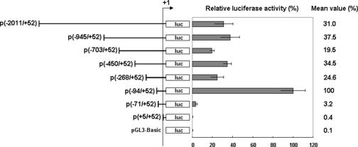
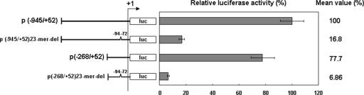
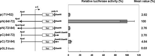
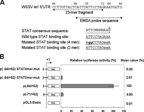
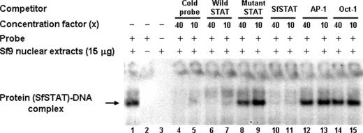
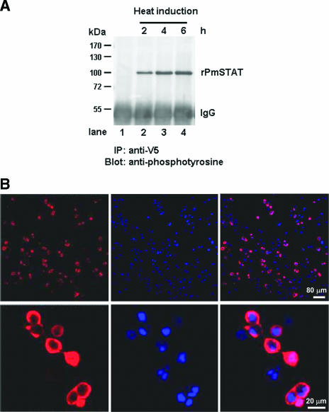
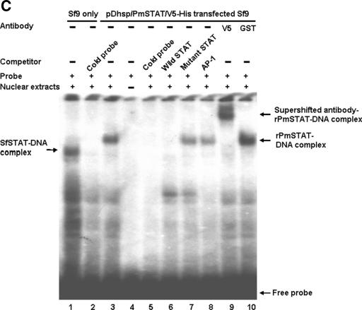
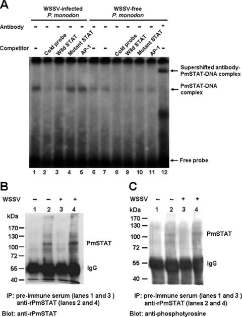
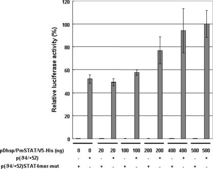
Although the Janus kinase-signal transducer and activator of transcription (JAK-STAT) signaling pathway is part of the antiviral response in arthropods such as Drosophila, here we show that white spot syndrome virus (WSSV) uses a shrimp STAT as a transcription factor to enhance viral gene expression in host cells. In a series of deletion and mutation assays using the WSSV immediate-early gene ie1 promoter, which is active in shrimp cells and also in insect Sf9 cells, an element containing a STAT binding motif was shown to be important for the overall level of WSSV ie1 promoter activity. In the Sf9 insect cell line, a specific protein-DNA complex was detected by using electrophoresis mobility shift assays (EMSA) with the 32P-labeled STAT binding motif of the WSSV ie1 promoter as the probe. When recombinant Penaeus monodon STAT (rPmSTAT) was overexpressed in Sf9 cells, EMSA with specific antibodies confirmed that the STAT was responsible for the formation of the specific protein-DNA complex. Another EMSA showed that in WSSV-infected P. monodon, levels of activated PmSTAT were higher than in WSSV-free P. monodon. A transactivation assay of the WSSV ie1 promoter demonstrated that increasing the level of rPmSTAT led to dose-dependent increases in ie1 promoter activity. These results show that STAT directly transactivates WSSV ie1 gene expression and contributes to its high promoter activity. We conclude that WSSV successfully annexes a putative shrimp defense mechanism, which it uses to enhance the expression of viral immediate-early genes.


Figures

FIG. 1.
FIG. 1.
Functional mapping of deletions of the WSSV ie1 promoter. Relative luciferase activity has been normalized to the activity of the p(−94/+52) vector, which was arbitrarily set to 100%. Data represent the means from triplicate experiments. Error bars show the SDs. The plasmid numbers in parentheses specify the beginning and end positions of the promoter fragments, and the arrow labeled +1 marks the transcription start site. The difference between the p(−94/+52) and p(−71/+52) plasmids is a 23-mer fragment that consists of an imperfect inverted repeat, −94CCTTGTTACTCATTTATTCCTAG−72.
FIG. 2.
FIG. 2.
Effect of deleting the 23-mer fragment from the WSSV ie1 promoter region. The two deletion constructs, p(−945/+52)23mer-del and p(−268/+52)23-mer-del, are both missing the −94/−72 region of the WSSV ie1 promoter. Data show the means of three repetitions, and error bars show the SDs.
FIG. 3.
FIG. 3.
Testing the enhancer activity of the 23-mer fragment. The solid arrows show the locations and orientations (sense or antisense) of the 23-mer fragment in two complementary plasmid pairs. In this figure, the (−94/+52) plasmid has been redesignated pN(−94/−72). Results are shown as means ± SDs from three independent experiments.
FIG. 4.
FIG. 4.
Site-directed mutagenesis of the STAT binding motif in the WSSV ie1 promoter region. (A) Locations and sequences of the 23-mer fragment and EMSA probe and the STAT consensus sequences. Sequences of the consensus, wild-type, and mutated STAT binding sites are shown. The boldface lowercase letters indicate the mutated nucleotides. (B) Relative luciferase activities of WSSV ie1 p(−94/+52) promoter constructs with wild-type or mutated STAT binding sites. Data represent the means ± SDs from three independent experiments.
FIG. 5.
FIG. 5.
EMSA of the −84/−64 region of the WSSV ie1 promoter with Sf9 cell nuclear extracts. Lane 1, 15 μg Sf9 cell nuclear extracts reacted with isotope-labeled probe (SfSTAT-DNA complex). Lane 2, isotope-labeled probe only. Lane 3, Sf9 cell nuclear extracts only. Lanes 4 to 15, SfSTAT-DNA complex competing with unlabeled probe, unlabeled wild type STAT oligonucleotide, 4-mer mutant STAT oligonucleotide, unlabeled SfSTAT oligonucleotide, unlabeled AP-1 oligonucleotide, and Oct-1 oligonucleotide. The relative concentrations of the unlabeled competitor oligonucleotides (40× or 10×) are indicated.
FIG.6.
FIG.6.
(A) Immunoprecipitation (IP) and Western blotting analysis of the phosphorylation status of rPmSTAT. Lane 1 (negative control), nuclear extract from Sf9 cells transfected with the empty plasmid pDhsp/V5-His. Lanes 2 to 4, phosphorylated rPmSTAT was detected in nuclear extracts from Sf9 cells transfected with pDhsp/PmSTAT/V5-His at 2 h after heat shock induction, and quantities of (pp)rPmSTAT increased through 4 to 6 h postinduction. IgG, immunoglobulin G. (B) Immunofluorescence staining of pDhsp/PmSTAT/V5-His-transfected Sf9 cells. Cells were probed with anti-V5 antibody coupled with Cy3-labeled secondary antibody (red) to detect STAT (left column) and counterstained with DAPI (blue) to show the location of the nuclei (middle column). The merged result (right column) shows that rPmSTAT is present in the cytoplasm and sometimes in the nuclei of the Sf9 cells. (C) EMSA and supershift EMSA to confirm that the WSSV ie1 promoter putative STAT binding region is binding to STAT. Lane 1, SfSTAT-DNA complex of 32P-labeled probe and untransfected Sf9 cell nuclear extract. Lane 2, SfSTAT-DNA complex competing with unlabeled probe. Lane 3, rPmSTAT-DNA complex of labeled probe and pDhsp/PmSTAT/V5-His-transfected Sf9 cell nuclear extract. Lane 4, labeled probe only. Lane 5, rPmSTAT-DNA complex competing with unlabeled probe. Lane 6, rPmSTAT-DNA complex competing with unlabeled wild-type STAT oligonucleotide. Lane 7, rPmSTAT-DNA complex competing with unlabeled mutant STAT oligonucleotide. Lane 8, rPmSTAT-DNA complex competing with unlabeled AP-1 oligonucleotide. Lane 9, binding of rPmSTAT-DNA complex with anti-V5 antibody directed against a V5 tag in rPmSTAT. Lane 10, an anti-GST antibody not specific for rPmSTAT failed to bind with and supershift the rPmSTAT-DNA complex. The concentration of the unlabeled competitors was in 40× molar excess relative to the labeled probe.
FIG.6.
FIG.6.
(A) Immunoprecipitation (IP) and Western blotting analysis of the phosphorylation status of rPmSTAT. Lane 1 (negative control), nuclear extract from Sf9 cells transfected with the empty plasmid pDhsp/V5-His. Lanes 2 to 4, phosphorylated rPmSTAT was detected in nuclear extracts from Sf9 cells transfected with pDhsp/PmSTAT/V5-His at 2 h after heat shock induction, and quantities of (pp)rPmSTAT increased through 4 to 6 h postinduction. IgG, immunoglobulin G. (B) Immunofluorescence staining of pDhsp/PmSTAT/V5-His-transfected Sf9 cells. Cells were probed with anti-V5 antibody coupled with Cy3-labeled secondary antibody (red) to detect STAT (left column) and counterstained with DAPI (blue) to show the location of the nuclei (middle column). The merged result (right column) shows that rPmSTAT is present in the cytoplasm and sometimes in the nuclei of the Sf9 cells. (C) EMSA and supershift EMSA to confirm that the WSSV ie1 promoter putative STAT binding region is binding to STAT. Lane 1, SfSTAT-DNA complex of 32P-labeled probe and untransfected Sf9 cell nuclear extract. Lane 2, SfSTAT-DNA complex competing with unlabeled probe. Lane 3, rPmSTAT-DNA complex of labeled probe and pDhsp/PmSTAT/V5-His-transfected Sf9 cell nuclear extract. Lane 4, labeled probe only. Lane 5, rPmSTAT-DNA complex competing with unlabeled probe. Lane 6, rPmSTAT-DNA complex competing with unlabeled wild-type STAT oligonucleotide. Lane 7, rPmSTAT-DNA complex competing with unlabeled mutant STAT oligonucleotide. Lane 8, rPmSTAT-DNA complex competing with unlabeled AP-1 oligonucleotide. Lane 9, binding of rPmSTAT-DNA complex with anti-V5 antibody directed against a V5 tag in rPmSTAT. Lane 10, an anti-GST antibody not specific for rPmSTAT failed to bind with and supershift the rPmSTAT-DNA complex. The concentration of the unlabeled competitors was in 40× molar excess relative to the labeled probe.
FIG. 7.
FIG. 7.
(A) EMSAs of nuclear extracts from WSSV-infected (72 hpi) and WSSV-free P. monodon, using a 32P-labeled ie1 promoter STAT binding sequence oligonucleotide as a probe. Competitors, when present, were in a 40× molar excess relative to the “hot” probe. Lane 7 used 20 μg of nuclear extract; all the other lanes used 10 μg. The antibody used in lane 12 was specifically directed against PmSTAT. (B and C) Immunoprecipitation (IP) and Western blot analysis of PmSTAT in nuclear extracts from WSSV-infected (+) and WSSV-free (−) P. monodon. The nuclear extracts (the same as those used in the EMSA) were immunoprecipitated with anti-rPmSTAT antiserum or preimmune serum (control), separated by gel electrophoresis, and probed with either anti-rPmSTAT antibody (B) or antiphosphotyrosine antibody (C). IgG, immunoglobulin G.
FIG. 8.
FIG. 8.
Dose-dependent transactivation of the WSSV ie1 promoter p(−94/+52) by recombinant PmSTAT. The data show relative luciferase activities at 6 h after heat shock in Sf9 cells that were cotransfected for 48 h with different concentrations of pDhsp/PmSTAT/V5-His and with either 500 ng of p(−94/+52) reporter plasmid or 500 ng of the 4-mer mutant plasmid. The relative luciferase activity of 500 ng of pDhsp/PmSTAT/V5-His was arbitrarily set to 100%. The means and standard deviations from three independent transfections are shown.

Similar articles

Cited by

References

    1. Agaisse, H., and N. Perrimon. 2004. The roles of JAK/STAT signaling in Drosophila immune responses. Immunol. Rev. 198:72-82. - PubMed
    1. Agaisse, H., U. M. Petersen, M. Boutros, B. Mathey-Prevot, and N. Perrimon. 2003. Signaling role of hemocytes in Drosophila JAK/STAT-dependent response to septic injury. Dev. Cell. 5:441-450. - PubMed
    1. Akira, S., Y. Nishio, M. Inoue, X. J. Wang, S. Wei, T. Matsusaka, K. Yoshida, T. Sudo, M. Naruto, and T. Kishimoto. 1994. Molecular cloning of APRF, a novel IFN-stimulated gene factor 3 p91-related transcription factor involved in the gp130-mediated signaling pathway. Cell 77:63-71. - PubMed
    1. Antonucci, T. K., and W. J. Rutter. 1989. Hepatitis B virus (HBV) promoters are regulated by the HBV enhancer in a tissue-specific manner. J. Virol. 63:579-583. - PMC - PubMed
    1. Barillas-Mury, C., Y.-S. Han, D. Seeley, and F. C. Kafatos. 1999. Anopheles gambiae Ag-STAT, a new insect member of the STAT family, is activated in response to bacterial infection. EMBO J. 18:959-967. - PMC - PubMed

Publication types

MeSH terms

Substances

LinkOut - more resources