. 2007 Feb;81(3):1350-9.
doi: 10.1128/JVI.01839-06. Epub 2006 Nov 1.

Role of V1V2 and other human immunodeficiency virus type 1 envelope domains in resistance to autologous neutralization during clade C infection

Affiliations

Role of V1V2 and other human immunodeficiency virus type 1 envelope domains in resistance to autologous neutralization during clade C infection

Rong Rong et al. J Virol. 2007 Feb.

Erratum in

  • J Virol. 2007 Nov;81(22):12715

Abstract

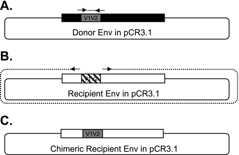
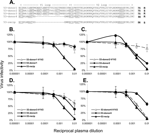
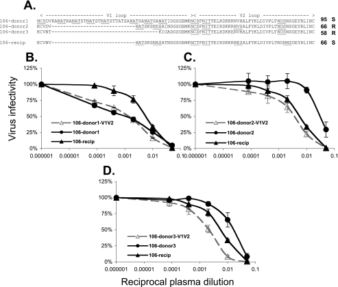
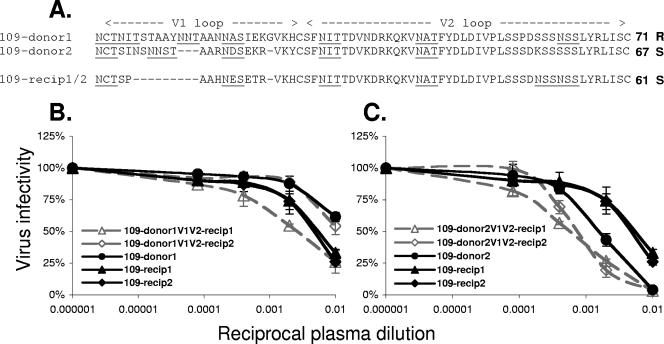
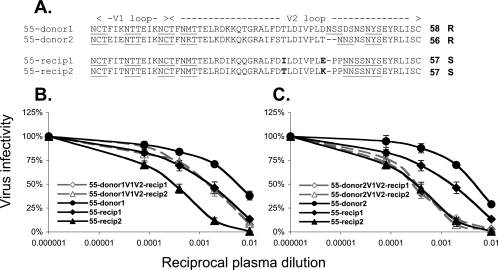
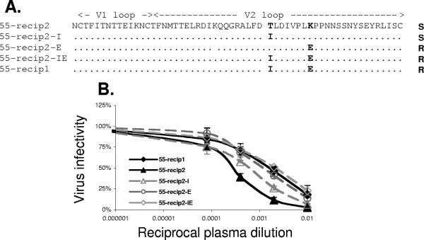
Biologically functional clade C envelope (Env) glycoproteins from the chronically (donor) and newly (recipient) infected partners of four heterosexual transmission pairs in Zambia were cloned and characterized previously. In each case, the donor viral quasispecies contained Envs that were resistant to autologous neutralization by contemporaneous plasma, while the recipient Envs were sensitive to neutralizing antibodies in this donor plasma sample. The donor Envs also varied in length, glycosylation, and amino acid sequence of the V1V2 hypervariable domain of gp120, while the recipient Envs were much more homogeneous. To assess the contribution of V1V2 to the neutralization phenotype of the donor Envs, V1V2 domains from neutralization-sensitive recipient Envs were replaced with donor V1V2 domains, and the autologous neutralization sensitivities of the chimeric Envs were evaluated using a virus-pseudotyping assay. Long donor V1V2 domains regulated sensitivity to autologous neutralization, although the effect was dependent on the Env background. Short donor V1V2 domains did not confer neutralization resistance. Primary sequence differences in V2 were also found to influence neutralization sensitivity in one set of recipient Envs. The results demonstrate that expansion of the V1V2 domain is one pathway to escape from autologous neutralization in subtype C Envs. However, V1V2-independent mechanisms of resistance also exist, suggesting that escape is multifaceted in chronic subtype C infection.


Figures

FIG. 1.
FIG. 1.
Construction of V1V2 chimeric Envs. V1V2 chimeric Envs were constructed using a domain exchange strategy. (A) Each donor V1V2 domain (gray box) was PCR amplified from the env gene using primers (arrows) that annealed to conserved sequences flanking V1V2 and amplified inward. (B) The recipient Env (open box) plus plasmid vector was PCR amplified using primers (arrows) that annealed to sites adjacent to those of the V1V2 primers and amplified outward. (C) The two fragments were blunt-end ligated together to produce the chimeric recipient Env in pCR3.1 containing a donor V1V2 domain (white and gray boxes).
FIG. 2.
FIG. 2.
Effect of donor Env V1V2 exchange on neutralization sensitivity for FTM transmission pair 53. (A) A predicted amino acid alignment of the V1V2 domain (residues HXB2 131 to 196) for donor (top) and recipient (bottom) Envs is shown. Each Env clone is designated by the transmission pair identifier, source (donor or recipient), and a number. All donor and recipient Envs were derived from uncultured patient peripheral blood mononuclear cells. Potential N-linked glycosylation sites (NXS or NXT, where X is any residue except proline) are underlined. Dashes indicate gaps in the sequence relative to the longest donor sequence. The length of each V1V2 domain from cysteine to cysteine is shown. “R” indicates that the Env was resistant to neutralization by donor plasma, while “S” indicates that the Env was sensitive. (B to E) Infectivity curves in the presence of donor plasma are shown for each set of parental donor (filled circles), recipient (filled triangles and diamonds), and V1V2-chimeric (open triangles and diamonds) Env pseudotypes. Virus infectivity (as a percentage of the control lacking plasma) is graphed against the reciprocal dilution factor of the contemporaneous donor plasma on a log10 scale. Each experiment was performed twice independently with duplicate wells. The error bars show the standard deviation for each data point.
FIG. 3.
FIG. 3.
Effect of donor Env V1V2 exchange on neutralization sensitivity for MTF transmission pair 106. See the legend to Fig. 2 for details.
FIG. 4.
FIG. 4.
Effect of donor Env V1V2 exchange on neutralization sensitivity for MTF transmission pair 109. See the legend to Fig. 2 for details.
FIG. 5.
FIG. 5.
Effect of donor Env V1V2 exchange on neutralization sensitivity for MTF transmission pair 55. See the legend to Fig. 2 for details.
FIG. 6.
FIG. 6.
Effects of V2 mutations on neutralization sensitivities of 55F recipient Envs. (A) A predicted amino acid alignment of the V1V2 domain with the mutations created in F28a (recip2) is shown. Potential N-linked glycosylation sites are underlined. Dots indicate identical sequence relative to recip2, and mutated residues are shown in boldface. (B) Infectivity curves are shown for recip2 (filled triangles), recip1 (filled diamonds), and mutants recip2-I (open triangles), recip2-E (open circles), and recip2-IE (open diamonds) Env pseudotypes against 55M donor plasma dilutions. Virus infectivity (as a percentage of the control lacking plasma) is graphed against the reciprocal plasma dilution on a log10 scale. Each experiment was performed at least twice independently with duplicate wells. The error bars show the standard deviation for each data point.

References

    1. Binley, J. M., T. Wrin, B. Korber, M. B. Zwick, M. Wang, C. Chappey, G. Stiegler, R. Kunert, S. Zolla-Pazner, H. Katinger, C. J. Petropoulos, and D. R. Burton. 2004. Comprehensive cross-clade neutralization analysis of a panel of anti-human immunodeficiency virus type 1 monoclonal antibodies. J. Virol. 78:13232-13252. - PMC - PubMed
    1. Bouma, P., M. Leavitt, P. F. Zhang, I. A. Sidorov, D. S. Dimitrov, and G. V. Quinnan, Jr. 2003. Multiple interactions across the surface of the gp120 core structure determine the global neutralization resistance phenotype of human immunodeficiency virus type 1. J. Virol. 77:8061-8071. - PMC - PubMed
    1. Carrillo, A., and L. Ratner. 1996. Cooperative effects of the human immunodeficiency virus type 1 envelope variable loops V1 and V3 in mediating infectivity for T cells. J. Virol. 70:1310-1316. - PMC - PubMed
    1. Chackerian, B., L. M. Rudensey, and J. Overbaugh. 1997. Specific N-linked and O-linked glycosylation modifications in the envelope V1 domain of simian immunodeficiency virus variants that evolve in the host alter recognition by neutralizing antibodies. J. Virol. 71:7719-7727. - PMC - PubMed
    1. Chen, B., E. M. Vogan, H. Gong, J. J. Skehel, D. C. Wiley, and S. C. Harrison. 2005. Structure of an unliganded simian immunodeficiency virus gp120 core. Nature 433:834-841. - PubMed

Publication types