. 2007 Jan;81(2):800-12.
doi: 10.1128/JVI.01756-06. Epub 2006 Nov 1.

Glycoprotein M of herpes simplex virus 1 is incorporated into virions during budding at the inner nuclear membrane

Affiliations

Glycoprotein M of herpes simplex virus 1 is incorporated into virions during budding at the inner nuclear membrane

Joel D Baines et al. J Virol. 2007 Jan.

Abstract

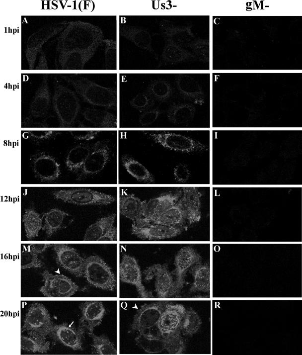
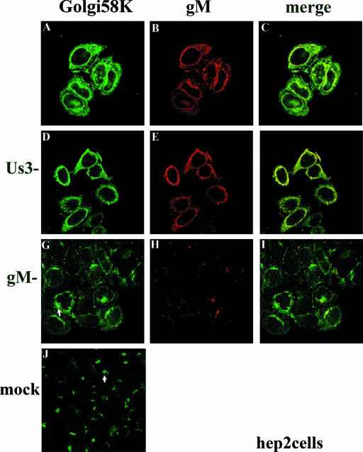
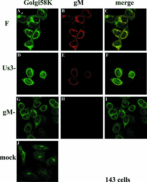
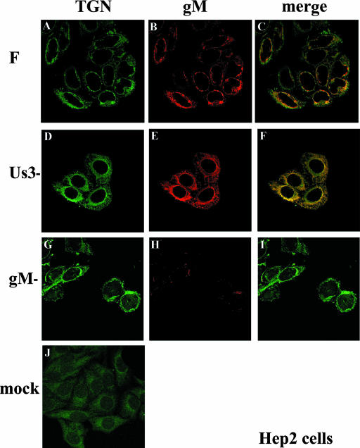
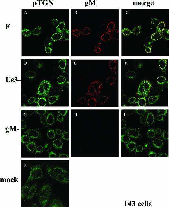
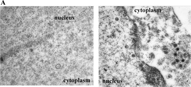
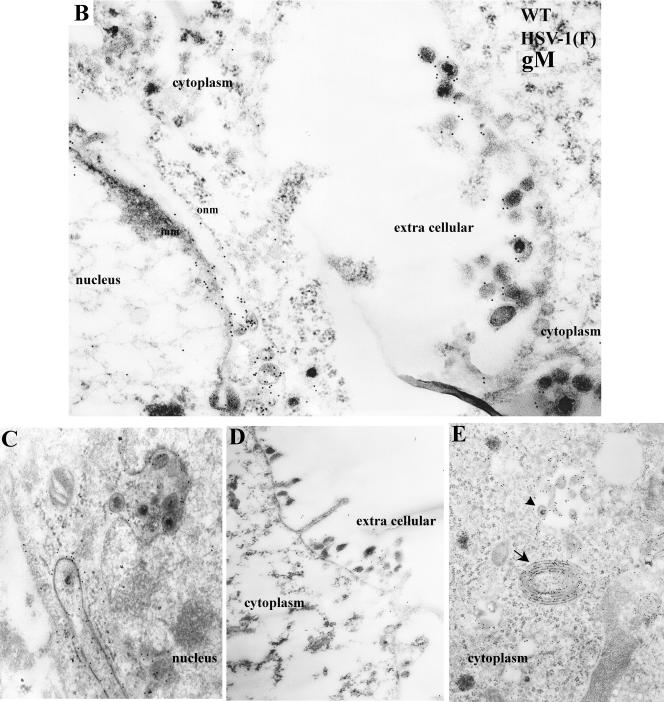
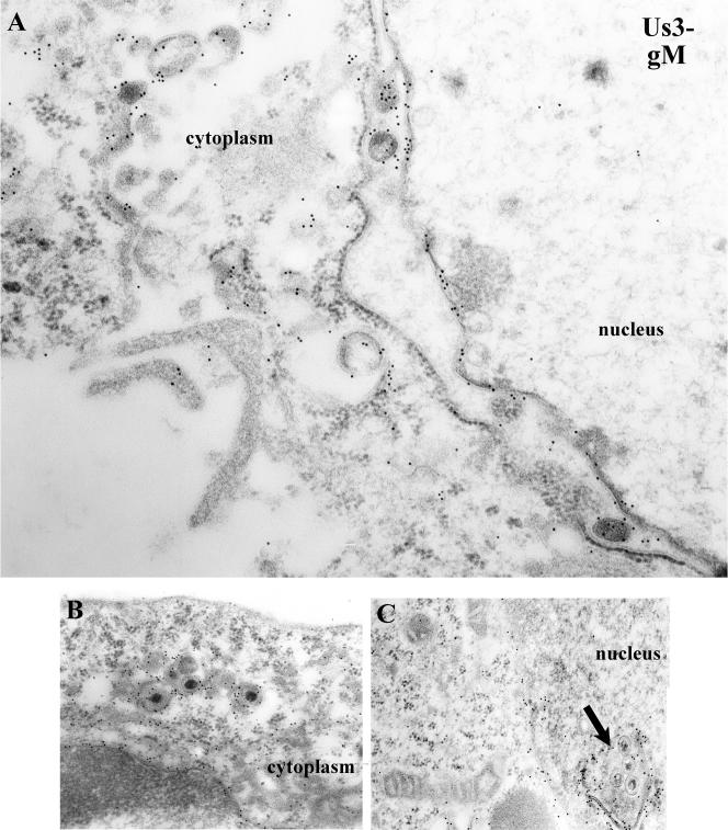
It is widely accepted that nucleocapsids of herpesviruses bud through the inner nuclear membrane (INM), but few studies have been undertaken to characterize the composition of these nascent virions. Such knowledge would shed light on the budding reaction at the INM and subsequent steps in the egress pathway. The present study focuses on glycoprotein M (gM), a type III integral membrane protein of herpes simplex virus 1 (HSV-1) that likely contains eight transmembrane domains. The results indicated that gM localized primarily at the perinuclear region, with especially bright staining near the nuclear membrane (NM). Immunogold electron microscopic analysis indicated that, like gB and gD (M. R. Torrisi et al., J. Virol. 66:554-561, 1992), gM localized within both leaflets of the NM, the envelopes of nascent virions that accumulate in the perinuclear space, and the envelopes of cytoplasmic and mature extracellular virus particles. Indirect immunofluorescence studies revealed that gM colocalized almost completely with a marker of the Golgi apparatus and partially with a marker of the trans-Golgi network (TGN), whether or not these markers were displaced to the perinuclear region during infection. gM was also located in punctate extensions and invaginations of the NM induced by the absence of a viral kinase encoded by HSV-1 U(S)3 and within virions located in these extensions. Our findings therefore support the proposition that gM, like gB and gD, becomes incorporated into the virion envelope upon budding through the INM. The localization of viral glycoproteins and Golgi and TGN markers to a perinuclear region may represent a mechanism to facilitate the production of infectious nascent virions, thereby increasing the amount of infectivity released upon cellular lysis.


Figures

FIG. 1.
FIG. 1.
Confocal indirect immunofluorescence localization of gM in HEp-2 cells infected with wild-type, gM-null mutant, or US3-null mutant viruses at various times after infection. Infected cells were fixed in methanol at the indicated times and reacted with gM-specific rabbit polyclonal antibody. Bound antibody was revealed by reaction with FITC-conjugated goat anti-rabbit immunoglobulin, followed by laser scanning confocal microscopy. Single optical sections are shown. The arrow in panel P indicates areas of nuclear fluorescence. The arrowheads in panels M and Q indicate fluorescence associated with the plasma membrane.
FIG. 2.
FIG. 2.
Indirect immunofluorescence of gM and Golgi localization in HEp-2 cells infected with wild type (top row), gM-null, or US3-null viruses at 16 h postinfection. HEp-2 cells were infected with the indicated viruses and were reacted with antibody to gM and mouse monoclonal antibody to Golgi 58K, a resident protein of the Golgi apparatus. Bound immunoglobulins were revealed by reaction with FITC conjugated goat anti-rabbit or Texas Red-conjugated anti-mouse immunoglobulin G followed by laser scanning confocal microscopy. Single optical sections are shown. Both the red channel and the green channel were scanned independently with only the fluorescence-stimulating laser powered on. The rightmost column shows scanned images of the left columns merged by using Adobe Photoshop software; a yellow color indicates regions where the two antibodies coincide. The arrows in panels G and J indicate different staining patterns of the Golgi marker in cells that were infected with the gM-null virus and mock infected, respectively.
FIG. 3.
FIG. 3.
Indirect immunofluorescence of infected and mock-infected 143 cells stained with gM or a Golgi-specific marker. 143 cells were infected with the indicated viruses. At 16 h postinfection, the cells were fixed and immunostained as described in the legend to Fig. 2. All images are of single optical sections collected using identical laser and photomultiplier tube (PMT) settings. The arrow in panel A indicates a cell with a Golgi 58K staining pattern that is reminiscent of that of mock-infected cells and different from the staining pattern of other infected cells in the same panel. F, HSV-1(F).
FIG. 4.
FIG. 4.
Indirect immunofluorescence of infected and mock-infected HEp-2 cells stained with TGN- or gM-specific antibodies. HEp-2 cells infected 16 h previously were fixed and reacted with gM specific antibody or a mouse monoclonal antibody to TGN38, a marker of the TGN. Confocal microscopy, image processing, and analyses were performed as described in the legend to Fig. 2. All images are of single optical sections taken at identical laser intensities and PMT settings. For illustrative purposes, the brightness of the lower left image (mock) was increased by 30% using Adobe Photoshop software. F, HSV-1(F).
FIG. 5.
FIG. 5.
Indirect immunofluorescence of infected and mock-infected 143 cells stained with TGN or gM-specific antibodies. 143 cells were mock infected or were infected with the indicated viruses and were fixed at 16 h after infection. The cells were immunostained with antibodies to TGN38 and gM, and bound immunoglobulin was revealed by reaction with appropriately conjugated antisera. Images of cells were taken at identical laser and PMT settings. Image collection, processing, and analysis were performed as in the legend to Fig. 2. F, HSV-1(F).
FIG.6.
FIG.6.
(A) Immune transmission electron microscopy of HEp-2 cells infected with the gM-null mutant and reacted with gM specific antibody. HEp-2 cells were infected with the gM-null virus R7216, fixed at 14 h after infection, and reacted with anti-gM rabbit polyclonal serum. Bound antibody was visualized by reaction with the F(ab)2 fragments of a goat anti-rabbit antibody conjugated to 12-nm colloidal gold particles. In this and subsequent electron photomicrographs, capsids of approximately 125 nm serve as a size standard. Magnifications: left panel, ×45,000; right panel, ×30,000. (B to E) Electron photomicrograph of a thin section of HEp-2 cells infected with HSV-1 (F) and stained with antibody to gM. HEp-2 cells were infected at 5.0 PFU per cell, harvested at 14 h after infection, fixed, embedded, sectioned, and reacted with rabbit gM-specific antibody and, subsequently, goat anti-rabbit IgG conjugated to 12 nm gold beads as described in Materials and Methods. In panel D, an arrow indicates heavily immunolabeled reduplicated membrane in cytoplasm. Arrowhead indicates a group of immunolabeled virions located within a cytoplasmic vacuole. Magnifications: C, D, and E, ×30,000; B, ×45,000.
FIG.6.
FIG.6.
(A) Immune transmission electron microscopy of HEp-2 cells infected with the gM-null mutant and reacted with gM specific antibody. HEp-2 cells were infected with the gM-null virus R7216, fixed at 14 h after infection, and reacted with anti-gM rabbit polyclonal serum. Bound antibody was visualized by reaction with the F(ab)2 fragments of a goat anti-rabbit antibody conjugated to 12-nm colloidal gold particles. In this and subsequent electron photomicrographs, capsids of approximately 125 nm serve as a size standard. Magnifications: left panel, ×45,000; right panel, ×30,000. (B to E) Electron photomicrograph of a thin section of HEp-2 cells infected with HSV-1 (F) and stained with antibody to gM. HEp-2 cells were infected at 5.0 PFU per cell, harvested at 14 h after infection, fixed, embedded, sectioned, and reacted with rabbit gM-specific antibody and, subsequently, goat anti-rabbit IgG conjugated to 12 nm gold beads as described in Materials and Methods. In panel D, an arrow indicates heavily immunolabeled reduplicated membrane in cytoplasm. Arrowhead indicates a group of immunolabeled virions located within a cytoplasmic vacuole. Magnifications: C, D, and E, ×30,000; B, ×45,000.
FIG. 7.
FIG. 7.
Electron photomicrograph of thin sections of HEp-2 cells infected with a US3 mutant of HSV-1(F) and stained with gM-specific antibody. Cells were infected with R7039, a viral mutant lacking US3. At 14 h postinfection, the cells were fixed and embedded. Thin sections were reacted with gM-specific antibody, followed by reaction with 12-nm gold-conjugated anti-rabbit IgG, and examined in a Phillips 201 electron microscope. In panel C, an arrow indicates a cross-section of a punctate extension or invagination of the nuclear membrane containing virions. This feature is peculiar to cells infected with the US3 deletion virus. Magnifications: A, ×45,000; B and C, ×30,000.

References

    1. Baines, J. D., R. J. Jacob, L. Simmerman, and B. Roizman. 1995. The UL11 gene products of herpes simplex virus 1 are present in the nuclear and cytoplasmic membranes and intranuclear dense bodies of infected cells. J. Virol. 69:825-833. - PMC - PubMed
    1. Baines, J. D., and B. Roizman. 1991. The open reading frames UL3, UL4, UL10 and UL16 are dispensable for the growth of herpes simplex virus 1 in cell culture. J. Virol. 65:938-944. - PMC - PubMed
    1. Baines, J. D., and B. Roizman. 1993. The UL10 gene of herpes simplex virus 1 encodes a novel glycoprotein, gM, which is present in the virion and in the plasma membrane of infected cells. J. Virol. 67:1441-1452. - PMC - PubMed
    1. Baines, J. D., P. L. Ward, G. Campadelli-Fiume, and B. Roizman. 1991. The UL20 gene of herpes simplex virus 1 encodes a function necessary for viral egress. J. Virol. 65:6414-6424. - PMC - PubMed
    1. Bloom, G. S., and T. A. Brashear. 1989. A novel 58-kDa protein associates with the Golgi apparatus and microtubules. J. Biol. Chem. 264:16083-16092. - PubMed

Publication types

MeSH terms

Substances

LinkOut - more resources