. 2007 Jan;81(2):558-67.
doi: 10.1128/JVI.01820-06. Epub 2006 Nov 1.

Hijacking components of the cellular secretory pathway for replication of poliovirus RNA

Affiliations

Hijacking components of the cellular secretory pathway for replication of poliovirus RNA

George A Belov et al. J Virol. 2007 Jan.

Abstract

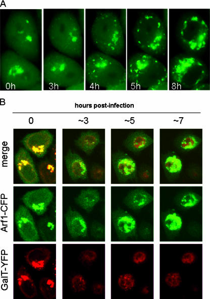
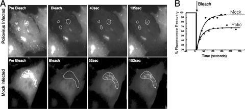
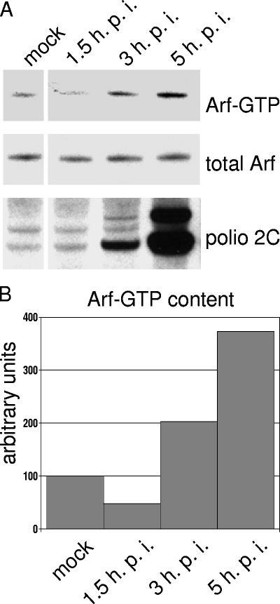
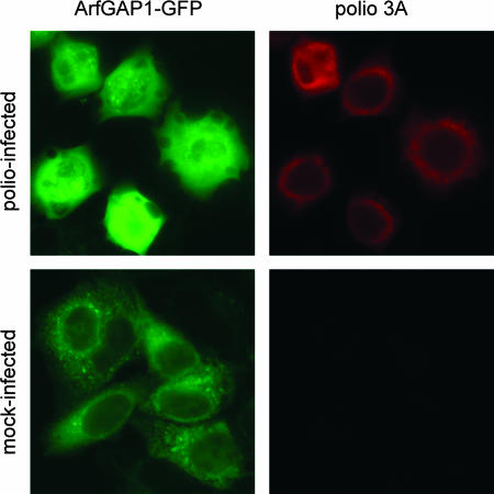
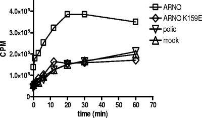
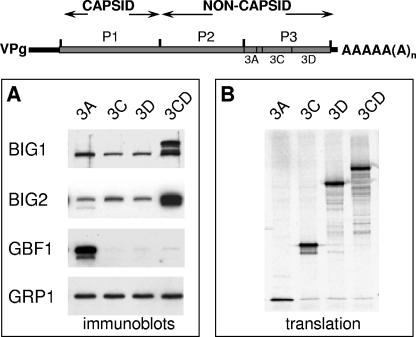
Infection of cells with poliovirus induces a massive intracellular membrane reorganization to form vesicle-like structures where viral RNA replication occurs. The mechanism of membrane remodeling remains unknown, although some observations have implicated components of the cellular secretory and/or autophagy pathways. Recently, we showed that some members of the Arf family of small GTPases, which control secretory trafficking, became membrane-bound after the synthesis of poliovirus proteins in vitro and associated with newly formed membranous RNA replication complexes in infected cells. The recruitment of Arfs to specific target membranes is mediated by a group of guanine nucleotide exchange factors (GEFs) that recycle Arf from its inactive, GDP-bound state to an active GTP-bound form. Here we show that two different viral proteins independently recruit different Arf GEFs (GBF1 and BIG1/2) to the new structures that support virus replication. Intracellular Arf-GTP levels increase approximately 4-fold during poliovirus infection. The requirement for these GEFs explains the sensitivity of virus growth to brefeldin A, which can be rescued by the overexpression of GBF1. The recruitment of Arf to membranes via specific GEFs by poliovirus proteins provides an important clue toward identifying cellular pathways utilized by the virus to form its membranous replication complex.


Figures

FIG. 1.
FIG. 1.
Relocalization of Arf1-GFP in infected cells. (A) HeLa cells transfected with pArf1-GFP for 18 h were infected with poliovirus and monitored every 6 min for 16 h. The figure shows selected images taken at the indicated times postinfection. (For the complete sequence, see the movie [and corresponding Web site] referred to in Results.) (B) HeLa cells were cotransfected with pGalT-YFP and pArf-CFP. After 18 h of incubation, the cells were infected with poliovirus and monitored for fluorescence of both of the expressed proteins.
FIG. 2.
FIG. 2.
Arf1-GFP dynamics in poliovirus-infected HeLa cells. (A) Regions of interest (dotted lines) were selected for photobleaching in cells ∼3 h postinfection. In mock-infected cells the Arf1-GFP-labeled Golgi apparatus was selected. A laser pulse was directed at the selected regions to bleach the fluorescence. Recovery of fluorescence was monitored with low intensity laser light every 5 s. (B) The recovery of fluorescence into the selected regions was quantified and plotted as described in Materials and Methods. Photobleaching was performed on 9 virus-infected and 12 mock-infected cells in two separate experiments. The mean rate of recovery (τ) for control cells was 32 ± 7.5 s−1, and for virus-infected cells it was 28 ± 4 s−1. The mean immobile fraction for control cells was 10% ± 7%, whereas for poliovirus-infected cells it was 35% ± 13%. Representative recovery curves from infected and mock-infected cells are plotted.
FIG. 3.
FIG. 3.
GTP-bound Arf in poliovirus-infected cells. HeLa cells were mock infected or infected with poliovirus. (A) At the indicated times postinfection, cells were lysed, and the Arf-GTP pull-down assay was performed as described in Materials and Methods. The upper panel shows the amount of GTP-bound Arf recovered from each cell lysates, and the middle panel shows the total amount of Arf in each lysates. Anti-polio-2C staining (bottom panel) was performed on the same membrane used for Arf-GTP blot after stripping it with Chemicon Re-Blot Plus Mild solution. (B) Quantitation was performed by using Total Lab 1D gel image analysis software.
FIG. 4.
FIG. 4.
ArfGAP1 in poliovirus-infected cells. HeLa cells transfected with an ArfGAP1-EGFP expression plasmid for 18 h were infected with poliovirus. Cells were fixed at 5 h postinfection and stained with anti-poliovirus 3A antibodies to visualize the replication complexes.
FIG. 5.
FIG. 5.
GEF activity in poliovirus-infected cells. Cytoplasmic extracts from poliovirus-infected cells were assayed for guanine nucleotide exchange activity on purified recombinant Arf1 protein in vitro as described in Materials and Methods. GEF activity in infected cells was compared to that in mock-infected cells or cells expressing ARNO or an inactive, mutant protein, ARNO E156K.
FIG. 6.
FIG. 6.
Recruitment of GEFs to membranes by poliovirus proteins in vitro. (A) Poliovirus proteins were synthesized in HeLa cell extracts, and the membrane fractions were collected and assayed by immunoblotting with antibodies to BIG1, BIG2, GBF1, and GRP1. (B) Translation of each poliovirus-specific RNA was monitored by labeling an aliquot with [35S]methionine and analyzing the translation products by SDS-PAGE.
FIG. 7.
FIG. 7.
Relocalization of BIG1 and GBF1 in HeLa cells upon poliovirus infection. (A) Mock-infected HeLa cells stained with anti-BIG1 antibodies. (B and C) Poliovirus-infected cells stained 3 h postinfection with antibodies against BIG1 and poliovirus nonstructural protein 2C, respectively. (D) Merged image of panels B and C. (E) Mock-infected cells stained with anti-GBF1 antibodies. (F and G) Poliovirus-infected cells stained 3 h postinfection with antibodies to GBF1 and poliovirus nonstructural protein 3A, respectively. (H) Merged image of panels F and G. Poliovirus proteins 2C and 3A both served as markers of viral RNA replication complexes.
FIG. 8.
FIG. 8.
Rescue of poliovirus RNA replication in the presence of BFA by GBF1. (A) Poliovirus replicon RNA containing the Renilla luciferase gene was introduced into HeLa cells previously transfected with expression plasmids for GBF1, BIG2, or both together. A total of 1 μg of BFA/ml was added where indicated. Each point is an average value from 16 wells of a 96-well plate. (B) Firefly luciferase activity was measured in the cells shown in panel A to evaluate transfection efficiencies. (C) Immunoblot showing expression of GBF1 and BIG2 in the transfected cells.

References

    1. Agol, V. I., G. A. Belov, K. Bienz, D. Egger, M. S. Kolesnikova, N. T. Raikhlin, L. I. Romanova, E. A. Smirnova, and E. A. Tolskaya. 1998. Two types of death of poliovirus-infected cells: caspase involvement in the apoptosis but not cytopathic effect. Virology 252:343-353. - PubMed
    1. Aldabe, R., and L. Carrasco. 1995. Induction of membrane proliferation by poliovirus proteins 2C and 2BC. Biochem. Biophys. Res. Commun. 206:64-76. - PubMed
    1. Barton, D. J., E. P. Black, and J. B. Flanegan. 1995. Complete replication of poliovirus in vitro: preinitiation RNA replication complexes require soluble cellular factors for the synthesis of VPg-linked RNA. J. Virol. 69:5516-5527. - PMC - PubMed
    1. Behnia, R., and S. Munro. 2005. Organelle identity and the signposts for membrane traffic. Nature 438:597-604. - PubMed
    1. Belov, G. A., M. H. Fogg, and E. Ehrenfeld. 2005. Poliovirus proteins induce membrane association of GTPase ADP-ribosylation factor. J. Virol. 79:7207-7216. - PMC - PubMed

Publication types

LinkOut - more resources