

Eradication of tumor colonization and invasion by a B cell-specific immunotoxin in a murine model for human primary intraocular lymphoma
- PMID: 17079483
- PMCID: PMC1931503
- DOI: 10.1158/0008-5472.CAN-06-1981
Eradication of tumor colonization and invasion by a B cell-specific immunotoxin in a murine model for human primary intraocular lymphoma
Abstract
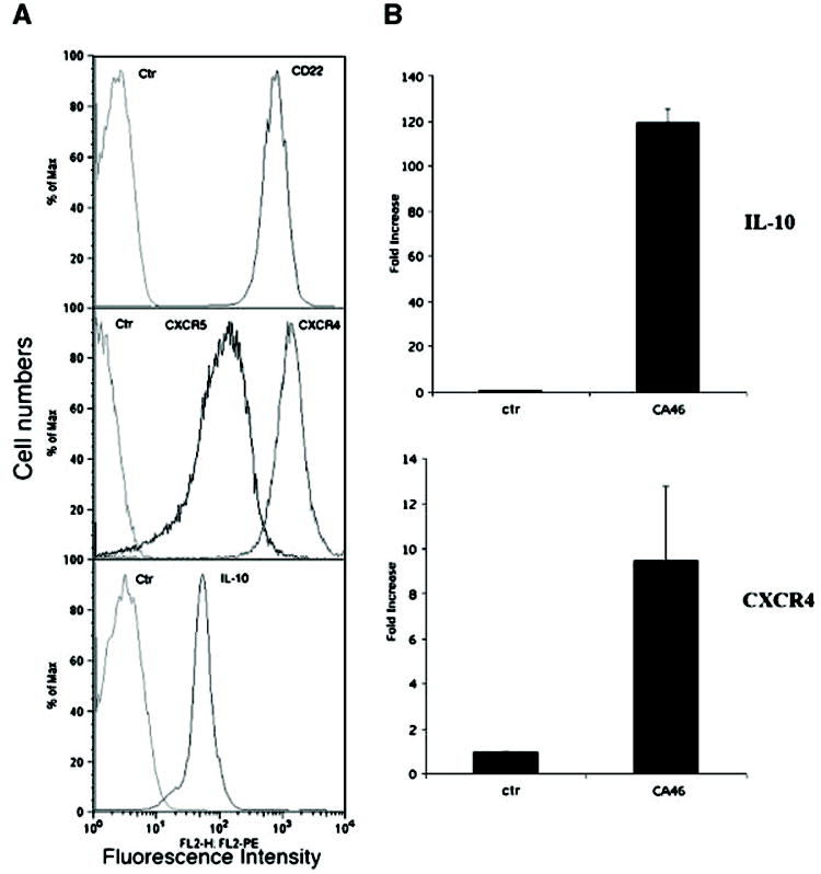
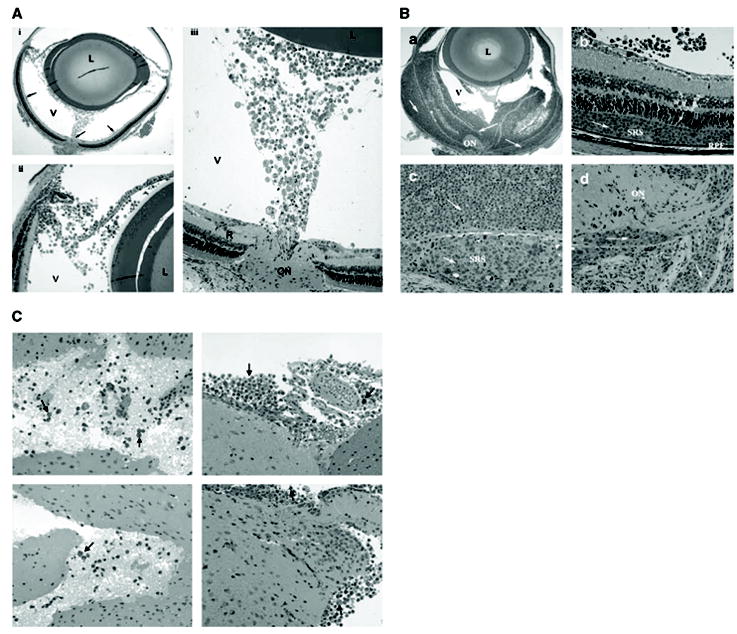
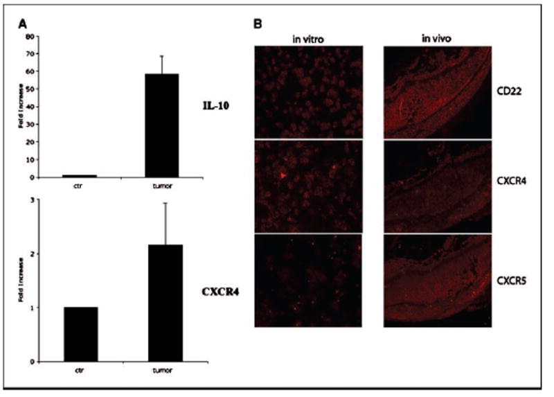
Human primary intraocular lymphoma (PIOL) is predominantly a B cell-originated malignant disease with no appropriate animal models and effective therapies available. This study aimed to establish a mouse model to closely mimic human B-cell PIOL and to test the therapeutic potential of a recently developed immunotoxin targeting human B-cell lymphomas. Human B-cell lymphoma cells were intravitreally injected into severe combined immunodeficient mice. The resemblance of this tumor model to human PIOL was examined by fundoscopy, histopathology, immunohistochemistry, and evaluated for molecular markers. The therapeutic effectiveness of immunotoxin HA22 was tested by injecting the drug intravitreally. Results showed that the murine model resembles human PIOL closely. Pathologic examination revealed that the tumor cells initially colonized on the retinal surface, followed by infiltrating through the retinal layers, expanding preferentially in the subretinal space, and eventually penetrating through the retinal pigment epithelium into the choroid. Several putative molecular markers for human PIOL were expressed in vivo in this model. Tumor metastasis into the central nervous system was also observed. A single intravitreal injection of immunotoxin HA22 after the establishment of the PIOL resulted in complete regression of the tumor. This is the first report of a murine model that closely mimics human B-cell PIOL. This model may be a valuable tool in understanding the molecular pathogenesis of human PIOL and for the evaluation of new therapeutic approaches. The results of B cell-specific immunotoxin therapy may have clinical implications in treating human PIOL.
Figures

Similar articles
-
Expression of chemokine receptors, CXCR4 and CXCR5, and chemokines, BLC and SDF-1, in the eyes of patients with primary intraocular lymphoma.Ophthalmology. 2003 Feb;110(2):421-6. doi: 10.1016/S0161-6420(02)01737-2. Ophthalmology. 2003. PMID: 12578791
-
Preclinical study of Ublituximab, a Glycoengineered anti-human CD20 antibody, in murine models of primary cerebral and intraocular B-cell lymphomas.Invest Ophthalmol Vis Sci. 2013 May 1;54(5):3657-65. doi: 10.1167/iovs.12-10316. Invest Ophthalmol Vis Sci. 2013. PMID: 23611989
-
Molecular pathology of primary intraocular lymphoma.Trans Am Ophthalmol Soc. 2003;101:275-92. Trans Am Ophthalmol Soc. 2003. PMID: 14971583 Free PMC article.
-
Molecular analysis of primary central nervous system and primary intraocular lymphomas.Curr Mol Med. 2001 May;1(2):259-72. doi: 10.2174/1566524013363915. Curr Mol Med. 2001. PMID: 11899075 Review.
-
Primary intraocular lymphoma: a review of the clinical, histopathological and molecular biological features.Graefes Arch Clin Exp Ophthalmol. 2004 Nov;242(11):901-13. doi: 10.1007/s00417-004-0973-0. Epub 2004 Oct 29. Graefes Arch Clin Exp Ophthalmol. 2004. PMID: 15565454 Review.
Cited by
-
CXCR4, CXCR5 and CD44 May Be Involved in Homing of Lymphoma Cells into the Eye in a Patient Derived Xenograft Homing Mouse Model for Primary Vitreoretinal Lymphoma.Int J Mol Sci. 2022 Oct 4;23(19):11757. doi: 10.3390/ijms231911757. Int J Mol Sci. 2022. PMID: 36233057 Free PMC article.
-
Murine models of B-cell lymphomas: promising tools for designing cancer therapies.Adv Hematol. 2012;2012:701704. doi: 10.1155/2012/701704. Epub 2012 Feb 12. Adv Hematol. 2012. PMID: 22400032 Free PMC article.
-
Intraocular Lymphoma Models.Ocul Oncol Pathol. 2015 Apr;1(3):214-22. doi: 10.1159/000370158. Epub 2015 Apr 9. Ocul Oncol Pathol. 2015. PMID: 27171354 Free PMC article. Review.
-
Diagnosis and management of primary intraocular lymphoma: an update.Clin Ophthalmol. 2007 Sep;1(3):247-58. Clin Ophthalmol. 2007. PMID: 19668478 Free PMC article.
-
Primary intraocular lymphoma.Arch Pathol Lab Med. 2009 Aug;133(8):1228-32. doi: 10.5858/133.8.1228. Arch Pathol Lab Med. 2009. PMID: 19653715 Free PMC article.
References
-
- Eby NL, Grufferman S, Flannelly CM, Schold SC, Vogel FS, Jr, Burger PC. Increasing incidence of primary brain lymphoma in the US. Cancer. 1988;62:2461–5. - PubMed
-
- Kadan-Lottick NS, Skluzacek MC, Gurney JG. Decreasing incidence rates of primary central nervous system lymphoma. Cancer. 2002;95:193–202. - PubMed
-
- Cote TR, Manns A, Hardy CR, Yellin FJ, Hartge P. Epidemiology of brain lymphoma among people with or without acquired immunodeficiency syndrome. AIDS/Cancer Study Group. J Natl Cancer Inst. 1996;88:675–9. - PubMed
-
-
Brain Tumor in the United States 1995-1999. Chicago (Illinois): Central Brain Tumor Registry of the United States, 2002-2003.
-
-
- Coupland SE, Heimann H, Bechrakis NE. Primary intraocular lymphoma: a review of the clinical, histo-pathological and molecular biological features. Graefes Arch Clin Exp Ophthalmol. 2004;242:901–13. - PubMed
Publication types
MeSH terms
Substances
Grants and funding
LinkOut - more resources
Full Text Sources
Other Literature Sources
Medical
