. 2006 Nov 14;103(46):17079-86.
doi: 10.1073/pnas.0607465103. Epub 2006 Nov 1.

Podocin and MEC-2 bind cholesterol to regulate the activity of associated ion channels

Affiliations

Podocin and MEC-2 bind cholesterol to regulate the activity of associated ion channels

Tobias B Huber et al. Proc Natl Acad Sci U S A. .

Abstract

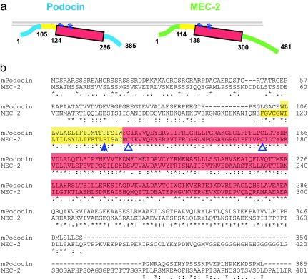
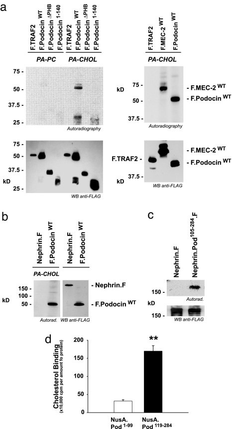
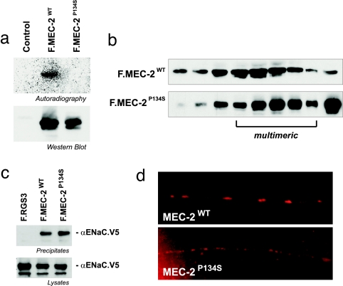
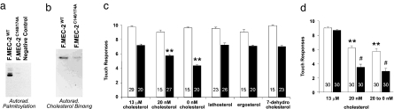
The prohibitin (PHB)-domain proteins are membrane proteins that regulate a variety of biological activities, including mechanosensation, osmotic homeostasis, and cell signaling, although the mechanism of this regulation is unknown. We have studied two members of this large protein family, MEC-2, which is needed for touch sensitivity in Caenorhabditis elegans, and Podocin, a protein involved in the function of the filtration barrier in the mammalian kidney, and find that both proteins bind cholesterol. This binding requires the PHB domain (including palmitoylation sites within it) and part of the N-terminally adjacent hydrophobic domain that attaches the proteins to the inner leaflet of the plasma membrane. By binding to MEC-2 and Podocin, cholesterol associates with ion-channel complexes to which these proteins bind: DEG/ENaC channels for MEC-2 and TRPC channels for Podocin. Both the MEC-2-dependent activation of mechanosensation and the Podocin-dependent activation of TRPC channels require cholesterol. Thus, MEC-2, Podocin, and probably many other PHB-domain proteins by binding to themselves, cholesterol, and target proteins regulate the formation and function of large protein-cholesterol supercomplexes in the plasma membrane.


Conflict of interest statement

Conflict of interest statement: Columbia University has filed a provisional patent application based on this research.

Figures

Fig. 1.
Fig. 1.
Structure and sequence of Podocin and MEC-2. (a) Membrane orientation of Podocin and MEC-2. The hydrophobic region (yellow) inserts into the inner leaflet of the plasma membrane, causing the remaining parts of the protein, including the PHB domain (red) to face the cytoplasm and the inner leaflet. Sites of palmitate attachment are indicated by blue wavy lines. (b) Alignment of mouse Podocin and C. elegans MEC-2, showing the hydrophobic region (yellow), PHB domain (red), palmitoylation sites (open blue triangles), and the site of the Pro-to-Ser mutation in the hydrophobic region that prevents cholesterol binding (filled blue triangle).
Fig. 2.
Fig. 2.
Podocin and MEC-2 bind cholesterol. (a) MEC-2 and Podocin expressed in HEK293T cells are labeled with photoactivatable [3H]photocholesterol (PA-CHOL) but not with photoactivatable [3H]phosphatidylcholine (PA-PC) (incubation time, 16 h). (Upper) Photolabeled proteins were resolved by SDS/PAGE and visualized by autoradiography. (Lower) Expression of proteins in the lysates. (b) The Podocin-interacting membrane protein Nephrin does not bind photoactivatable cholesterol. (c) A fusion protein with a C-terminal fusion of the PHB domain and the hydrophobic region of Podocin (amino acids 105–284) with the extracellular and transmembrane domains of Nephrin binds photoactivatable cholesterol, indicating that this domain can convey cholesterol binding. (d) Direct binding of cholesterol to Podocin; in vitro binding of cholesterol. NusA fused to the N-terminal domain of Podocin (amino acids 1–99) or NusA fused to the cholesterol-binding domain of Podocin (amino acids 119–284) was incubated with radioactively labeled cholesterol, washed extensively, and subjected to scintillation counting.
Fig. 3.
Fig. 3.
MEC-2(P134S) does not bind cholesterol but can bind DEG/ENaC channels and multimerize. (a) Photoaffinity cholesterol labels FLAG-tagged wild-type MEC-2 but not MEC-2(P134S). (Upper) Photolabeled proteins were immunoprecipitated with anti-FLAG antibody, resolved by SDS/PAGE, and visualized by autoradiography. (Lower) The expression of proteins in the lysates. (b) Velocity-gradient centrifugation shows that both wild type and MEC-2(P134S) multimerize. (c) Wild-type MEC-2 and MEC-2(P134S) equivalently coimmunoprecipitate V5-tagged rat αENaC from HEK293T cells (the second transmembrane domain of αENaC can substitute for that of MEC-4 in vivo) (33). The rat channel protein was used because we were unable to express MEC-4 to sufficient levels. This experiment demonstrates that MEC-2 can bind to other DEG/ENaC proteins and that the mutant binding does not depend on cholesterol. (d) Localization of wild-type and mutant MEC-2 in processes of touch-receptor neurons in C. elegans. Both proteins are found in the process (suggesting that both localize to the plasma membrane), but the P134S protein is not found in the characteristic puncta formed by the mechanosensory-channel complex. Proteins were visualized in C. elegans with a MEC-2-specific antibody (9).
Fig. 4.
Fig. 4.
Cholesterol dependence of touch sensitivity in C. elegans. Substitution of Ala for Cys in two predicted palmitoylation sites results in the loss of palmitoylation (a) and a reduction of cholesterol binding (b). HEK293T cells were transfected with wild-type MEC-2 or MEC-2(C140/174A) and labeled with [3H]palmitic acid (a) or [3H]photoaffinity cholesterol (b). Equal expression of proteins in the lysates was confirmed on Western blots (data not shown). (c) Touch sensitivity in MEC-2(C140/174A) mutants (black bars) requires cholesterol or its derivatives. Responses of wild-type animals (white bars) are also shown and are not affected by limited cholesterol depletion. mec-2-null worms were transformed with the mec-2(C140/174A) gene and grown on plates with defined sterol concentrations before analysis of touch sensitivity. Depicted is the mean ± SEM (number of animals tested is indicated. ∗∗, P < 0.001 as compared with mec-2(C140/174A) at high cholesterol). (d) Severe cholesterol depletion lowers the sensitivity of wild-type animals (white bars) and mec-2(C140/174A) mutants (black bars). Worms grown on plates containing 20 nM cholesterol for three generations were either maintained on 20 nM cholesterol for another generation or placed on cholesterol-free plates before analysis of response to gentle touch (number of animals tested is indicated; ∗∗, P < 0.001 as compared with wild-type worms on 13 μM cholesterol; #, P < 0.001 as compared with mutants on 13 μM cholesterol).
Fig. 5.
Fig. 5.
Podocin interacts and colocalizes with TRPC6. (a) Mouse TRPC6 coimmunoprecipitates with FLAG-tagged Podocin (F.Podocin) but not with a control protein (F.GFP). (Top) Coprecipitated TRPC6 channel after immunoprecipitation of Podocin or GFP. (Middle and Bottom) Expression of the proteins in the lysates. (b) TRPC6 is located at the slit diaphragm (SD) of podocyte foot processes (FP) near the glomerular basement membrane (GBM). This localization mimics that of Podocin. Rat kidneys were perfused with ice-cold PBS, fixed in situ, and subjected to immunogold electron microscopy. Arrows indicate the localization of gold particles in the electron micrograph.
Fig. 6.
Fig. 6.
Activation of TRPC-channel activity by Podocin. (a) Podocin, but not PodocinΔPHB, enhances TRPC6 currents in Xenopus oocytes stimulated with 10 μM OAG. Expression of TRPC6 induces an inward Na+ current in a Ca2+-free bath solution that is further augmented by stimulation with OAG. The OAG-induced currents were significantly augmented in oocytes coexpressing TRPC6 and Podocin but were not increased in oocytes coexpressing TRPC6 and PodocinΔPHB. (b) Podocin increases the effect of OAG (10 μM 1-oleoyl-2-acetyl-sn-glycerol; black bars) on NMDG-sensitive conductance (GNMDG) of TRPC6 channels in Xenopus oocytes, but mutant Podocins do not. Currents in control oocytes (white bars) were not affected. The Podocin mutants used were PodocinΔPHB, PodocinP120S, and PodocinC126/160A (see Fig. 1). The number of oocytes examined is given in parentheses. ∗, P < 0.05 as compared with water-injected oocytes; #, P < 0.05 as compared with TRPC6 coexpressed with PodocinΔPHB. (c) Wild-type, but not mutant, Podocin increases histamine-induced calcium influx (measured as a change in fluorescence, ΔF/F) in HeLa Cx43 cells. Cells were transiently mock transfected (filled circles) or transfected with DNA coding for wild-type Podocin (filled triangles), PodocinΔPHB (open triangles), and PodocinC126/160A (inverted filled triangles) and measured simultaneously in the same experiment by using a FLIPR. Data are means ± SD from three to five independent experiments. Measurements are taken at the shoulder of the response (line in the inset, which shows the calcium responses of the cells challenged with 10 μM histamine). Vertical scale, 104 arbitrary fluorescence units; horizontal scale, 2 min. (d) The stimulation by Podocin is abolished when cells are treated with MBCD to deplete cholesterol.

References

    1. Ponting CP, Schultz J, Milpetz F, Bork P. Nucleic Acids Res. 1999;27:229–232. - PMC - PubMed
    1. Morrow IC, Parton RG. Traffic. 2005;6:725–740. - PubMed
    1. Benzing T. J Am Soc Nephrol. 2004;15:1382–1391. - PubMed
    1. Ernstrom GG, Chalfie M. Annu Rev Genet. 2002;36:411–453. - PubMed
    1. Stewart GW. Curr Opin Hematol. 2004;11:244–250. - PubMed

Publication types

MeSH terms