Comparative Study
. 2006 Nov 1;26(44):11403-12.
doi: 10.1523/JNEUROSCI.2592-06.2006.

Weakened center-surround interactions in visual motion processing in schizophrenia

Affiliations
Comparative Study

Weakened center-surround interactions in visual motion processing in schizophrenia

Duje Tadin et al. J Neurosci. .

Abstract

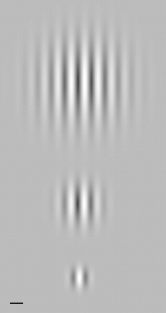
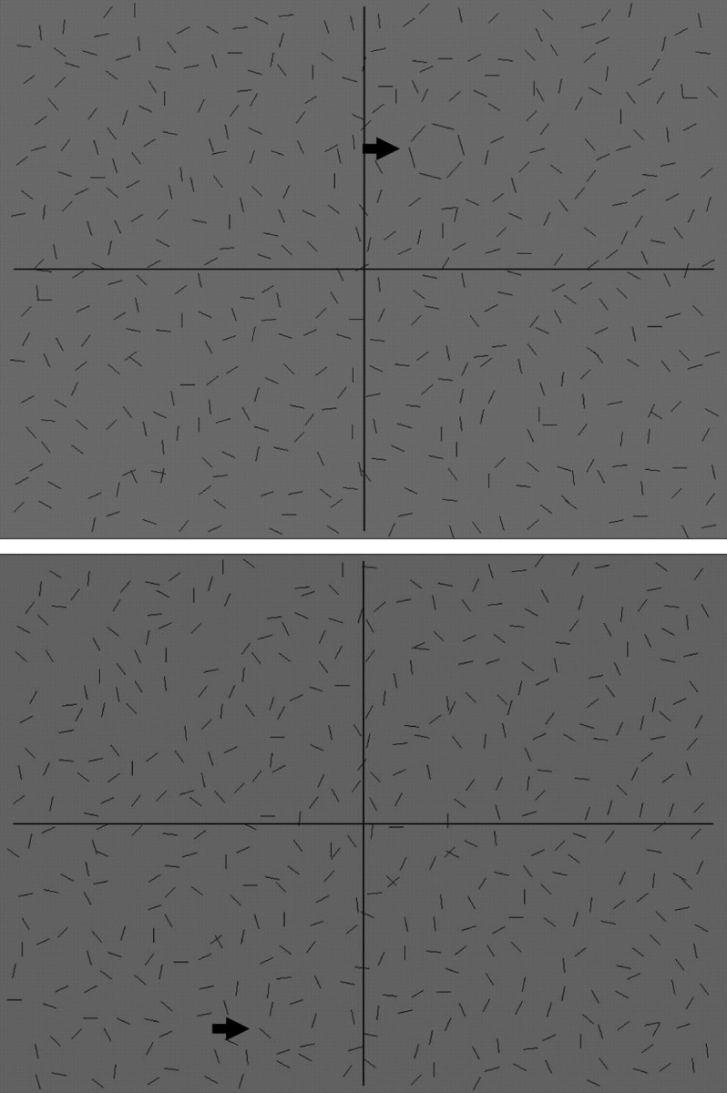
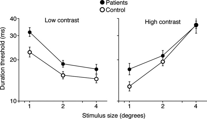
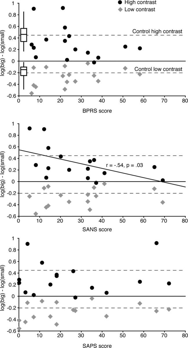
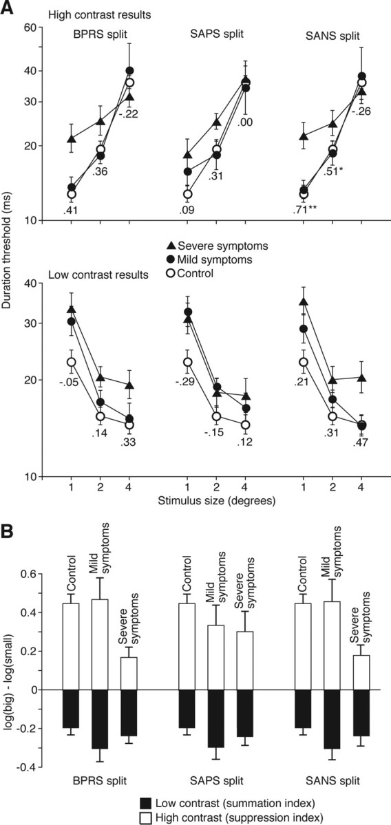
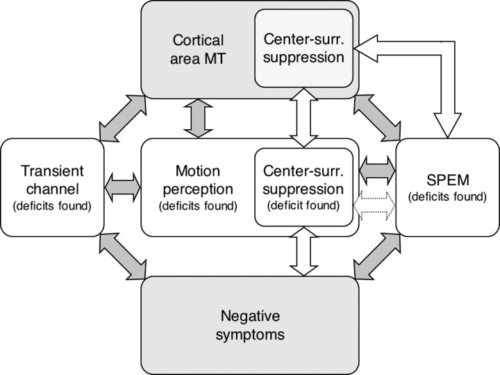
Schizophrenia is often accompanied by a range of visual perception deficits, with many involving impairments in motion perception. The presence of perceptual abnormalities may impair neural processes that depend on normal visual analysis, which in turn may affect overall functioning in dynamic visual environments. Here, we examine the integrity of suppressive center-surround mechanisms in motion perception of schizophrenic patients. Center-surround suppression has been implicated in a range of visual functions, including figure-ground segregation and pursuit eye movements, visual functions that are impaired in schizophrenia. In control subjects, evidence of center-surround suppression is found in a reduced ability to perceive motion of a high-contrast stimulus as its size increases. This counterintuitive finding is likely a perceptual correlate of center-surround mechanisms in cortical area MT. We now show that schizophrenic patients exhibit abnormally weak center-surround suppression in motion, an abnormality that is most pronounced in patients with severe negative symptoms. Interestingly, patients with the weakest surround suppression outperformed control subjects in motion discriminations of large high-contrast stimuli. This enhanced motion perception of large high-contrast stimuli is consistent with an MT abnormality in schizophrenia and has a potential to disrupt smooth pursuit eye movements and other visual functions that depend on unimpaired center-surround interactions in motion.


Figures

Figure 1.
Figure 1.
Examples of three different stimulus sizes used in the motion discrimination task. Scale bar, 1°. These Gabor patches consisted of a stationary Gaussian spatial window and dark/white bars that drifted either rightward or leftward.
Figure 2.
Figure 2.
Examples of stimuli used for the global form task. The target was a quasicircular shape formed by six nearby lines. Task difficulty was adjusted by varying line jitter (the angular deviations among target contours from the canonical values). In the top example, there is no line jitter, and the target is easily detected (see arrow). The bottom example shows a difficult trial in which the target is hard to find because of substantial line jitter (see arrow).
Figure 3.
Figure 3.
Effects of size and contrast on motion discrimination thresholds in patients and controls. The left panel shows the effects of stimulus size on the discriminability of low-contrast moving stimuli. The right panel depicts the effects increasing stimulus size has on high-contrast moving stimuli. Error bars are SEM.
Figure 4.
Figure 4.
Relationship between the effect of stimulus size and symptom severity in schizophrenia. Positive y-axis values indicate that the psychophysical threshold for perceiving motion direction of the large stimulus was higher than that for perceiving motion direction of the small moving stimulus, a result suggesting surround suppression. Negative y-axis values indicate that motion of the large moving stimulus was easier to perceive than that of the small moving stimulus, a result suggesting spatial summation. The black circles depict high-contrast results for individual patients, whereas the gray diamonds show low-contrast results for each patient tested. The dashed lines in each panel show average high- and low-contrast results for control subjects. The box plots are added to better illustrate the range of the control results (with the middle horizontal line representing the median, the horizontal box boundaries representing the quartiles, and the “whiskers” stretching out to the minimum and the maximum values).
Figure 5.
Figure 5.
Median split analysis results. A, Duration thresholds as a function of stimulus size in control subjects and patients subgroups split around median BPRS, SAPS, and SANS scores. The top and bottom panels show results from high- and low-contrast conditions, respectively. The numbers under data symbols indicate correlations between BPRS, SAPS, and SANS scores and duration thresholds for given stimulus size. For example, the leftmost number in the top panel (r = 0.41) indicates the correlation between BPRS scores and duration thresholds for small, high-contrast moving stimuli. Error bars indicate SEM. B, Effect of stimulus size on motion perception of control subjects and patients subgroups split around median BPRS, SAPS, and SANS scores. The y-axis convention is the same as in Figure 4, in which higher positive numbers indicate strong center-surround suppression and negative numbers indicate spatial summation.
Figure 6.
Figure 6.
Transient channel perceptual deficits in schizophrenia. The presented framework is intended as an overview of the main trends in the literature and not as a comprehensive model. Some of the presented links pertain to the experimental questions that are still actively investigated (e.g., a link between negative symptoms and SPEM), and thus, there is a possibility that those links might need to be altered or perhaps even eliminated. Area MT links (top row of arrows), Numerous neurophysiological, psychophysical, and clinical results link cortical area MT with motion perception, the transient (magnocellular) channel, and center-surround suppression of moving stimuli (Orban, 1997; Born and Bradley, 2005). Moreover, neural processing within area MT is critically involved in SPEM (Wurtz et al., 1990). Thus, deficits in these perceptual and oculomotor motor functions suggest a possible MT deficit in schizophrenia. Links among perceptual and oculomotor deficits (horizontal arrows), Motion perception is one of the key functions of the transient channel. Thus, nearly all motion perception deficits can be considered transient channel deficits. Motion perception is also a critical first step in SPEM generation (Ilg, 1997). For example, the magnitude of motion perception deficits in schizophrenia predicts the severity of the SPEM deficit, suggesting a link between SPEM and motion perception deficits (Stuve et al., 1997; Chen et al., 1999b,c). More research is required to elucidate the connections among various perceptual deficits in schizophrenia and to test a possible perceptual origin of the SPEM deficit. Links between negative symptoms and perceptual/oculomotor abnormalities in schizophrenia (bottom row of arrows), Several studies have found that patients with predominantly negative symptoms have more pronounced transient channel deficits (Cadenhead et al., 1997; Slaghuis and Bishop, 2001; Butler et al., 2003). Moreover, a connection between negative or deficit symptoms and SPEM deficit has been found with a variety of experimental designs (Sweeney et al., 1994; Ross et al., 1996, 1997; Roitman et al., 1997; Slaghuis et al., 2005) (but see Kelly et al., 1990; Nkam et al., 2001). There is, however, an inherent difficulty in comparing a stable, trait-like characteristic such as SPEM defect and clinical symptom severity that varies over time. Therefore, depending on when patients are examined, correlation between symptoms and SPEM deficit may or may not be observed. What is relevant for our purpose is that a large majority of studies that examined the relationship between clinical symptoms and SPEM found a link with negative symptoms. The existence of a link between negative symptoms and motion perception deficits (e.g., velocity discrimination, perception of random-dot motion) has yet to be fully investigated. Nevertheless, based on the central role of motion perception in the transient visual channel, we suspect that such a connection exists. Center-surround suppression links (white arrows), The remaining connections pertain to the results of this paper. The task used in the present study is believed to reflect cortical center-surround mechanisms, particularly those within area MT (Tadin et al., 2003). The activity of MT center-surround neurons has been shown to directly affect SPEM (Born et al., 2000), suggesting a functional link between SPEM and surround suppression. The remaining full arrow depicts the link between surround suppression and negative symptoms (Figs. 4, 5). The present finding suggests the possibility of a connection between center-surround suppression abnormality in schizophrenia and SPEM deficit (dotted arrow). Such a link is consistent with other connections shown in the figure.

Similar articles

Cited by

References

    1. Albright TD, Desimone R. Local precision of visuotopic organization in the middle temporal area (MT) of the macaque. Exp Brain Res. 1987;65:582–592. - PubMed
    1. Allman J, Meizin F, McGuiness E. Direction- and velocity-specific responses from beyond the classical receptive field in the middle temporal visual area (MT) Perception. 1985;14:105–126. - PubMed
    1. Andreasen NC, Olsen S. Negative v positive schizophrenia: definition and validation. Arch Gen Psychiatry. 1982;39:789–794. - PubMed
    1. Andreasen NC, O'Leary DS, Flaum M, Nopoulos P, Watkins GL, Boles Ponto LL, Hichwa RD. Hypofrontality in schizophrenia: distributed dysfunctional circuits in neuroleptic-naive patients. Lancet. 1997;349:1730–1734. - PubMed
    1. Betts LR, Taylor CP, Sekuler AB, Bennett PJ. Aging reduces center-surround antagonism in visual motion processing. Neuron. 2005;45:361–366. - PubMed

Publication types