Comparative Study
. 2006 Nov 1;26(44):11442-53.
doi: 10.1523/JNEUROSCI.3561-06.2006.

Noradrenergic inputs to the bed nucleus of the stria terminalis and paraventricular nucleus of the hypothalamus underlie hypothalamic-pituitary-adrenal axis but not hypophagic or conditioned avoidance responses to systemic yohimbine

Affiliations
Comparative Study

Noradrenergic inputs to the bed nucleus of the stria terminalis and paraventricular nucleus of the hypothalamus underlie hypothalamic-pituitary-adrenal axis but not hypophagic or conditioned avoidance responses to systemic yohimbine

Layla Banihashemi et al. J Neurosci. .

Abstract

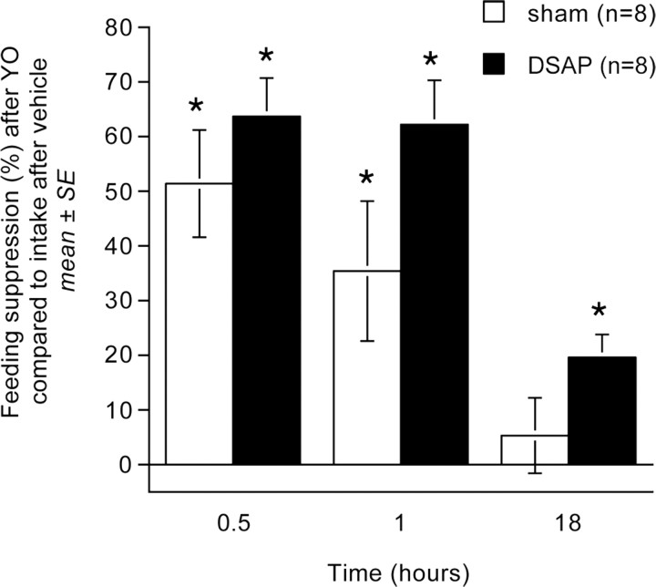
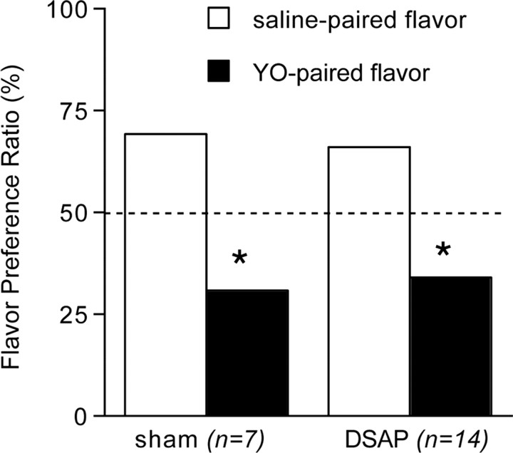
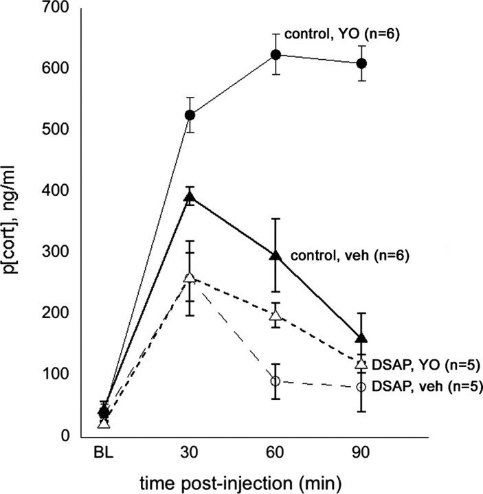
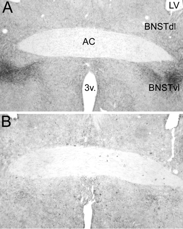
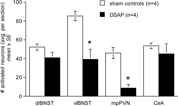
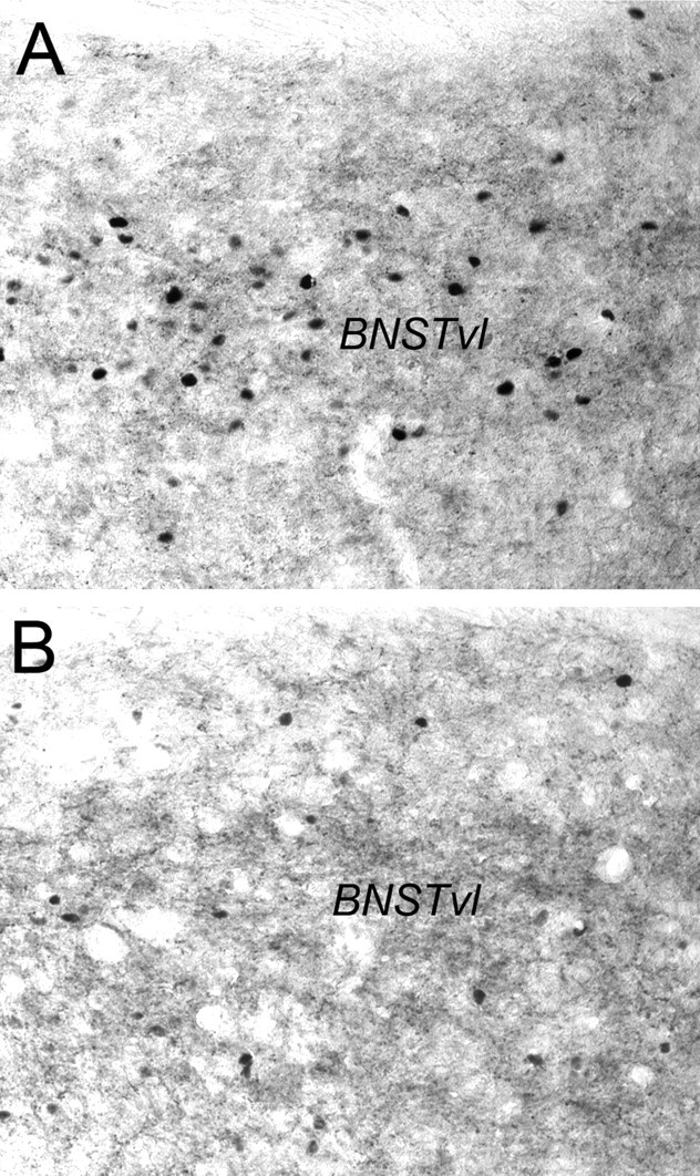
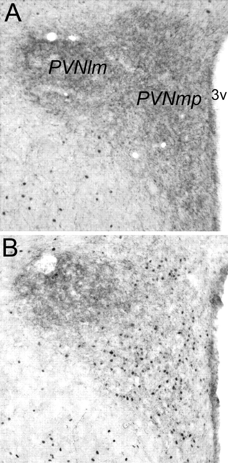
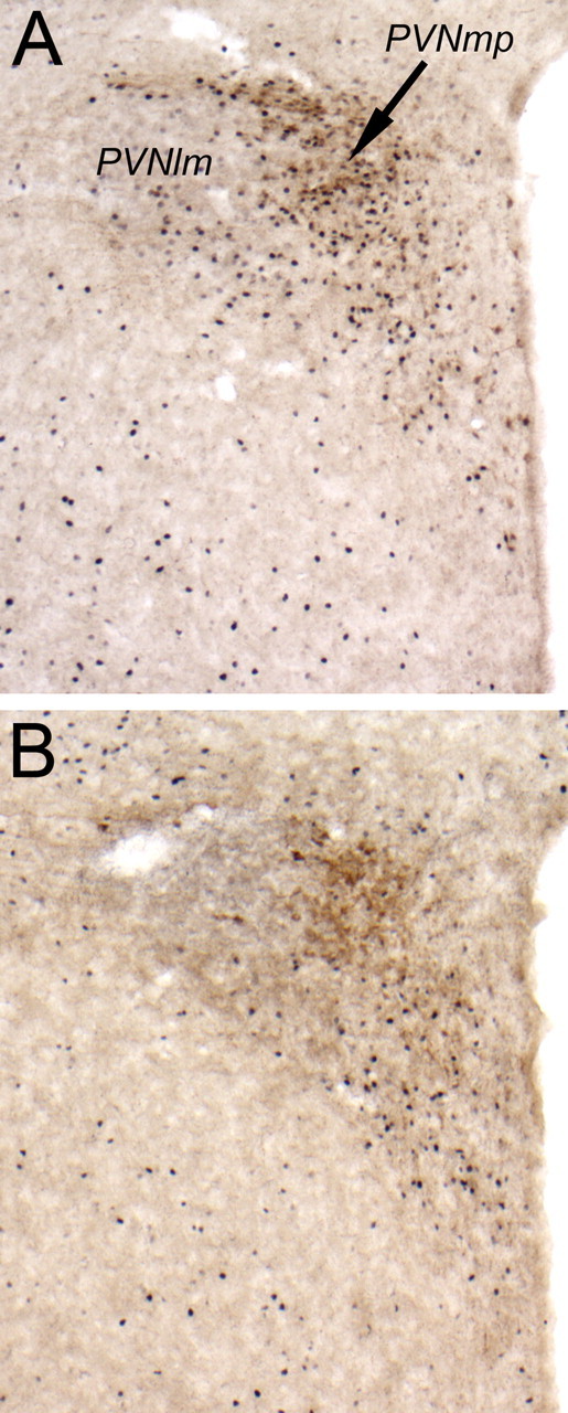
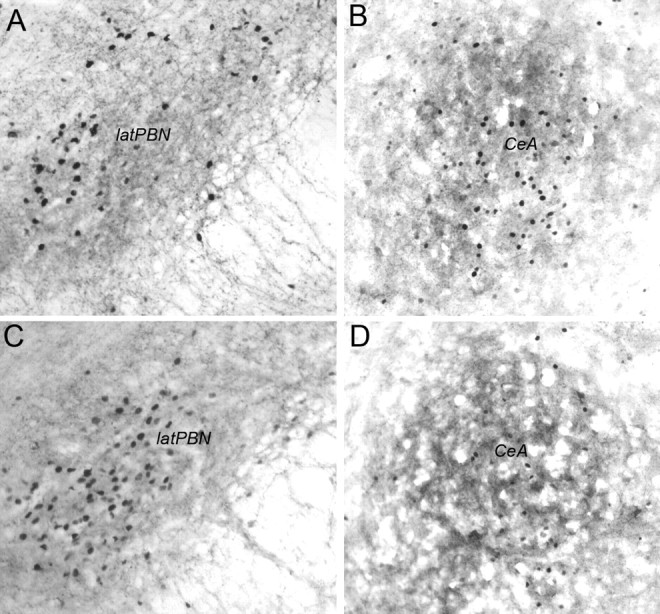
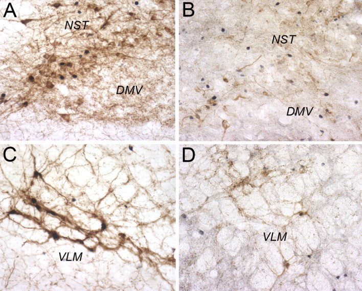
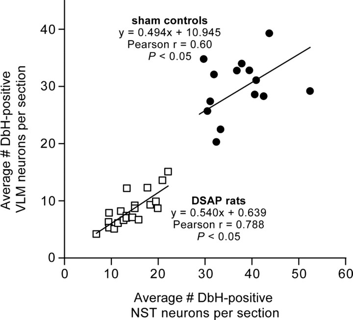
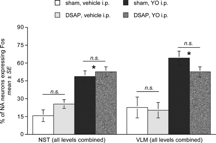
The alpha2 adrenoceptor antagonist yohimbine (YO) increases transmitter release from adrenergic/noradrenergic (NA) neurons. Systemic YO activates the hypothalamic-pituitary-adrenal (HPA) axis, inhibits feeding, and supports conditioned flavor avoidance (CFA) in rats. To determine whether these effects require NA inputs to the bed nucleus of the stria terminalis (BNST), vehicle or saporin toxin conjugated to an antibody against dopamine beta hydroxylase (DSAP) was microinjected bilaterally into the BNST to remove its NA inputs. Subsequent tests failed to reveal any lesion effect on the ability of YO (5.0 mg/kg, i.p.) to inhibit food intake or to support CFA. Conversely, HPA axis responses to YO were significantly blunted in DSAP rats. In a terminal experiment, DSAP and control rats were perfused 90-120 min after intraperitoneal injection of YO or vehicle. Brains were processed to reveal Fos immunolabeling and lesion extent. NA fibers were markedly depleted in the BNST and medial parvocellular paraventricular hypothalamus (PVNmp) in DSAP rats, evidence for collateralized NA inputs to these regions. DSAP rats displayed significant loss of caudal medullary NA neurons, and markedly blunted Fos activation in the BNST and in corticotropin-releasing hormone-positive PVNmp neurons after YO. We conclude that a population of medullary NA neurons provides collateral inputs to the BNST and PVNmp, and that these inputs contribute importantly to Fos expression and HPA axis activation after YO treatment. Conversely, NA-mediated activation of BNST and PVNmp neurons is unnecessary for YO to inhibit food intake or support CFA, evidence for the sufficiency of other intact neural pathways in mediating those effects.


Figures

Figure 1.
Figure 1.
Suppression of deprivation-induced food intake at three time points in sham control and DSAP rats after YO treatment (5.0 mg/kg BW, i.p.). Bars depict the magnitude of feeding suppression relative to intake by the same rats after deprivation and vehicle treatment (0.15 m NaCl, i.p.). YO-induced feeding suppression did not differ significantly between DSAP and sham control rats at any time point. *Significantly suppressed compared with intake at the same time point after vehicle treatment (p < 0.05). Error bars indicate SEM.
Figure 2.
Figure 2.
Average group preference ratios (mean ± SE) for novel flavors in two-bottle choice tests after flavors were previously paired with vehicle treatment (0.15 m NaCl, i.p.) or YO (5.0 mg/kg BW, i.p.). The dashed line indicates the expected preference ratio of 50%:50% if there was no effect of flavor pairing condition. Sham control and DSAP rats show a significantly lower preference for YO-paired flavors compared with vehicle-paired flavors. Error bars are too small to be visible. *Significantly reduced preference for YO-paired flavors compared with intake of saline-paired flavors (p < 0.05).
Figure 3.
Figure 3.
Plasma corticosterone levels (p[cort], ng/ml) measured at baseline (BL; preinjection) and at three subsequent time points (i.e., 30, 60, and 90 min) after intraperitoneal injection of 0.15 m NaCl vehicle (veh) or after intraperitoneal YO (5 mg/kg BW) in control rats (n = 6; solid symbols) and in DSAP rats (n = 5; open symbols). Symbols represent averaged group data (mean ± SE).
Figure 4.
Figure 4.
Representative low-magnification photomicrographs of dual DbH and Fos immunolabeling within the BNSTdl and BNSTvl. A, Sham control rat treated intraperitoneally with vehicle. B, DSAP rat treated intraperitoneally with YO. AC, Anterior commissure; 3v, third ventricle; LV, lateral ventricle. See Figure 6 for a higher-magnification view of YO-induced Fos immunolabeling within the BNSTvl.
Figure 5.
Figure 5.
Comparison of YO-induced Fos expression within the BNST (dl and vl), PVNmp, and CeA in sham control and DSAP rats. *Significantly fewer activated neurons than in sham controls (p < 0.05).
Figure 6.
Figure 6.
Representative photomicrographs of dual CRH and Fos immunolabeling within the BNSTvl. A, Sham control rat treated intraperitoneally with YO. B, DSAP rat treated intraperitoneally with YO. See Figure 4 for regional orientation.
Figure 7.
Figure 7.
Representative photomicrographs of dual DbH and Fos immunolabeling within the PVN (lm, lateral magnocellular subdivision; mp, medial parvocellular subdivision). A, Sham control rat treated intraperitoneally with vehicle. B, DSAP rat treated intraperitoneally with YO. 3v, Third ventricle.
Figure 8.
Figure 8.
Representative color photomicrographs of dual CRH and Fos immunolabeling within the PVN (lm, lateral magnocellular subdivision; mp, medial parvocellular subdivision). A, Sham control rat treated intraperitoneally with YO. B, DSAP rat treated intraperitoneally with YO. 3v, Third ventricle.
Figure 9.
Figure 9.
Representative photomicrographs of dual DbH and Fos immunolabeling in the lateral PBN (A, C) and dual CRH and Fos immunolabeling in the CeA (B, D). A, B, Sham control rat treated intraperitoneally with YO. C, D, DSAP rat treated intraperitoneally with YO. latPBN, Lateral parabrachial nucleus.
Figure 10.
Figure 10.
Representative color photomicrographs of dual DbH and Fos immunolabeling within the dorsomedial medulla (A, B) and VLM (C, D) at the rostrocaudal level of the area postrema. A, C, Sham control rat treated intraperitoneally with YO. B, D, DSAP rat treated intraperitoneally with YO. DMV, Dorsal motor nucleus of the vagus.
Figure 11.
Figure 11.
Scatter plot showing correlations between counts of DbH-positive NA neurons within the VLM and NST in sham control and DSAP rats. Symbols represent individual rats.
Figure 12.
Figure 12.
Similar proportions of DbH-immunopositive NA neurons present within the NST and VLM are activated to express Fos in sham control and DSAP rats after YO treatment. All three analyzed levels of the NST and VLM are combined (for breakdown by rostrocaudal level, see Table 3). *Significantly more activation in both surgical groups after YO treatment compared with activation after intraperitoneal vehicle (p < 0.05). n.s., No significant difference between surgical groups.

References

    1. Alden M, Besson JM, Bernard JF. Organization of the efferent projections from the pontine parabrachial area to the bed nucleus of the stria terminalis and neighboring regions: a PHA-L study in the rat. J Comp Neurol. 1994;341:289–314. - PubMed
    1. Aston-Jones G, Delfs JM, Druhan J, Zhu Y. The bed nucleus of the stria terminalis: a target site for noradrenergic actions in opiate withdrawal. Ann NY Acad Sci. 1999;877:486–498. - PubMed
    1. Baldwin HA, Johnston AL, File SE. Antagonistic effects of caffeine and yohimbine in animal tests of anxiety. Eur J Pharmacol. 1989;159:211–215. - PubMed
    1. Callahan MF, Beales M, Oltmans GA. Yohimbine and rauwolscine reduce food intake of genetically obese (ob/ob) and lean mice. Pharmacol Biochem Behav. 1984;20:591–599. - PubMed
    1. Cecchi M, Khoshbouei H, Morilak DA. Modulatory effects of norepinephrine, acting on alpha1 receptors in the central nucleus of the amygdala, on behavioral and neuroendocrine responses to acute immobilization stress. Neuropharmacology. 2002a;43:1139–1147. - PubMed

Publication types

MeSH terms

LinkOut - more resources