Comparative Study
. 2006 Nov 1;26(44):11454-61.
doi: 10.1523/JNEUROSCI.2260-06.2006.

Emotional modulation of pain: is it the sensation or what we recall?

Affiliations
Comparative Study

Emotional modulation of pain: is it the sensation or what we recall?

Fabio Godinho et al. J Neurosci. .

Abstract

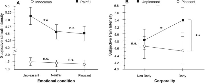
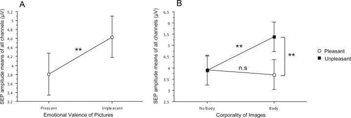
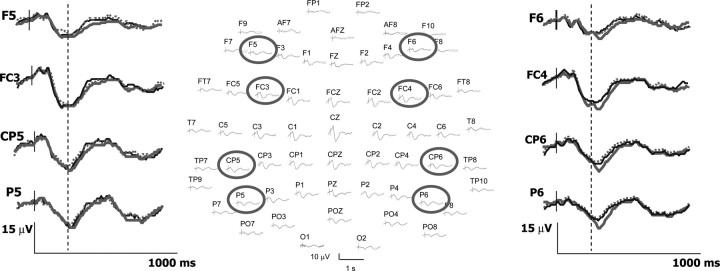
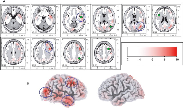
Emotions modulate pain perception, although the mechanisms underlying this phenomenon remain unclear. In this study, we show that intensity reports significantly increased when painful stimuli were concomitant to images showing human pain, whereas pictures with identical emotional values but without somatic content failed to modulate pain. Early somatosensory responses (<200 ms) remained unmodified by emotions. Conversely, late responses showed a significant enhancement associated with increased pain ratings, localized to the right prefrontal, right temporo-occipital junction, and right temporal pole. In contrast to selective attention, which enhances pain ratings by increasing sensory gain, emotions triggered by seeing other people's pain did not alter processing in SI-SII (primary and second somatosensory areas), but may have biased the transfer to, and the representation of pain in short-term memory buffers (prefrontal), as well as the affective assignment to this representation (temporal pole). Memory encoding and recall, rather than sensory processing, appear to be modulated by empathy with others' physical suffering.


Figures

Figure 1.
Figure 1.
Valence and arousal values of emotional pictures. Valence (1, very unpleasant; 5, neutral; 9, very pleasant) and arousal (1, calm; 9, excited) ratings were plotted for each image in men's and women's paradigms. Values assigned to unpleasant, pleasant, and neutral sets were taken from IAPS data. Additionally, images from several internet sources were included in the pleasant group to strengthen valence of women's pleasant subset and to introduce pleasant pictures explicitly related to human somatic experience. Valence and arousal ratings for these pictures were obtained in a pilot study with 10 men and 10 women. As a result, arousal and valence dimensions were matched, and comparable emotional manipulation was obtained in both genders.
Figure 2.
Figure 2.
Structure of the block design paradigm. A, Internal structure of one block. Three images with same emotional valence and two electrical stimuli of same intensity composed each block of 13 s duration. Electrical stimuli were delivered after the first and the third pictures. B, The whole paradigm had a sequence of 100 blocks (25 min time length).
Figure 3.
Figure 3.
Effects of emotional context on subjective stimulus intensity. A, Significant emotional modulation of perceived intensity was only observed for painful stimuli (black squares), but not for innocuous stimuli (white circles) whose perception remained stable despite changing the emotional context. Note also that only unpleasant pictures modulated pain, whereas pleasant pictures did not change significantly the perception of stimuli relative to neutral images. B, ANOVA interaction plot shows that the subjective pain ratings were significantly different between pleasant and unpleasant conditions containing explicit references to human body sensations (body). Differences were not significant between pleasant and unpleasant pictures not depicting body sensations (nonbody). Moreover, pain scores were significantly higher during the unpleasant-body than the unpleasant-nonbody condition. Values are expressed as mean ± SE (**p < 0.01; *p < 0.05). Values for neutral pictures were never significant and are not illustrated for graph clarity.
Figure 4.
Figure 4.
Effects of emotional context on cortical responses to painful stimuli. A, Main effect of the emotional condition on cortical responses to painful stimuli. SEP amplitudes were significantly higher during the unpleasant than during the pleasant condition. B, ANOVA interaction plot shows that cortical SEP were significantly increased when unpleasant-body pictures were displayed relative to either pleasant-body or unpleasant-nonbody. No significant differences were observed when comparing unpleasant and pleasant conditions without human somatic contents (nonbody). Note that this emotional effect is symmetrical to that described on behavioral pain ratings. Values are expressed as mean ± SE (**p < 0.01).
Figure 5.
Figure 5.
Cortical responses to painful stimuli associated or not to emotional modulation of pain. The 64-channel SEPs at their scalp positions (nose up) are represented in the center. Positive voltages are represented downward. Traces from eight selected electrodes (circles) are enlarged in the left and in the right parts to illustrate that SEP amplitudes to painful stimuli were significantly enhanced during the unpleasant-body condition (gray traces) relative to both unpleasant-nonbody (dotted) and pleasant-body (black) conditions. Note also that amplitude differences occurred only after 270 ms poststimulus (dotted line) and concerned exclusively the electrodes located on the right side of the scalp. See Results for details.
Figure 6.
Figure 6.
Anatomical (LORETA and BESA) modeling of cortical regions showing the earliest activity associated with emotional modulation of pain. A, The difference (subtraction) between SEP traces obtained during unpleasant- and pleasant-body conditions, as well as between unpleasant-body and unpleasant-nonbody conditions, were assessed by LORETA within 270–400 ms, when significant differences of SEP amplitudes were observed. Cortical areas of maximal activity are illustrated on normalized Talairach slices. These areas were lateralized toward the right hemisphere and concerned the DLPFC, the temporal pole, and the temporo-occipital junction (blue circles). Only regions whose differential activities exceeded by at least 3 SD of the mean prestimulus baseline are illustrated. The Z-levels in the color scale correspond to the number of SDs from the mean prestimulus baseline. Additionally, dipole analysis was performed on the same epoch (green circles). Dipolar sources lay close to the most active areas showed by LORETA. No sources were found over SI/SII or posterior insula with either technique. B, Three-dimensional representation of both hemispheres showing the cortical regions aforementioned (blue circles).

References

    1. Arntz A, Dreessen L, Merckelbach H. Attention, not anxiety, influences pain. Behav Res Ther. 1991;29:41–50. - PubMed
    1. Baddeley A. Working memory: looking back and looking forward. Nat Rev Neurosci. 2003;4:829–839. - PubMed
    1. Bantick SJ, Wise RG, Ploghaus A, Clare S, Smith SM, Tracey I. Imaging how attention modulates pain in humans using functional MRI. Brain. 2002;125:310–319. - PubMed
    1. Barba C, Valeriani M. Assessing somatosensory evoked potential (SEP) generators by human intracranial recordings. Clin Neurophysiol. 2004;115:488–489. - PubMed
    1. Botvinick M, Jha AP, Bylsma LM, Fabian SA, Solomon PE, Prkachin KM. Viewing facial expressions of pain engages cortical areas involved in the direct experience of pain. NeuroImage. 2005;25:312–319. - PubMed

Publication types