. 2006 Nov 1;20(21):3010-21.
doi: 10.1101/gad.1493506.

The order of expression of transcription factors directs hierarchical specification of hematopoietic lineages

Affiliations

The order of expression of transcription factors directs hierarchical specification of hematopoietic lineages

Hiromi Iwasaki et al. Genes Dev. .

Abstract

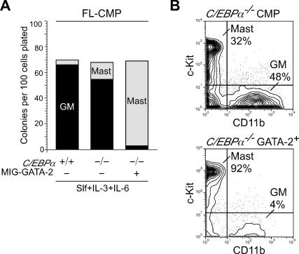
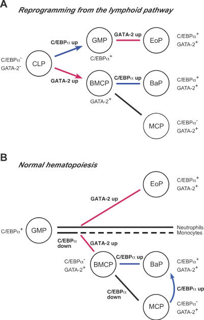
The mechanism of lineage specification in multipotent stem cells has not been fully understood. We recently isolated progenitors with the eosinophil, basophil, or mast cell lineage potential, all of which originate from granulocyte/monocyte progenitors (GMPs). By using these prospectively purified progenitors, we show here that the expression timing of GATA-2 and CCAAT enhancer-binding protein alpha (C/EBPalpha) can differentially control their lineage commitment. The expression of GATA-2 instructed C/EBPalpha-expressing GMPs to commit exclusively into the eosinophil lineage, while it induced basophil and/or mast cell lineage commitment if C/EBPalpha was suppressed at the GMP stage. Furthermore, simply by switching the order of C/EBPalpha and GATA-2 transduction, even lymphoid-committed progenitors recaptured these developmental processes to be reprogrammed into each of these lineages. We propose that the order of expression of key transcription factors is critical for their interplay to selectively drive lineage specification programs, by which stem cells could generate multiple lineage cells in a hierarchical manner.


Figures

Figure 1.
Figure 1.
Expression profiles of transcription factors and lineage-related genes in purified progenitor populations. (A) A developmental scheme of lineage-restricted progenitor populations used in this study. (HSC) Hematopoietic stem cell; (CLP) common lymphoid progenitor; (CMP) common myeloid progenitor; (MEP) megakaryocyte/erythrocyte progenitor; (GMP) granulocyte/monocyte progenitor; (BMCP) basophil/mast cell progenitor; (MCP) mast cell progenitor; (BaP) basophil progenitor; (EoP) eosinophil progenitor. (B) Semiquantitative RT–PCR analyses of transcription factors and lineage-related genes in purified progenitor populations. (mMCP-5) Mouse mast cell protease-5; (MBP) major basic protein (EoPO) eosinophil peroxidase; (HPRT) hypoxanthine–guanine phosphoribosyl transferase. (*) MCPs were purified from GMP cultures on day 3. (C) Real-time PCR assays of transcription factor expression in purified stem and progenitor cells. ΔCt values for each sample were standardized by GAPDH Ct values. PCR assays were performed using three sets of cDNA samples prepared independently, resulting in the same pattern of relative expression.
Figure 2.
Figure 2.
Enforced GATA-2 instructs GMPs to exclusively select the eosinophil fate. (A) Clonogenic analyses of GMPs with or without GATA-2 transduction. Purified GMPs were transduced with a GFP-tagged retrovirus carrying a murine GATA-2 cDNA or an empty control retro-virus. GFP-positive GMPs were isolated 36 h after the retroviral infection and subjected to methylcellulose assays. Types of colonies were evaluated at day 5 by May-Giemsa staining. Cytokines added are indicated. (B) Progeny of GMPs transduced with the GATA-2 retrovirus. An image of fluorescence microscopy (GFP filter, ×1000, upper panel), and that of May-Giemsa staining (×1000, bottom panel) are shown. Cells positive for GFP possessed eosinophilic granules. (C) The phenotypic analysis of progeny of GMPs transduced with GATA-2. Although BMCPs gave rise to FcεRIα+ basophils and mast cells in the culture, GATA-2+ GMPs did not give rise to FcεRIα+ cells. (D) Gene expression analyses of GMPs immediately after the completion of GATA-2 transduction. (E) Real-time PCR analysis of GATA-2 mRNA before and after GATA-2 transduction. ΔCt values for each samples were standardized by GAPDH Ct values. GATA-2 transduction experiment was performed three times using GMPs purified independently, and the same results were obtained.
Figure 3.
Figure 3.
The effect of enforcement or reduction of C/EBPα expression on the basophil/mast cell development from myeloid progenitors. (A) FACS analyses of day 3 progeny of GMPs with or without C/EBPα transduction. Purified GMPs were transduced with a hCD4-tagged retrovirus carrying a murine C/EBPα cDNA or an empty retrovirus as control. hCD4-positive GMPs were isolated 36 h after the retroviral infection and cultured for an additional 72 h in the presence of Slf, IL-3, and IL-6. (Right) By maintaining C/EBPα expression, GMPs became incapable of producing FcεRIαloc-Kithi MCPs or FcεRIαhic-Kit BaPs. FACS plots are representative of three independent experiments. (B) Limiting dilution analysis of mast cell or basophil development from C/EBPαΔ/Δ GMPs. GMPs were purified from C/EBPαF/F mice (Zhang et al. 2004) and transduced with a GFP-tagged retrovirus carrying a Cre cDNA or an empty retrovirus as control. GFP-positive GMPs were isolated 36 h after the retroviral infection and subjected to limiting dilution assays in the presence of Slf, IL-3, and IL-6. A complete excision of floxed C/EBPα alleles after Cre transduction was determined by PCR assay as previously reported (Zhang et al. 2004) (not shown). The development of basophils and/or mast cells in each cultures was determined by May-Giemsa and Toluidine Blue staining. The frequency of mast cell read-out from C/EBPαΔ/Δ GMPs was fourfold higher than that from control C/EBPαF/F GMPs (left), whereas mature basophil development was blocked completely by the C/EBPα disruption (right). (C) Reduction of C/EBPα by an RNAi technique using shRNA expression vector by retrovirus transduction. GMPs transduced with three different shRNAs targeting C/EBPα (see Materials and Methods) were measured for their expression of C/EBPα by a real-time PCR analysis standardized by a GAPDH expression level (left), and also analyzed for their frequency of mast cell readout by a limiting dilution assay (right). The expression level of C/EBPα in GMPs was inversely correlated with the frequency of mast cell lineage readout.
Figure 4.
Figure 4.
Enforced expression of GATA-2 in C/EBPα−/− myeloid progenitors CMPs purified from E14 C/EBPα−/− FL were transduced with a GFP-tagged retrovirus carrying a murine GATA-2 cDNA or an empty retrovirus as control. GFP-positive CMPs were isolated 36 h after the retroviral infection and cultured for 7 d on methylcellulose (A) or in suspension (B) in the presence of Slf, IL-3, and IL-6. Types of colonies were determined by May-Giemsa and Toluidine Blue staining. The enforced expression of GATA-2 instructed C/EBPα−/− FL-CMPs to select the mast cell fate. Representative data of three independent experiments are shown.
Figure 5.
Figure 5.
The ordered expression of C/EBPα and/or GATA-2 can convert CLPs to either GMPs, EoPs, BMCPs, or BaPs. (A) Expression profile of hCD4 and GFP reporters after the two-step transduction. Note that the expression level of GFP and hCD4 was consistent irrespective of the order of transduction. (B) Methylcellulose assays for lineage outcomes of CLPs transduced with C/EBPα and/or GATA-2. R1–R4 represent sorting regions defined in A. Cytokines added were Slf, IL-3, IL-5, IL-6, IL-7, GM-CSF, Epo, and Tpo. Types of colonies were determined at day 7 by May-Giemsa and Toluidine Blue staining. (C) Phenotypic and morphological analyses of progeny of CLPs transduced with C/EBPα and/or GATA-2 in a different order. CLPs were purified according to sorting regions (R1–R4) defined in A and cultured in suspension for 7 d in the presence of Slf, IL-3, IL-5, IL-6, IL-7, GM-CSF, Epo, and Tpo. FACS analyses and morphologies (May-Giemsa staining, ×1000) of day 7 progeny are shown. Representative data of three independent experiments are shown.
Figure 6.
Figure 6.
Expression profiles of transcription factors and lineage-related genes in CLPs after two-step retroviral transduction of C/EBPα and GATA-2. (A)RT–PCR analyses of lineage-related gene expression in CLPs purified immediately after the completion of two-step retroviral transduction. R1–R4 represent sorting regions defined in Figure 5. (B) Change in the expression level of C/EBPα and GATA-2 mRNA in CLPs after tranduction of control-GFP, C/EBPα, or GATA-2, determined by a real-time PCR assay. ΔCt values for each samples were standardized by GAPDH Ct values. (C) Change in the expression level of lymphoid-affiliated EBF, Pax-5, and GATA-3 in CLPs after tranduction of control-GFP, C/EBPα, or GATA-2. Representative data of three independent experiments are shown.
Figure 7.
Figure 7.
Schematic presentation of roles of transcription factors in lineage specification to eosinophils, basophils, and mast cells during reprogramming of CLPs and in normal hematopoiesis. (A) Lineage-instructive signals for each myeloid lineage on the basis of reprogramming of CLPs. CLPs can be converted to GMPs and BMCPs by the enforced expression of C/EBPα and GATA-2, respectively. In this model, the order of C/EBPα and GATA-2 expression is critical for CLPs to differentiate into EoPs via GMPs, or into BaPs via BMCPs. (B) Hematopoietic progenitor development downstream from GMPs in a physiological setting. Up-regulation of GATA-2 instructs GMPs to differentiate into eosinophil, basophil, and mast cell lineages. Upon GATA-2 up-regulation, if GMPs maintain C/EBPα expression, they become EoPs, whereas if GMPs down-regulate C/EBPα, they become BMCPs. For BaP development, C/EBPα needs to be reactivated after the BMCP stage. The mechanism of C/EBPα down-regulation in GMPs is not clear at this time, but since C/EBPα is not required for generation of mature neutrophils and monocytes after the GMP stage (Zhang et al. 2004), a fraction of GMPs may naturally down-regulate C/EBPα. (Red lines) Up-regulation of GATA-2; (blue lines) up-regulation of C/EBPα; (arrows) reprogramming of progenitors by enforced expression of transcription factors.

Similar articles

Cited by

References

    1. Akashi, K., Traver, D., Miyamoto, T., Weissman, I.L. A clonogenic common myeloid progenitor that gives rise to all myeloid lineages. Nature. 2000;404:193–197. - PubMed
    1. Arinobu, Y., Iwasaki, H., Gurish, M.F., Mizuno, S.I., Shigematsu, H., Ozawa, H., Tenen, D.G., Austen, K.F., Akashi, K. Developmental checkpoints of the basophil/ mast cell lineages in adult murine hematopoiesis. Proc. Natl. Acad. Sci. 2005;102:18105–18110. - PMC - PubMed
    1. Baba, Y., Garrett, K.P., Kincade, P.W. Constitutively active β-catenin confers multilineage differentiation potential on lymphoid and myeloid progenitors. Immunity. 2005;23:599–609. - PMC - PubMed
    1. Boyce, J.A., Friend, D., Matsumoto, R., Austen, K.F., Owen, W.F. Differentiation in vitro of hybrid eosinophil/basophil granulocytes: Autocrine function of an eosinophil developmental intermediate. J. Exp. Med. 1995;182:49–57. - PMC - PubMed
    1. Brummelkamp, T.R., Bernards, R., Agami, R. A system for stable expression of short interfering RNAs in mammalian cells. Science. 2002;296:550–553. - PubMed

Publication types

MeSH terms

Substances

Associated data