. 2007 Jan;18(1):166-75.
doi: 10.1091/mbc.e06-08-0664. Epub 2006 Nov 1.

The vacuolar transporter chaperone (VTC) complex is required for microautophagy

Affiliations

The vacuolar transporter chaperone (VTC) complex is required for microautophagy

Andreas Uttenweiler et al. Mol Biol Cell. 2007 Jan.

Abstract

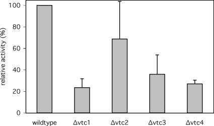
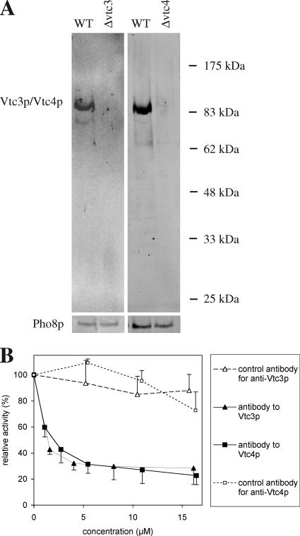
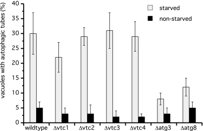
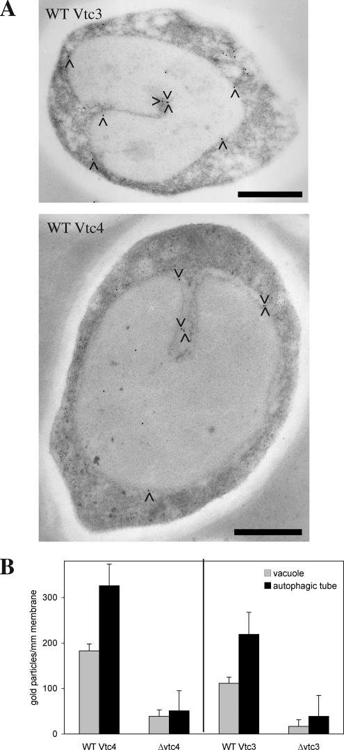
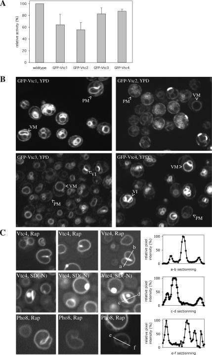
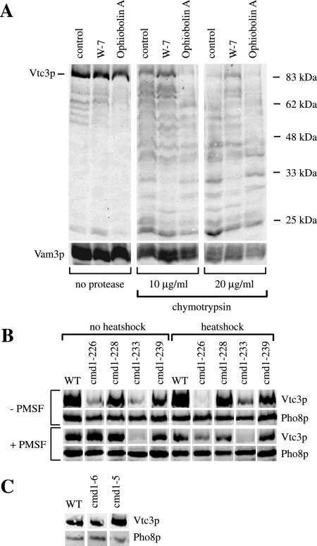
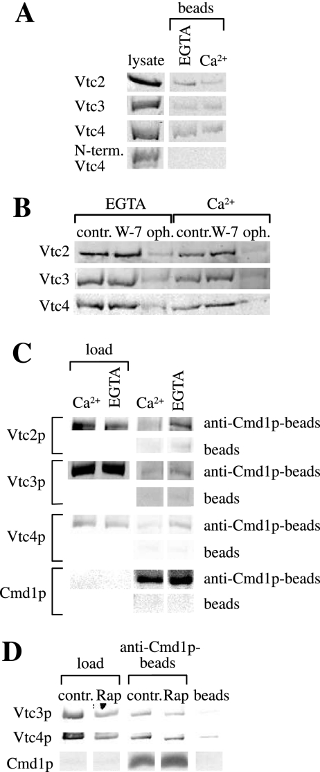
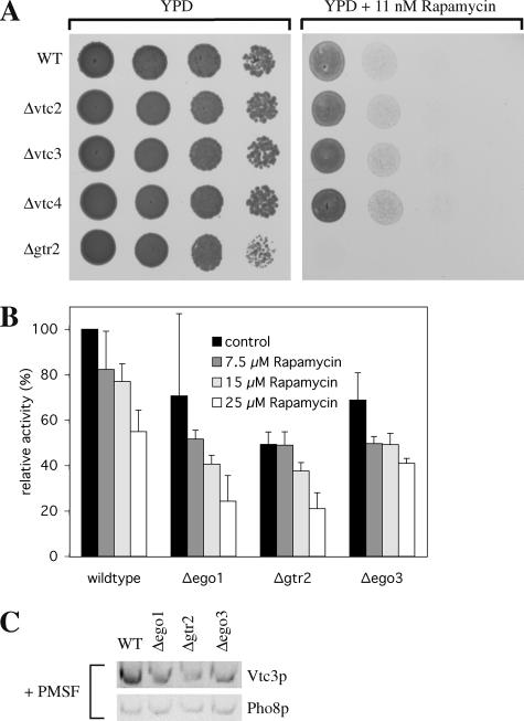
Microautophagy involves direct invagination and fission of the vacuolar/lysosomal membrane under nutrient limitation. This occurs by an autophagic tube, a specialized vacuolar membrane invagination that pinches off vesicles into the vacuolar lumen. In this study we have identified the VTC (vacuolar transporter chaperone) complex as required for microautophagy. The VTC complex is present on the ER and vacuoles and at the cell periphery. On induction of autophagy by nutrient limitation the VTC complex is recruited to and concentrated on vacuoles. The VTC complex is inhomogeneously distributed within the vacuolar membranes, showing an enrichment on autophagic tubes. Deletion of the VTC complex blocks microautophagic uptake into vacuoles. The mutants still form autophagic tubes but the production of microautophagic vesicles from their tips is impaired. In line with this, affinity-purified antibodies to the Vtc proteins inhibit microautophagic uptake in a reconstituted system in vitro. Our data suggest that the VTC complex is an important constituent of autophagic tubes and that it is required for scission of microautophagic vesicles from these tubes.


Figures

Figure 1.
Figure 1.
Microautophagic activity of Δvtc vacuoles in vitro. Yeast cells were grown in log phase at 30°C in YPD and vacuoles were prepared at 30°C. In vitro microautophagic reactions were run with cytosol from starved wild-type cells (K91-1A) at 27°C. Data from four to nine independent experiments were averaged. Error bars, SD.
Figure 2.
Figure 2.
(A) Specificity of affinity-purified antibodies (goat) to Vtc3p and Vtc4p. Vacuolar preparations from wild type or from the respective deletion strains were analyzed by SDS-PAGE and Western blotting with the affinity purified antibodies to Vtc3p or Vtc4p. (B) Sensitivity of in vitro microautophagy to Vtc antibodies. In vitro microautophagic reactions were run with various concentrations of affinity-purified antibodies (goat) to the indicated proteins. Vacuoles were preincubated with antibodies for 10 min on ice before starting the uptake reaction. Control antibodies are total IgG extracted from the sera by adsorption to protein A agarose after the specific antibodies to Vtc3p or Vtc4p had been completely depleted by affinity purification. Data from three independent experiments were averaged. Error bars, SD.
Figure 3.
Figure 3.
Frequency of autophagic tubes in vtc mutants in vivo. Yeast cells were grown in log phase at 30°C in YPD. Vacuoles were stained with 10 μM FM4-64 for 1 h, washed twice with SD(-N), and starved for 3.5 h in SD(-N) at 30°C. Tube frequencies were determined by fluorescence microscopy. Data from four determinations (200 cells) were averaged.
Figure 4.
Figure 4.
Localization of Vtc proteins by immunoelectron microscopy in vivo. (A) Immunogold labeling. Yeast cells were grown in log phase at 30°C in YPD, washed twice with SD(-N), and starved for 3.5 h in SD(-N) at 30°C. Cells were prepared for electron microscopy by high-pressure freezing and freeze-substitution. Thin sections showing vacuolar invaginations (n = 41 for wild-type Vtc3 and n = 30 for ΔVtc3, n = 31 for wild-type Vtc4 and Δvtc4) were stained with antibodies and protein A-gold. The pictures show two examples of labeled thin sections. (B) Quantitation. Gold particles on the pictures from A were counted along invaginated membranes and on the vacuolar boundary membranes (statistical significances: p ≥ 95% for Vtc3p and p ≥99% for Vtc4p). SD for each bar was determined from three data sets of equal size (randomly chosen pictures). Black bars, 1 μm.
Figure 5.
Figure 5.
GFP-Vtc proteins. (A) Microautophagic activity of GFP-Vtc vacuoles in vitro. Individual Vtc open reading frames in BY4742 yeast cells were N-terminally tagged by integrating GFP sequences into the genomic locus (Janke et al., 2004), making the tagged proteins the only source of the respective Vtc protein. The tagged open reading frames were expressed under control of a cointegrated ADH promotor. The cells were grown in log phase at 30°C in YPD and vacuoles were prepared at 30°C. In vitro microautophagic reactions were run with cytosol from starved wild-type cells (K91-1A) at 27°C. Data from four to six independent experiments were averaged. Error bars, SD (B and C). Confocal fluorescence microscopy. Yeast cells expressing GFP-Vtc or GFP-Pho8 proteins were analyzed in exponential growth in rich YPD media (B) or after induction of a starvation response (C) by 3 h of incubation in YPD/rapamycin (200 nM) or SD(-N) starvation medium. The GFP signals across the boundary membrane and autophagic tubes were quantified using ImageJ software. PM, plasma membrane; VM, vacuolar membrane; VI, vacuolar interface.
Figure 6.
Figure 6.
(A) Limited proteolysis of Vtc3p. In vitro microautophagic reactions were run without any inhibitor (control) or with 400 μM W-7 or 100 μM ophiobolin A. After 1 h at 27°C, the reactions were chilled on ice and stopped by dilution with 600 μl 150 mM KCl in PS buffer. Vacuoles were sedimented (6500 × g, 6 min, 2°C) and resuspended in 500 μl 150 mM KCl in PS buffer. Chymotrypsin was added to final concentrations as indicated and incubated for 10 min on ice. Digestion was stopped by chloroforme-methanol precipitation. Proteins were analyzed by SDS-PAGE and Western blotting against Vtc3p and the vacuolar SNARE protein Vam3p (control). (B) Temperature-sensitive mutants. Yeast cells were grown in log phase at 25°C in YPD. For heat-shock treatment part of the cultures were transferred to 37°C for 1 h. Cells were harvested and spheroplasted at 30°C (no heat shock) or at 37°C (heat shock) in presence or absence of 1 mM PMSF, and vacuoles were prepared. Vacuoles (50 μg protein) were sedimented (20,000 × g, 5 min, 2°C), resuspended in 25 μl of sample buffer, heated to 95°C for 10 min, and analyzed by SDS-PAGE on a 10% gel and Western blotting against the N-terminal SPX-domain of Vtc3p or against the lumenal vacuolar protein Pho8p (alkaline phosphatase) as a loading control. (C) Mutants in the Ca2+-binding sites. Yeast cells were grown in log phase at 30°C in YPD, and vacuoles were prepared at 30°C. Vacuoles (30 μg of protein) were sedimented (20,000 × g, 5 min, 2°C), resuspended in 25 μl of sample buffer, heated to 95°C for 10 min, and analyzed as in B.
Figure 7.
Figure 7.
(A) Binding of Vtc central domains to Cmd1p-Sepharose. E. coli lysates containing His6-GST-Vtc domains were incubated with Cmd1p-Sepharose in TBS buffer containing 1 mM EGTA or 1 mM Ca2+. Beads were washed once in binding buffer, and proteins were eluted by heating in sample buffer and analyzed by SDS-PAGE and Western blotting using an antibody to the His6-tag. The “lysate” lane shows 25% (Vtc2p), 12.5% (Vtc4p and N-term. Vtc4p), or 6.5% (Vtc3p) of the amounts incubated with the beads. (B) Inhibitor sensitivity of Cmd1p binding. E. coli lysates containing the central His6-GST-Vtc domains were incubated with Cmd1p-Sepharose in the presence of 1 mM EGTA or 1 mM Ca2+ as in A. The beads had been preincubated for 10 min at room temperature with TBS buffer (control), 400 μM W-7 or 200 μM ophiobolin A (final inhibitor concentrations during adsorption). Proteins bound to the beads were analyzed as in A. (C) Coimmunoprecipitation from native membranes. Total cellular membranes (1–2 mg/ml) derived from a BY4742 wild-type strain were prepared and solubilized in CHAPS. Samples were incubated with 20 μl of protein A agarose beads alone (beads) or protein A agarose beads and 20 μg affinity-purified anti-Cmd1p antibodies (anti-Cmd1p-beads) in the presence of 1 mM Ca2+ or 2 mM EGTA for 1.5 h at 4°C on a rotator. The beads were washed and proteins were eluted by heating in sample buffer. Vtc proteins were analyzed by SDS-PAGE and Western blot. Loads are 0.5% (Vtc2p) and 1% (Vtc3p and Vtc4p) of the material incubated with the beads. (D) Coimmunoprecipitation of VTC complex with calmodulin in dependence of rapamycin. Yeast cells were cultured in the presence of 200 nM rapamycin for 3 h at 30°C (control: without rapamycin) and membranes were prepared. Immunoprecipitation was carried out as described in C in the presence of 2 mM EGTA. Vtc proteins were analyzed by SDS-PAGE and Western blot. Loads are 0.5% of the material incubated with the beads.
Figure 8.
Figure 8.
EGO complex mutants. (A) Hypersensitivity to rapamycin. Yeast cells were grown in log phase at 30°C in YPD. Cells were washed twice with H2O, spotted onto agar plates (series of 10-fold dilutions) containing no inhibitor (YPD) or subinhibitory concentrations (11 nM) of rapamycin and incubated for 2 d at 30°C. (B) Microautophagic activity in vitro. In vitro microautophagy reactions were run without inhibitor (control) or in the presence of different concentrations of rapamycin. Data from three to seven independent experiments were averaged. Error bars, SD. (C) Vtc3p in vacuolar preparations. Yeast cells were grown in log phase at 30°C in YPD and vacuoles were prepared with 1 mM PMSF in the spheroplasting buffer. Vacuoles (33 μg protein) were pelleted (20,000 × g, 5 min, 2°C) and analyzed as in Figure 6B.

References

    1. Baba M., Takeshige K., Baba N., Ohsumi Y. Ultrastructural analysis of the autophagic process in yeast: detection of autophagosomes and their characterization. J. Cell Biol. 1994;124:903–913. - PMC - PubMed
    1. Babu Y. S., Sack J. S., Greenhough T. J., Bugg C. E., Means A. R., Cook W. J. Three-dimensional structure of calmodulin. Nature. 1985;315:37–40. - PubMed
    1. Bachs O., Lanini L., Serratosa J., Coll M. J., Bastos R., Aligue R., Rius E., Carafoli E. Calmodulin-binding proteins in the nuclei of quiescent and proliferatively activated rat liver cells. J. Biol. Chem. 1990;265:18595–18600. - PubMed
    1. Bergamini E., Cavallini G., Donati A., Gori Z. The anti-ageing effects of caloric restriction may involve stimulation of macroautophagy and lysosomal degradation, and can be intensified pharmacologically. Biomed. Pharmacother. 2003;57:203–208. - PubMed
    1. Burgoyne R. D., Clague M. J. Calcium and calmodulin in membrane fusion. Biochim. Biophys. Acta. 2003;1641:137–143. - PubMed

Publication types

MeSH terms

Substances