. 2007 Jan;18(1):142-52.
doi: 10.1091/mbc.e06-05-0453. Epub 2006 Nov 1.

Kidins220/ARMS is transported by a kinesin-1-based mechanism likely to be involved in neuronal differentiation

Affiliations

Kidins220/ARMS is transported by a kinesin-1-based mechanism likely to be involved in neuronal differentiation

Aurora Bracale et al. Mol Biol Cell. 2007 Jan.

Abstract

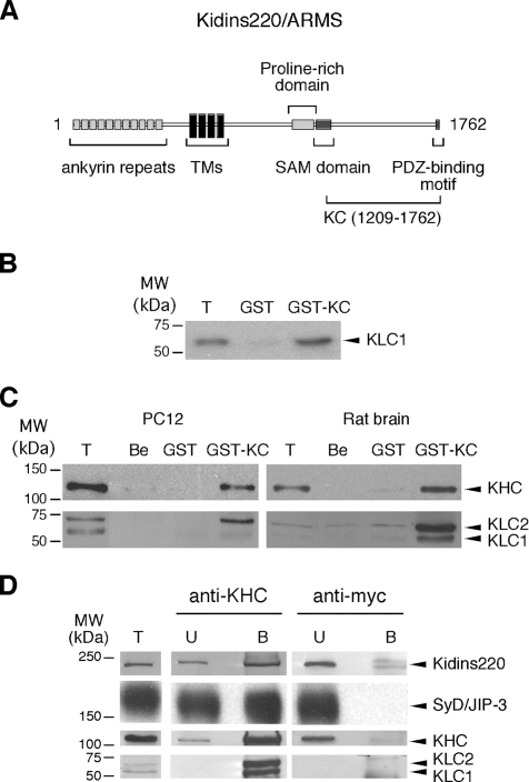
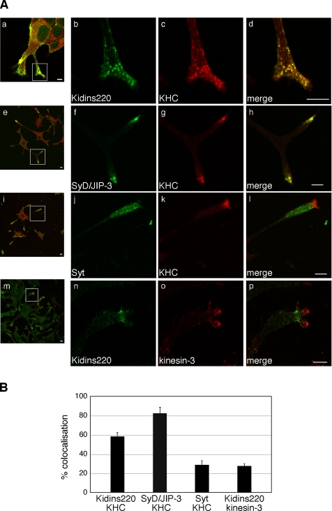
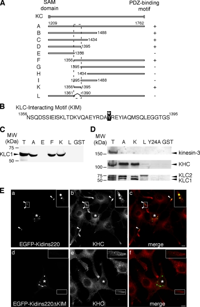
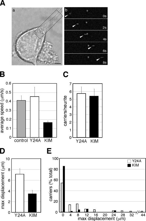
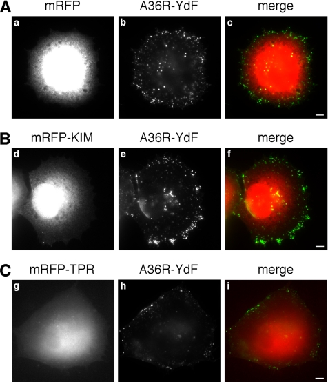
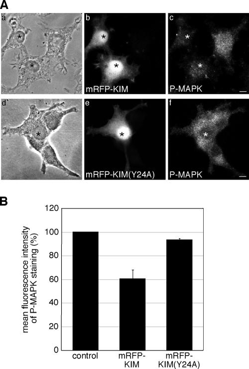
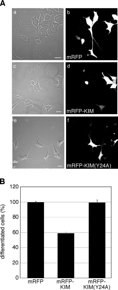
Kinase D-interacting substrate of 220 kDa/ankyrin repeat-rich membrane spanning (Kidins220/ARMS) is a conserved membrane protein mainly expressed in brain and neuroendocrine cells, which is a downstream target of the signaling cascades initiated by neurotrophins and ephrins. We identified kinesin light chain 1 (KLC1) as a binding partner for Kidins220/ARMS by a yeast two-hybrid screen. The interaction between Kidins220/ARMS and the kinesin-1 motor complex was confirmed by glutathione S-transferase-pull-down and coimmunoprecipitation experiments. In addition, Kidins220/ARMS and kinesin-1 were shown to colocalize in nerve growth factor (NGF)-differentiated PC12 cells. Using Kidins220/ARMS and KLC1 mutants, we mapped the regions responsible for the binding to a short sequence of Kidins220/ARMS, termed KLC-interacting motif (KIM), which is sufficient for the interaction with KLC1. Optimal binding of KIM requires a region of KLC1 spanning both the tetratricopeptide repeats and the heptad repeats, previously not involved in cargo recognition. Overexpression of KIM in differentiating PC12 cells impairs the formation and transport of EGFP-Kidins220/ARMS carriers to the tips of growing neurites, leaving other kinesin-1 dependent processes unaffected. Furthermore, KIM overexpression interferes with the activation of the mitogen-activated protein kinase signaling and neurite outgrowth in NGF-treated PC12 cells. Our results suggest that Kidins220/ARMS-positive carriers undergo a kinesin-1-dependent transport linked to neurotrophin action.


Figures

Figure 1.
Figure 1.
Direct interaction between Kidins220/ARMS and kinesin-1. (A) Schematic representation of Kidins220/ARMS. The KC domain of Kidins220/ARMS (residues 1209-1762), which was used as bait in the yeast two-hybrid screen, is indicated. TMs, transmembrane domains. (B) KLC1 binds GST-KC in an in vitro pull-down assay using 35S-labeled KLC1. The Coomassie staining of the SDS-PAGE gel corresponding to this experiment is available in Supplemental Figure S3Aa. (C) The kinesin-1 motor complex binds to GST-KC. PC12 cells and rat brain extracts were incubated with GST-KC, GST prebound to glutathione beads, or with empty beads (Be). The bound material was analyzed by Western blot by using anti-KHC and anti-KLC antibodies. The Ponceau staining of the nitrocellulose membranes corresponding to this experiment is available in Supplemental Figure S3Ab. (D) Native Kidins220/ARMS and kinesin-1 form a complex in PC12 cells. PC12 cell lysates were incubated with either anti-KHC or anti-myc monoclonal antibodies, and the immunoprecipitated material was subsequently analyzed by Western blot by using anti-Kidins220/ARMS, anti-SyD/JIP-3, anti-KHC, and anti-KLC antibodies. Lanes T show 1/10 (B) or 1/100 (C and D) of the starting material for comparison. In lanes U and B, ∼1 and 80% of the unbound (U) and bound (B) material are loaded, respectively. The amount of SyD/JIP-3 associated with kinesin-1 is about fivefold the amount of Kidins220/ARMS. The nonlinear detection of KLC versus KHC in C and D is likely to be dependent on the properties of the anti-KLC antibody, which does not allow a concentration-dependent recognition of KLC in the same range of KHC and Kidins220/ARMS. The limitations of the anti-KLC antibody for the quantitative visualization of KLC have been documented previously (Pfister et al., 1989; Stenoien and Brady, 1997).
Figure 2.
Figure 2.
Kidins220/ARMS and kinesin-1 colocalize in PC12 cells. (A) PC12 cells treated for 3 d with NGF were stained with polyclonal (b) or monoclonal (n) anti-Kidins220/ARMS, polyclonal anti-SyD/JIP-3 (f), polyclonal anti-Syt (j), monoclonal anti-KHC (c, g, and k), or polyclonal anti-kinesin-3 (o) antibodies and analyzed by confocal microscopy. Bars, 5 μm. (B) The percentage of colocalization of the indicated proteins in neurites is shown. Kidins220/ARMS:KHC, n = 16 neurites; SyD/JIP-3:KHC, n = 11 neurites; Syt:KHC, n = 16 neurites; for Kidins220/ARMS:kinesin-3, n = 22 neurites. Error bars represent SEM.
Figure 3.
Figure 3.
The region 83–296 of KLC1 mediates the binding to Kidins220/ARMS. (A) Scheme of the KLC1 mutants used in this study. The empty box corresponds to the heptad repeats region, whereas the gray ovals represent the TPR motifs. The putative inhibitory region for the Kidins220/ARMS–KLC interaction (aa 1-63) is marked by a dashed line. The ability of each mutant to bind Kidins220/ARMS is reported on the right: +, ≅100%; ++, ≅200%; +/−, ≅ <25%; and −, no binding. (B) The ability of in vitro 35S-labeled KLC1 mutants to bind GST-KC is shown. Intensity of the bands was quantified as described in Materials and Methods. 1/25 of the starting in vitro transcription/translation mixes is loaded in T for comparison. The Coomassie staining of the SDS-PAGE gels corresponding to this experiment is available in Supplemental Figure S3B.
Figure 4.
Figure 4.
KIM is sufficient for the binding of Kidins220/ARMS to KLC. (A) Scheme of the Kidins220/ARMS mutants used in this study. The ability of each mutant to bind KLC1 is reported on the right (+, binding, −, no binding). The position of the KIM motif is highlighted by the dashed line. (B) Sequence of the KIM motif (KC-K). The asterisk indicates the tyrosine (residue 1379 of the full-length protein) that is mutated to alanine in KIM(Y24A). (C) The ability of some of the KC mutants expressed as GST-fusion proteins to bind in vitro 35S-labeled KLC1 is shown. We loaded 1/25 of the starting in vitro transcription/translation mix in T for comparison. The Coomassie staining of the SDS-PAGE gel corresponding to this experiment is available in Supplemental Figure S3Ca. (D) The KIM motif is sufficient to bind the native kinesin-1 motor complex. PC12 cells lysates were incubated with either GST or GST-fusion proteins prebound to glutathione-beads, as indicated. The bound material was analyzed by Western blot by using anti-kinesin-3, anti-KHC, and anti-KLC antibodies. T corresponds to 10 μg of total lysate. The Ponceau staining of the nitrocellulose membrane corresponding to this experiment is available in Supplemental Figure S3Cb. (E) PC12 cells were transfected with EGFP-Kidins220/ARMS (a–c) or EGFP-Kidins220/ARMS-ΔKIM (d–f), and stained with anti-KHC antibody (b and e). Full-length Kidins220/ARMS-positive puncta are positive for KHC (c; arrowheads and inset), whereas there is no significant overlapping between KHC and the mutant lacking the KIM domain (f; inset). Asterisks indicate transfected cells. Bars, 5 μm.
Figure 5.
Figure 5.
KIM overexpression impairs Kidins220/ARMS trafficking in PC12 cells. (A) EGFP-Kidins220/ARMS is transported along neurites in PC12 cells. (a) Phase image of a microinjected cell. (b) Time series of the boxed area. Arrowheads indicate an EGFP-Kidins220/ARMS–positive moving carrier. Bar, 5 μm. (B–E) NGF-treated PC12 cells were coinjected with plasmids driving the expression of EGFP-Kidins220/ARMS alone or in combination with either mRFP-KIM, or mRFP-KIM(Y24A). (B) The average speed of EGFP-Kidins220/ARMS–positive carriers is decreased by mRFP-KIM expression. (C) The average number of EGFP-Kidins220/ARMS–positive carriers per neurite is not affected by the overexpression of mRFP-KIM. n = 13 neurites were analyzed for both conditions. (D) The average maximum displacement of EGFP-Kidins220/ARMS–positive carriers is reduced in mRFP-KIM–expressing cells. The analysis of the movement of EGFP-Kidins220/ARMS positive carriers is shown in E (see text and Materials and Methods for details). A 4-μm binning starting from 0 was applied. n = 24 carriers for EGFP-Kidins220/ARMS–expressing cells, n = 70 carriers for EGFP-Kidins220/ARMS– and mRFP-KIM–expressing cells; n = 74 for EGFP-Kidins220/ARMS– and mRFP-KIM(Y24A)–expressing cells. Error bars represent SEM.
Figure 6.
Figure 6.
The translocation of vaccinia virus to the plasma membrane is unaffected by KIM. HeLa cells were infected with the A36R-YdF vaccinia virus strain, and 4 h later they were transfected with mRFP (A), mRFP-KIM (B), or mRFP-TPR (C). After a further 4 h, cells were processed for immunofluorescence in absence of permeabilization, and extracellular virus particles were visualized. The distribution of the virus on the plasma membrane is independent of the overexpression of mRFP-KIM (compare b and e), whereas it is reduced by the overexpression of the mRFP-TPR domain (compare b and h). Bars, 5 μm.
Figure 7.
Figure 7.
Overexpression of KIM reduces the phosphorylation of MAPK in PC12 cells. (A) PC12 cells were transfected with mRFP-KIM (a–c) or mRFP-KIM(Y24A) (d–f) and then fixed and immunostained for P-MAPK (c and f). mRFP-KIM–expressing cells show a reduced P-MAPK staining compared with nontransfected cells and mRFP-KIM(Y24A)–expressing cells. Transfected cells are marked with asterisks. Bars, 5 μm. (B) The mean fluorescence intensities of untransfected cells and cells overexpressing mRFP-KIM or mRFP-KIM(Y24A) were determined as described in Materials and Methods. mRFP-KIM, n = 74 cells; mRFP-KIM(Y24A), n = 63 cells; nontransfected cells, n = 204 derived from three independent experiments. Error bars represent SEM.
Figure 8.
Figure 8.
KIM overexpression inhibits neurite outgrowth in PC12 cells. (A) Undifferentiated PC12 cells were transfected with mRFP (a and b), mRFP-KIM (c and d), or mRFP-KIM(Y24A) (e and f); treated for 3 d with NGF; and analyzed by confocal microscopy. Bars, 20 μm. (B) Percentage of differentiated cells in each condition is shown. Differentiation in mRFP-expressing cells was set to 100%. Between 50 and 140 cells were counted for each experiment (n = 3). Error bars represent SEM.

References

    1. Aravind L., Iyer L. M., Leipe D. D., Koonin E. V. A novel family of P-loop NTPases with an unusual phyletic distribution and transmembrane segments inserted within the NTPase domain. Genome Biol. 2004;5:R30. - PMC - PubMed
    1. Arevalo J. C., Pereira D. B., Yano H., Teng K. K., Chao M. V. Identification of a switch in neurotrophin signaling by selective tyrosine phosphorylation. J. Biol. Chem. 2006;281:1001–1007. - PubMed
    1. Arevalo J. C., Yano H., Teng K. K., Chao M. V. A unique pathway for sustained neurotrophin signaling through an ankyrin-rich membrane-spanning protein. EMBO J. 2004;23:2358–2368. - PMC - PubMed
    1. Bibel M., Barde Y. A. Neurotrophins: key regulators of cell fate and cell shape in the vertebrate nervous system. Genes Dev. 2000;14:2919–2937. - PubMed
    1. Bohnert S., Schiavo G. Tetanus toxin is transported in a novel neuronal compartment characterized by a specialized pH regulation. J. Biol. Chem. 2005;280:42336–42344. - PubMed

Publication types

MeSH terms