. 2007 Apr;36(4):409-17.
doi: 10.1165/rcmb.2006-0214OC. Epub 2006 Nov 1.

Mitochondrial localization and function of heme oxygenase-1 in cigarette smoke-induced cell death

Affiliations

Mitochondrial localization and function of heme oxygenase-1 in cigarette smoke-induced cell death

Dirk-Jan Slebos et al. Am J Respir Cell Mol Biol. 2007 Apr.

Retraction in

Abstract

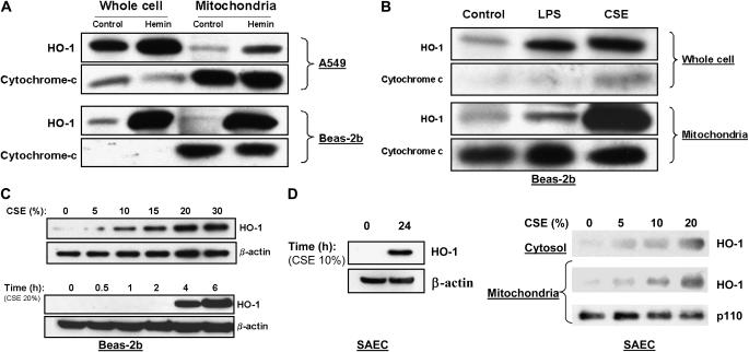
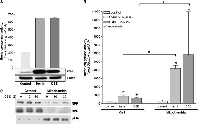
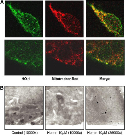
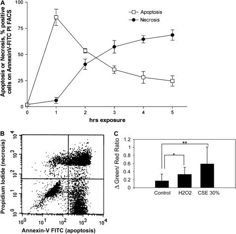
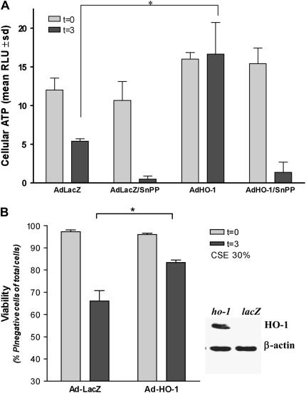
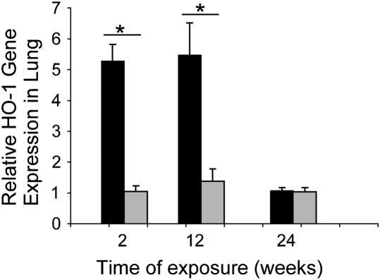
Cigarette smoke-induced apoptosis and necrosis contribute to the pathogenesis of chronic obstructive pulmonary disease. The induction of heme oxygenase-1 provides cytoprotection against oxidative stress, and may protect in smoking-related disease. Since mitochondria regulate cellular death, we examined the functional expression and mitochondrial localization of heme oxygenase-1 in pulmonary epithelial cells exposed to cigarette smoke extract (CSE), and its role in modulating cell death. Heme oxygenase-1 expression increased dramatically in cytosolic and mitochondrial fractions of human alveolar (A549), or bronchial epithelial cells (Beas-2b) exposed to either hemin, lipopolysaccharide, or CSE. Mitochondrial localization of heme oxygenase-1 was also observed in a primary culture of human small airway epithelial cells. Furthermore, heme oxygenase activity increased dramatically in mitochondrial fractions, and in whole cell extracts of Beas-2b after exposure to hemin and CSE. The mitochondrial localization of heme oxygenase-1 in Beas-2b was confirmed using immunogold-electron microscopy and immunofluorescence labeling on confocal laser microscopy. CSE caused loss of cellular ATP and rapid depolarization of mitochondrial membrane potential. Apoptosis occurred in Beas-2b at low concentrations of cigarette smoke extract, whereas necrosis occurred at high concentrations. Overexpression of heme oxygenase-1 inhibited CSE-induced Beas-2b cell death and preserved cellular ATP levels. Finally, heme oxygenase-1 mRNA expression was elevated in the lungs of mice chronically exposed to cigarette smoke. We demonstrate the functional compartmentalization of heme oxygenase-1 in the mitochondria of lung epithelial cells, and its potential role in defense against mitochondria-mediated cell death during CSE exposure.


Figures

<b>Figure 1.</b>
Figure 1.
CSE and stress treatments result in mitochondrial accumulation of HO-1 protein in epithelial cells. (A) A549 cells or Beas-2b cells were treated in the absence (control) or presence of hemin (10 μM) for 18 h. (B) Beas-2b cells were treated with LPS (0.1 μg/ml) or CSE (15%) for 18 h. (C) Beas-2b cells were starved overnight in growth factor–free media and then treated with varying concentrations of CSE (0–30%) for 4 h, or with a fixed dose (20%) for 0–6 h. (D) Primary small airway epithelial cells (SAEC) were cultured as described in Materials and Methods, and treated with varying concentrations of CSE (10% for 0–24 h, left panel) or (0–20% for 24 h, right panel). Cells were harvested and fractionated into either whole cell or mitochondrial fractions as described in Materials and Methods. Samples containing equivalent amounts of protein were subjected to SDS-PAGE and Western immunoblot analysis for expression of HO-1 (A–D). Cytochrome-c (A, B) or mitochondrial p110 (D) served as a marker of the relative content of mitochondrial protein. β-Actin served as a standard for protein loading in whole cell experiments (C, D).
<b>Figure 2.</b>
Figure 2.
Cellular or mitochondrial HO-1 induced by CSE and heme is functionally active. (A) Beas-2b cells were subjected to heme (10 μM) or CSE (20%) for 18 h. Corresponding whole cell extracts were analyzed for HO activity and HO-1 protein by Western immunoblot analysis. β-Actin served as a standard for protein loading. Western blots are representative of three independent experiments. (B) Beas-2b were subjected to heme (10 μM) or CSE (15%) for 18 h. Corresponding whole cell and mitochondrial extracts were analyzed for relative HO activity. Activity data represent mean ± SD of three independent experiments (n = 3), each with triplicate determinations *P < 0.05, #relative to corresponding whole cell extract. Units represent pmol bilirubin/mg protein (cellular or mitochondrial)/h (A, B). (C) Beas-2b treated with CSE (10–20%) or media were harvested and fractionated into either whole cell or mitochondrial fractions as described in Materials and Methods. Samples containing equivalent amounts of protein were subjected to SDS-PAGE and Western immunoblot analysis for expression of NADPH cytochrome p450 reductase (NPR) or NAD(P)H biliverdin reductase (BVR). Mitochondrial p110 served as a marker of the relative content of mitochondrial protein.
<b>Figure 3.</b>
Figure 3.
HO-1 co-localizes with mitochondria. Beas-2b cells were starved overnight in growth factor–free media, and then treated with hemin (10 μM) for 18 h. (A) Cells were stained with mitotracker Red, and immunostained with HO-1 antibody as described in Materials and Methods. Upper and lower panels are two representative series from different cells. (B) Alternatively, cells were immunogold-stained for HO-1 and analyzed with scanning electron microscopy. The arrows show the position of the mitochondrial membrane.
<b>Figure 4.</b>
Figure 4.
CSE induces apoptotic and necrotic cell death in Beas-2b cells. (A) Beas-2b cells were starved overnight in growth factor–free media, and then treated with 50% CSE for various time intervals (0–5 h). The percentage of cells positive for apoptosis or necrosis was determined by Annexin V–FITC/PI fluorescence-activated cell sorter analysis. Each data point represents the mean ± SD of three independent experiments. (B) A representative quadrant from cells exposed to 50% CSE for 3 h is displayed. (C) Normal Beas-2b at 50% confluence were loaded with JC-1 and then exposed to CSE (30%) or H2O2 (1 mM) as described in Materials and Methods. The green:red fluorescence ratio, an indicator of mitochondrial depolarization, was taken at 30-s intervals at five different positions (20 cells each). Data represent the difference between the ratio at 20 min exposure and time of reagent addition, normalized to average baseline. Data are representative of the mean and SD, the average green:red ratio change of 20 cells at three to five positions per well. **P < 0.01, *P < 0.05.
<b>Figure 5.</b>
Figure 5.
HO-1 protects against CSE-induced cell death and loss of ATP. Beas-2b cells were starved overnight in growth factor–free media, and then infected with Ad-HO-1 or Ad-LacZ for 6 h. At 24 h after transfection, cells were treated with 30% CSE for an additional 3 h, or sham treatment, in the absence or presence of SnPPIX (20 μM). After treatments, cells were analyzed for total ATP content (A), or assayed for cell viability by PI exclusion (B). Expression of HO-1 from the adenovirus was confirmed by Western immunoblot analysis (B, insert). β-Actin served as a standard for protein loading. *P < 0.01.
<b>Figure 6.</b>
Figure 6.
AKR/J mice were exposed to 24 wk of CS exposure (black bars) or air (gray bars) as described in Materials and Methods. At 2, 12, and 24 wk exposure lungs were excised. Relative HO-1 gene expression in lung tissue was determined at each indicated exposure time, by real-time PCR as described in Materials and Methods (n = 3–8 animals per time point per exposure condition). *P < 0.01.

Similar articles

Cited by

References

    1. Pauwels RA, Rabe KF. Burden and clinical features of chronic obstructive pulmonary disease (COPD). Lancet 2004;364:613–620. - PubMed
    1. Barnes PJ, Shapiro SD, Pauwels RA. Chronic obstructive pulmonary disease: molecular and cellular mechanisms. Eur Respir J 2003;22:672–688. - PubMed
    1. Rahman I, MacNee W. Role of oxidants/antioxidants in smoking-induced lung diseases. Free Radic Biol Med 1996;21:669–681. - PubMed
    1. Rustemeier K, Stabbert R, Haussmann HJ, Roemer E, Carmines EL. Evaluation of the potential effects of ingredients added to cigarettes. Part 2: chemical composition of mainstream smoke. Food Chem Toxicol 2002;40:93–104. - PubMed
    1. Pauwels RA, Buist AS, Calverley PM, Jenkins CR, Hurd SS. GOLD Scientific Committee. Global strategy for the diagnosis, management, and prevention of chronic obstructive pulmonary disease. NHLBI/WHO Global Initiative for Chronic Obstructive Lung Disease (GOLD) Workshop summary. Am J Respir Crit Care Med 2001;163:1256–1276. - PubMed

Publication types

MeSH terms