

Review
doi: 10.1172/JCI29894.
Myasthenia gravis: past, present, and future
Affiliations
- PMID: 17080188
- PMCID: PMC1626141
- DOI: 10.1172/JCI29894
Item in Clipboard
Review
Myasthenia gravis: past, present, and future
J Clin Invest.
2006 Nov.
Abstract
Myasthenia gravis (MG) is an autoimmune syndrome caused by the failure of neuromuscular transmission, which results from the binding of autoantibodies to proteins involved in signaling at the neuromuscular junction (NMJ). These proteins include the nicotinic AChR or, less frequently, a muscle-specific tyrosine kinase (MuSK) involved in AChR clustering. Much is known about the mechanisms that maintain self tolerance and modulate anti-AChR Ab synthesis, AChR clustering, and AChR function as well as those that cause neuromuscular transmission failure upon Ab binding. This insight has led to the development of improved diagnostic methods and to the design of specific immunosuppressive or immunomodulatory treatments.
Figures
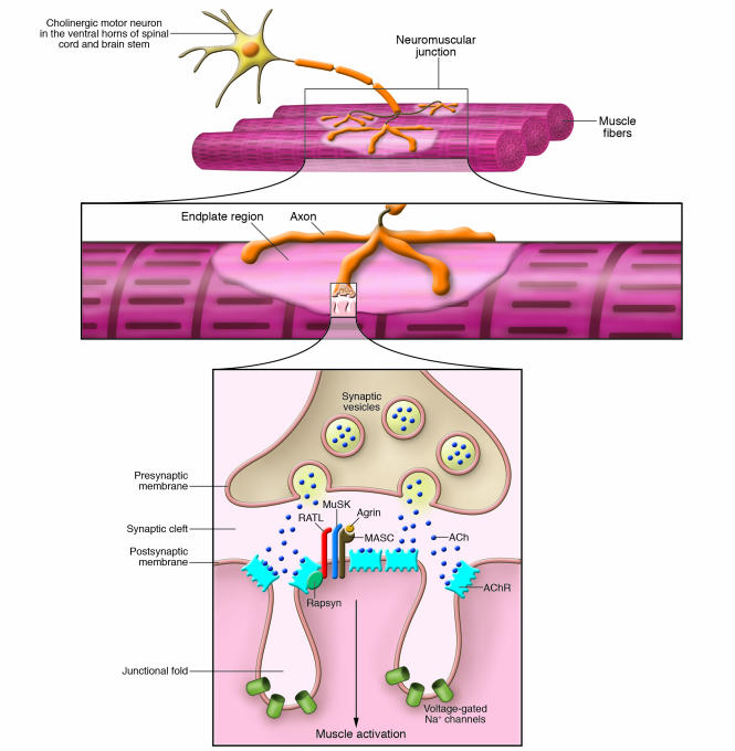
As it enters the muscle and approaches its target fibers, each α–motor neuron axon divides into branches that innervate many individual muscle fibers. Each branch loses its myelin sheath and further subdivides into many presynaptic boutons, which contain ACh-loaded synaptic vesicles and face the surface of the muscle fiber at the NMJ. The synaptic bouton and the muscle surface are separated by the synaptic cleft, which contains AChE and proteins and proteoglycans involved in stabilizing the NMJ structure. The NMJ postsynaptic membrane has characteristic deep folds, and the AChR is densely packed at the fold top. When the nerve action potential reaches the synaptic bouton, ACh is released into the synaptic cleft, where it diffuses to reach and bind the AChR. ACh binding triggers the AChR ion channel opening, permitting influx of Na+ into the muscle fiber. The resulting EPP activates voltage-gated Na+ channels at the bottom of the folds, leading to further Na+ influx and spreading of the action potential along the muscle fiber. Other proteins, including Rapsyn, MuSK, and agrin, which are involved in AChR clustering, are also present on the muscle membrane in close proximity to the AChR. MASC, myotube-associated specificity component; RATL, rapsyn-associated transmembrane linker. Figure modified with permission from Lippincott Williams and Wilkins (126).
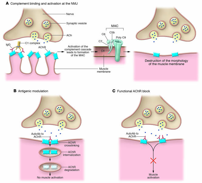
(A) Ab binding to the AChR activates the complement cascade, resulting in the formation of membrane attack complex (MAC) and localized destruction of the postsynaptic NMJ membrane. This ultimately leads to a simplified, altered morphology of the postsynaptic membrane of the NMJ of MG patients, which lacks the normal deep folds and has a relatively flat surface. (B) Abs cross-link AChR molecules on the NMJ postsynaptic membrane, causing endocytosis of the cross-linked AChR molecules and their degradation (antigenic modulation). This ultimately leads to a reduced number of AChR molecules on the postsynaptic membrane. (C) Ab binding the ACh-binding sites of the AChR causes functional block of the AChR by interfering with binding of ACh released at the NMJ. This results in failure of neuromuscular transmission.
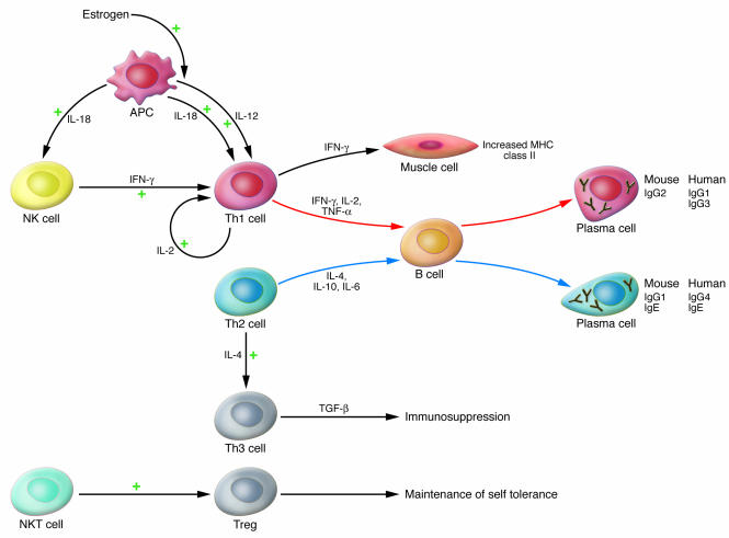
Th1 cytokines stimulate production of IgG subclasses that bind and activate complement effectively, whereas Th2 cytokines stimulate the production of Ig classes and IgG subclasses that do not. The Th2 cytokine IL-4 is also a differentiation factor for Th3 cells, immunosuppressive cells that secrete TGF-β. The Th1 cytokine IFN-γ stimulates expression of MHC class II molecules on the muscle cell membrane, thus facilitating presentation of muscle AChR. The IL-18 secreted by APCs favors the differentiation of Th1 cells both directly and indirectly through the action of NK cells. CD1-d–restricted NKT cells can activate Tregs, thereby inhibiting autoimmune processes. See text for further details.
References
-
- Robertson D.N. Enumerating neurology. Brain. 2000;123:663–664. - PubMed
-
- Marsteller H.B. The first American case of myasthenia gravis. Arch. Neurol. 1988;45:185–187. - PubMed
-
- Pascuzzi R.M. The history of myasthenia gravis. Neurol. Clin. 1994;12:231–242. - PubMed
-
- Keesey J.C. “Crisis” in myasthenia gravis: an historical perspective. Muscle Nerve. 2002;26:1–3. - PubMed
Publication types
MeSH terms
Substances
Grants and funding
LinkOut - more resources
Full Text Sources
Other Literature Sources
Medical
Miscellaneous
