. 2006 Nov;116(11):2985-94.
doi: 10.1172/JCI29224.

Mutation of beta-glucosidase 2 causes glycolipid storage disease and impaired male fertility

Affiliations

Mutation of beta-glucosidase 2 causes glycolipid storage disease and impaired male fertility

Yildiz Yildiz et al. J Clin Invest. 2006 Nov.

Abstract

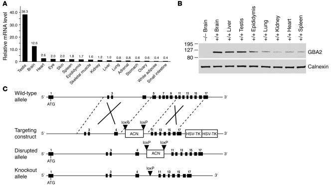
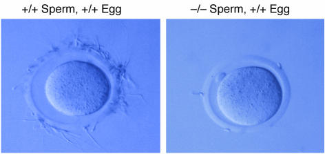
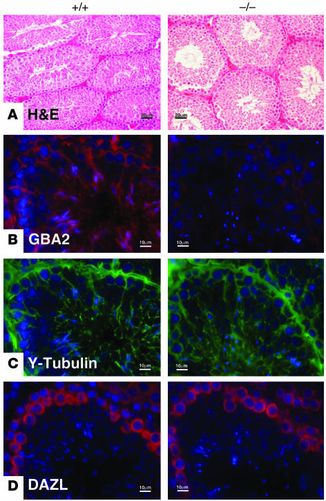
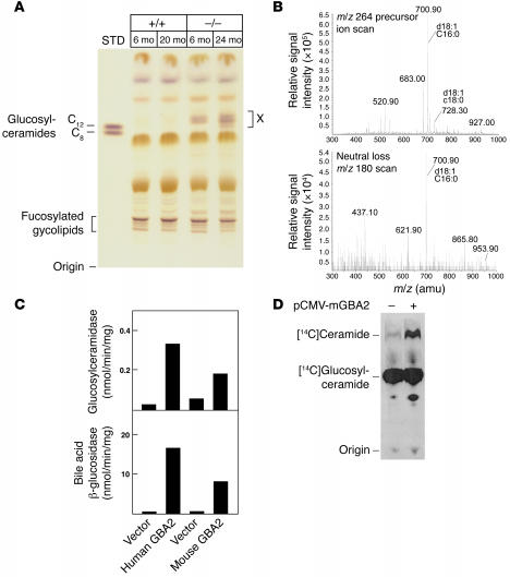
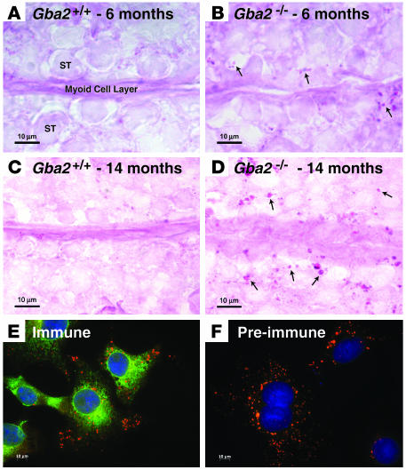
beta-Glucosidase 2 (GBA2) is a resident enzyme of the endoplasmic reticulum thought to play a role in the metabolism of bile acid-glucose conjugates. To gain insight into the biological function of this enzyme and its substrates, we generated mice deficient in GBA2 and found that these animals had normal bile acid metabolism. Knockout males exhibited impaired fertility. Microscopic examination of sperm revealed large round heads (globozoospermia), abnormal acrosomes, and defective mobility. Glycolipids, identified as glucosylceramides by mass spectrometry, accumulated in the testes, brains, and livers of the knockout mice but did not cause obvious neurological symptoms, organomegaly, or a reduction in lifespan. Recombinant GBA2 hydrolyzed glucosylceramide to glucose and ceramide; the same reaction catalyzed by the beta-glucosidase acid 1 (GBA1) defective in subjects with the Gaucher's form of lysosomal storage disease. We conclude that GBA2 is a glucosylceramidase whose loss causes accumulation of glycolipids and an endoplasmic reticulum storage disease.


Figures

Figure 1
Figure 1. Tissue distribution of GBA2 mRNA and protein and strategy to knock out Gba2.
(A) The relative levels of GBA2 mRNA in the indicated pooled tissues of male mice (n = 6) were determined by real-time RT-PCR. Data were normalized to the threshold cycle value determined in the liver (CT = 28). (B) Microsomal membranes were prepared and pooled from the indicated tissues (+/+, wild-type; –/–, homozygous) isolated from male mice (n = 6). Aliquots of protein (30 μg) were resolved by SDS-PAGE, transferred to nitrocellulose, and then blotted with an antibody raised against amino acids 1–508 of the GBA2 protein. To ensure equal amounts of protein were examined in each lane, bound GBA2 antibody complexes were removed and the filter was probed a second time with an antiserum raised against calnexin. (C) Schematics of the Gba2 wild-type allele, the targeting construct used to induce a deletion mutation by homologous recombination in ES cells, the predicted disrupted allele, and the final knockout allele are shown. Numbered black boxes indicate exons, and ATG represents the initiation codon specified within exon 1. Black triangles indicate positions of loxP sites recognized and cleaved by Cre recombinase. HSV-TK, herpes simplex virus thymidine kinase. ACN, angiotensinogen converting enzyme promoter-Cre recombinase-neomycin resistance.
Figure 2
Figure 2. Characterization of Gba2–/– mice.
(A) PCR genotyping of Gba2 wild-type, heterozygous (+/–), and homozygous genomic DNA. A 640-bp product was generated from the mutant allele, while a 530-bp product was produced from the wild-type allele. (B) Immunoblotting of GBA2 protein levels in pooled brains, testes, and livers from Gba2 wild-type, heterozygous, and homozygous mice (n = 6 per genotype). The filter of the upper blot was stripped of antibody-antigen complexes and reprobed with an antiserum recognizing calnexin to serve as a loading control. (C) Bile acid glucosidase and transferase enzyme activities were determined in the brains, testes, and livers of Gba2 wild-type, heterozygous, and homozygous mice. “X” in the reaction shown is either dolichyl phosphoglucose or octyl β-glucoside.
Figure 3
Figure 3. Sperm morphology in Gba2 wild-type and knockout mice.
(A) Photomicrographs taken on light microscope showing normal sickle-headed sperm in wild-type mice and round-headed sperm in knockout mice. Original magnification, ×400. (B) Fluorescent staining for cell nuclei with DAPI (blue), immunofluorescent staining of acrosomes with peanut lectin (green), and fluorescent staining of mitochondria (red). Original magnification, ×630. (C) Electron microscopic analyses of sperm showing abnormal head shape, nuclear organization, and distribution of mitochondria in knockout sperm. Scale bars are indicated in individual panels.
Figure 4
Figure 4. Failure of sperm from Gba2–/– mice to bind the zona pellucida.
Cumulus enclosed oocytes from Gba2+/+ mice were incubated for 5 hours with either Gba2+/+ sperm (left panel) or Gba2–/– sperm (right panel) and then examined by light microscopy. Sperm from knockout mice were deficient in zona pellucida binding. Original magnification, ×160.
Figure 5
Figure 5. Cell type specific expression patterns of GBA2, tyrosine-tubulin, and DAZL in testes from Gba2 wild-type and knockout mice.
(A) H&E staining of testis showing near-normal cellular organization of the gonads. (B) Immunofluorescent staining of GBA2 in Sertoli cells of the organ. (C) Immunofluorescent staining with antibodies that recognize tyrosine-tubulin, a Sertoli cell marker protein. (D) Immunofluorescent staining with antibodies that recognize DAZL, a germ cell marker protein. Scale bars are indicated in individual panels.
Figure 6
Figure 6. Analysis of glycolipids in Gba2 wild-type and knockout mice.
(A) Glycolipids were extracted from testes of 6-, 20-, and 24-month-old mice, resolved by 1-dimensional TLC, and visualized by staining with orcinol. The positions of the origin and glucosylceramide standards are indicated at left. A glycolipid that accumulated with age in knockout tissue is marked “X” at right. The positions to which several fucosylated glycolipids migrated to are indicated at left. (B) Structural analysis of glycolipid X shown in A. Glycolipids were extracted from the testes of Gba2–/– mice and resolved by 1-dimensional TLC, and glycolipid X was analyzed by mass spectrometry. An m/z 264 precursor ion scan revealed compounds of molecular masses (700.9 and 728.3) characteristic of monohexosylceramides containing sphingosine (d18:1) and either 16:0 or 18:0 fatty acyl groups. A neutral loss scan for m/z 180 diagnostic for glucose- or galactose-containing monohexosylceramides revealed a major compound of mass 700.9. (C) Expression of mouse and human GBA2 in COS cells and measurement of glucosylceramidase and bile acid glucosidase enzyme activities. Cells were transfected with the indicated plasmid DNA, and lysates were assayed for enzyme activity using fluorescently labeled glucosylceramide or bile acid glucoside substrates. (D) Expression of mouse GBA2 (mGBA2) in human embryonic kidney 293 cells and measurement of glucosylceramidase activity. Cells were transfected and assayed for enzyme activity using [14C]glucosylceramide. The positions of the origin and to which authentic glucosylceramide and ceramide standards migrated to are indicated at left. pCMV-mGBA2, expression plasmid encoding mouse GBA2 enzyme.
Figure 7
Figure 7. Glycolipid accumulation in Gba2–/– mice and subcellular localization of GBA2.
Glycolipids were visualized in the testes of 6- and 14-month-old wild-type (A and C) and knockout (B and D) mice by periodic acid Schiff staining of 8-μm sections. Arrows indicate small, intensely stained deposits of glycolipids that accumulated with age in the knockout mice. ST, seminiferous tubule. (E) The subcellular localization of GBA2 (green) was determined by immunofluorescent staining of simian COS cells. Lysosomes (red) were identified by staining with LysoTracker. (F) COS cells stained with a preimmune serum and LysoTracker. Scale bars are indicated in individual panels.

Comment in

References

    1. Simons K., Toomre D. Lipid rafts and signal transduction. Nat. Rev. Mol. Cell Biol. 2000;1:31–39. - PubMed
    1. Anderson R.G., Jacobson K. A role for lipid shells in targeting proteins to caveolae, rafts, and other lipid domains. Science. 2002;296:1821–1825. - PubMed
    1. Menaldino D.S., et al. Sphingoid bases and de novo ceramide synthesis: enzymes involved, pharmacology, and mechanisms of action. Pharmacol. Res. 2003;47:373–381. - PubMed
    1. Merrill J.A.H. De novo sphingolipid biosynthesis: a necessary, but dangerous pathway. J. Biol. Chem. 2002;277:25843–25846. - PubMed
    1. Perry R.J., Ridgway N.D. Molecular mechanisms of ceramide transport. Biochim. Biophys. Acta. 2005;1734:220–234. - PubMed

Publication types

MeSH terms