. 2006 Nov;116(11):3060-9.
doi: 10.1172/JCI27341.

Deficiency in neuronal TGF-beta signaling promotes neurodegeneration and Alzheimer's pathology

Affiliations

Deficiency in neuronal TGF-beta signaling promotes neurodegeneration and Alzheimer's pathology

Ina Tesseur et al. J Clin Invest. 2006 Nov.

Abstract

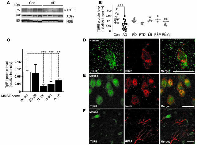
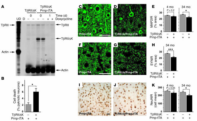
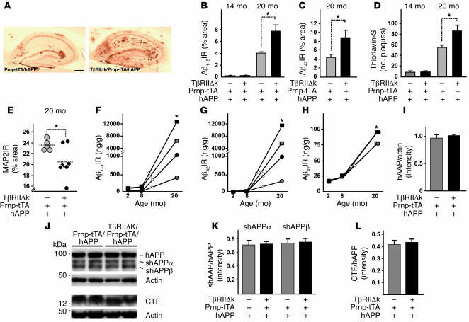
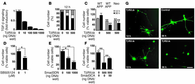
Alzheimer's disease (AD) is characterized by progressive neurodegeneration and cerebral accumulation of the beta-amyloid peptide (Abeta), but it is unknown what makes neurons susceptible to degeneration. We report that the TGF-beta type II receptor (TbetaRII) is mainly expressed by neurons, and that TbetaRII levels are reduced in human AD brain and correlate with pathological hallmarks of the disease. Reducing neuronal TGF-beta signaling in mice resulted in age-dependent neurodegeneration and promoted Abeta accumulation and dendritic loss in a mouse model of AD. In cultured cells, reduced TGF-beta signaling caused neuronal degeneration and resulted in increased levels of secreted Abeta and beta-secretase-cleaved soluble amyloid precursor protein. These results show that reduced neuronal TGF-beta signaling increases age-dependent neurodegeneration and AD-like disease in vivo. Increasing neuronal TGF-beta signaling may thus reduce neurodegeneration and be beneficial in AD.


Figures

Figure 1
Figure 1. TβRII protein levels are decreased in the human AD brain.
(A) Representative immunoblots of lysates from AD and age-matched control (Con) parietal corteices probed with antibodies against TβRII, actin, and neuron-specific enolase (NSE). (B) TβRII levels of nondemented control, AD, Parkinson’s disease (PD), progressive frontotemporal dementia (FTD), lewy body dementia (LB), progressive supranuclear palsy (PSP), and Pick’s disease (Pick’s) cases were quantified and normalized against protein amounts loaded. Each symbol represents an individual case. (C) TβRII protein levels grouped per mini-mental state exam (MMSE) score (white bar, nondemented controls; black bars, AD). (D and E) TβRII immunofluorescence localized to neurons in the mid-frontal gyrus of the brain from a 69-year-old human (D) and the cortex of a 3-month-old wild-type mouse (E). (F) Little TβRII staining localized to astrocytes in the mouse brain. Scale bars: 10 μm. **P < 0.05, ***P < 0.005; Student’s t test.
Figure 2
Figure 2. Reduced neuronal TGF-β signaling increases neurodegeneration in vivo.
(A) RNase protection assay shows TβRIIΔk mRNA expression and inhibition of transgene expression in transgenic mice caused by feeding doxycycline-containing food pellets. Arrows on the left indicate undigested (UD) riboprobes; arrows on the right indicate the corresponding protected fragments. TβRII represents the wild-type receptor. D, digested riboprobe. Each lane represents 1 animal. (B) TβRIIΔk-expressing primary neurons show reduced survival in culture. Primary neuron cultures were stained with BODIPY-FL-Fallacidin and Hoechst after 4 days in culture, and the number of viable and dead cells was counted based on the absence or presence of pyknotic nuclei. Data are mean ± SEM of triplicate wells of 3 mice per genotype. (CK) Sagittal brain sections of 34-month-old (CK) as well as 4-month-old (E, H, and K) TβRIIΔk/Prnp-tTA or Prnp-tTA mice were immunolabeled for MAP2 (CE), synaptophysin (FH), and NeuN (IK), and percent immunoreactive area (IR) of the neuropil (CH) or mean number of labeled cells (IK) in the neocortex was determined by confocal microscopy and computer-aided image analysis. Scale bars: 50 μm. Images are examples from cases with clearly visible degeneration. Data are mean ± SEM (n = 5–7 mice per genotype). *P < 0.05, ***P < 0.005; Student’s t test.
Figure 3
Figure 3. Decreased neuronal TGF-β signaling in old hAPP mice increases Aβ levels, amyloid deposition, and dendritic degeneration.
(A) Representative images of hippocampi of 20-month-old TβRIIΔk/Prnp-tTA/hAPP and Prnp-tTA/hAPP mice stained with an antibody against Aβ42. Scale bar: 500 μm. (BD) Quantification of immunoreactive area occupied by staining with antibody against Aβ1–5 (B), antibody against Aβ42 (C), and thioflavin-S (D) of sagittal sections from 14- and 20-month-old TβRIIΔk/Prnp-tTA/hAPP and Prnp-tTA/hAPP mice (n = 5–7 mice per genotype). (E) Expression of TβRIIΔk in neurons of aged hAPP mice reduced MAP2 immunoreactivity in the hippocampus. Sagittal brain sections of 20-month-old mice were stained with MAP2, and percent immunoreactive area of the neuropil was determined by confocal microscopy and computer-aided image analysis. Each symbol represents 1 mouse. (FH) Quantification of Aβ1–x (F), Aβ42 (G), and percent Aβ42 (H) via ELISA in hippocampus (squares) and cortex (circles) of TβRIIΔk/Prnp-tTA/hAPP (black symbols) and Prnp-tTA/hAPP (gray symbols) mice aged 2, 8, and 20 months (n = 5–7 mice per genotype). Note the cutoff in the y axes. (IL) Signal intensities of total APP (I), shAPPα and shAPPβ (K), and CTFs (L) were quantified and normalized against total APP levels (K and L) or protein amounts loaded (I). Representative Western blots (J) of cell pellets (hAPP, CTF, and actin) or supernatants (shAPPα and shAPPβ) probed with antibodies against total APP, CTFs, actin, shAPPα, and shAPPβ. *P < 0.05; Student’s t test (BD and FH), Tukey-Kramer test (E).
Figure 4
Figure 4. Inhibition of endogenous TGF-β signaling increases neuritic degeneration and Aβ production in neuroblastoma cells.
(A) Transient transfection of TβRIIΔk and a TGF-β reporter plasmid (p800 luciferase) into B103-APPwt cells dose-dependently inhibited signaling 24 hours after transfection. (B) Quantification of the percentage of B103-APPwt cells transiently transfected with TβRIIΔk that showed healthy (black), beaded (white), or no processes (gray) 12 hours after transfection. (C) Expression of TβRIIΔk in B103-APPwt (APPwt) or B103 cells lacking APP (neo) significantly reduced the percentage of cells with healthy processes 12 and 36 hours after transfection. (DF) Downstream inhibition of TGF-β signaling via treatment with 1 μM SB505124 (D) or expression of dominant-negative Smad3 (Smad3DN; E) reduced the percentage of cells with healthy processes 36 hours after treatment. (F) Expression of constitutively active Smad3 (Smad3CA) partially rescued the phenotype in cells coexpressing TβRIIΔk. (G) B103-APPwt cells expressing TβRIIΔk showed beading and retraction of neurites (12 h), followed by rounding up of the cell body (36 h). Control cells were transfected with filler plasmid and GFP. Scale bar: 50 μm. *P < 0.05, **P < 0.01 versus respective controls; Student’s t test.
Figure 5
Figure 5. Inhibition of neuronal TGF-β signaling increases Aβ, shAPP and CTF in neuroblastoma cells.
(A and B) B103-APPwt cells were transiently transfected with TβRIIΔk and GFP and sorted via fluorescence-activated cell sorting (A) or treated with 1 μM SB505124 to inhibit ALK5 signaling (B). Total secreted Aβ was measured via ELISA 5 days after treatment. (C) Representative Western blots of cell pellets (hAPP, CTFα, CTFβ) or supernatants (shAPPα, shAPPβ) of B103-APPwt cells or B103 cells lacking APP treated with 0 or 1 μM SB505124 and probed with antibodies against total hAPP, CTFα, CTFβ, shAPPα, and shAPPβ. (DF) Signal intensities of total hAPP (D), shAPPα and shAPPβ (E), and CTFα and CTFβ (F) were quantified and normalized against hAPP levels (E and F), and the ratio between shAPPα and shAPPβ was calculated (E). Data are mean ± SEM of triplicate wells of 1 representative experiment. **P < 0.01, ***P < 0.001; Student’s t test.

Comment in

References

    1. Selkoe D.J. Alzheimer’s disease: genes, proteins, and therapy. Physiol. Rev. 2001;81:741–766. - PubMed
    1. Connor B., Dragunow M. The role of neuronal growth factors in neurodegenerative disorders of the human brain. Brain Res. Brain Res. Rev. 1998;27:1–39. - PubMed
    1. Murer M.G., Yan Q., Raisman-Vozari R. Brain-derived neurotrophic factor in the control human brain, and in Alzheimer’s disease and Parkinson’s disease. Prog. Neurobiol. 2001;63:71–124. - PubMed
    1. Allen S.J., Wilcock G.K., Dawbarn D. Profound and selective loss of catalytic TrkB immunoreactivity in Alzheimer’s disease. Biochem. Biophys. Res. Commun. 1999;264:648–651. - PubMed
    1. Ferrer I., et al. BDNF and full-length and truncated TrkB expression in Alzheimer disease. Implications in therapeutic strategies. J. Neuropathol. Exp. Neurol. 1999;58:729–739. - PubMed

Publication types

MeSH terms

Substances