. 2007 May 1;61(9):1049-61.
doi: 10.1016/j.biopsych.2006.08.005. Epub 2006 Nov 1.

The striatal-enriched protein tyrosine phosphatase gates long-term potentiation and fear memory in the lateral amygdala

Affiliations

The striatal-enriched protein tyrosine phosphatase gates long-term potentiation and fear memory in the lateral amygdala

Surojit Paul et al. Biol Psychiatry. .

Abstract

Background: Formation of long-term memories is critically dependent on extracellular-regulated kinase (ERK) signaling. Activation of the ERK pathway by the sequential recruitment of mitogen-activated protein kinases is well understood. In contrast, the proteins that inactivate this pathway are not as well characterized.

Methods: Here we tested the hypothesis that the brain-specific striatal-enriched protein tyrosine phosphatase (STEP) plays a key role in neuroplasticity and fear memory formation by its ability to regulate ERK1/2 activation.

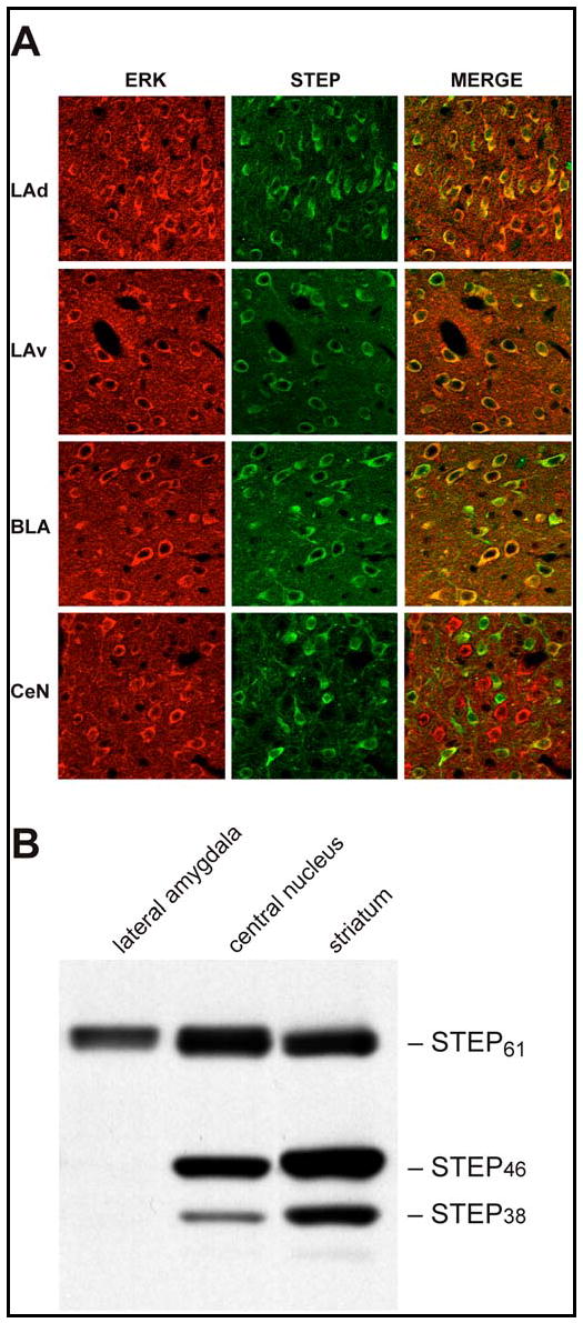
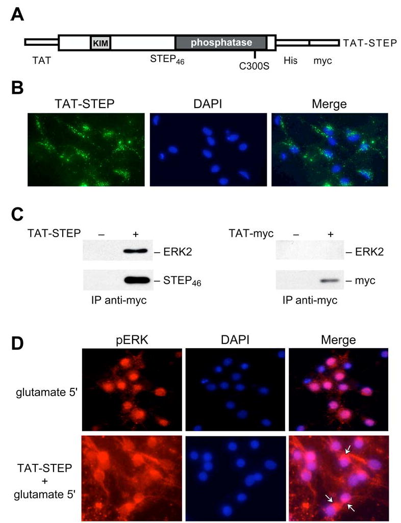
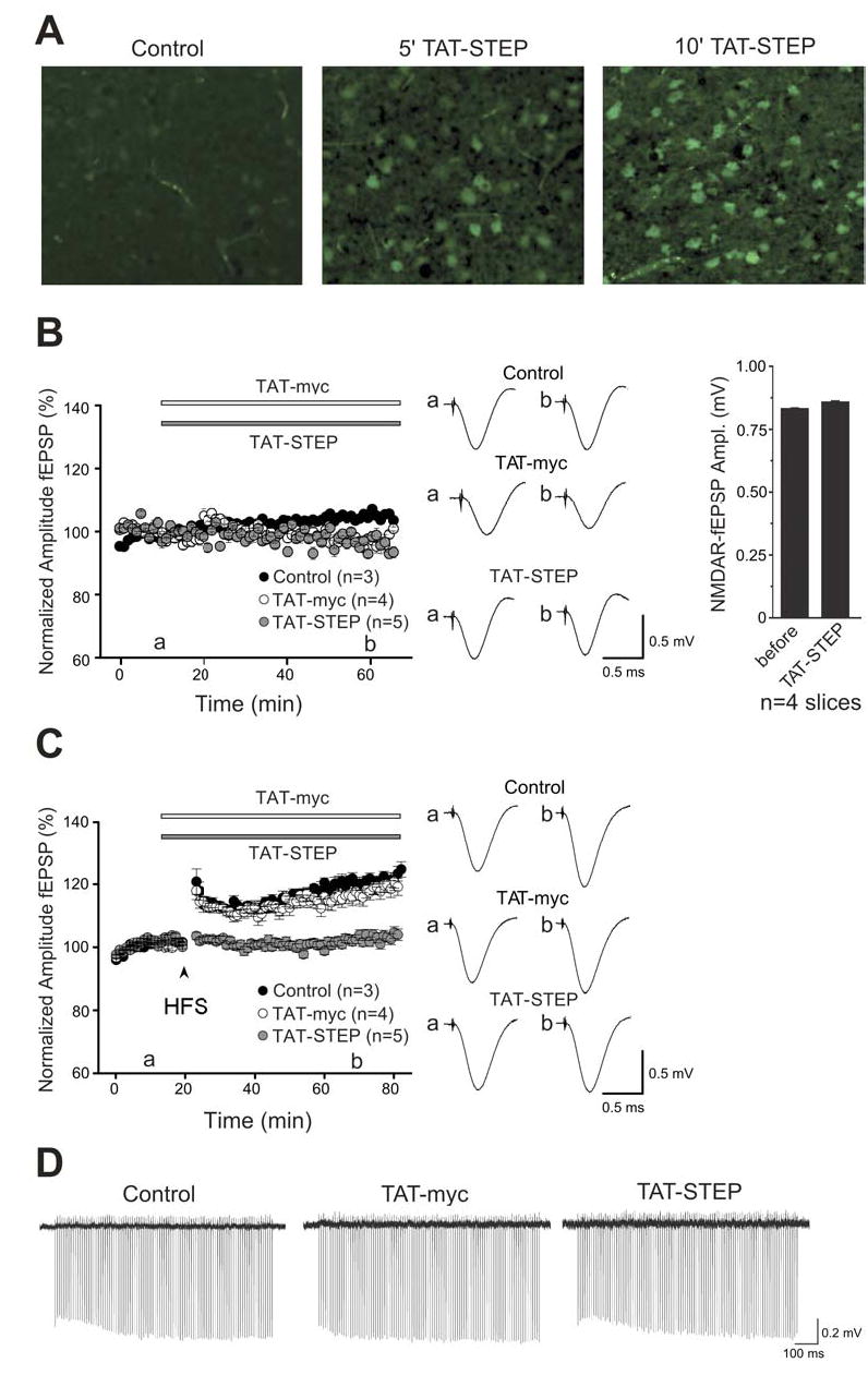
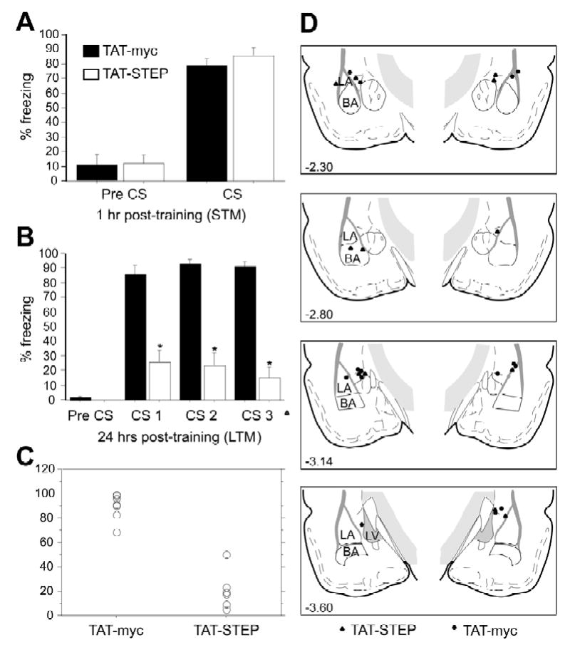
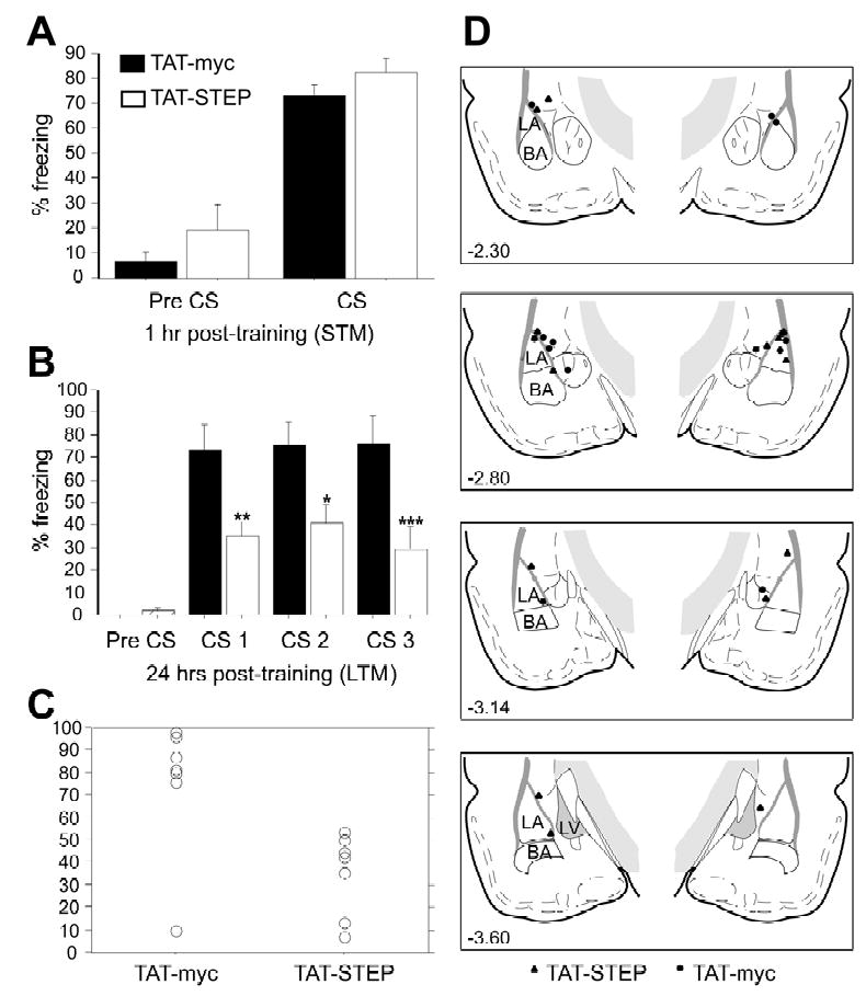
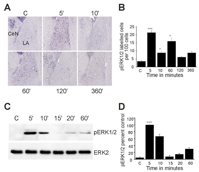
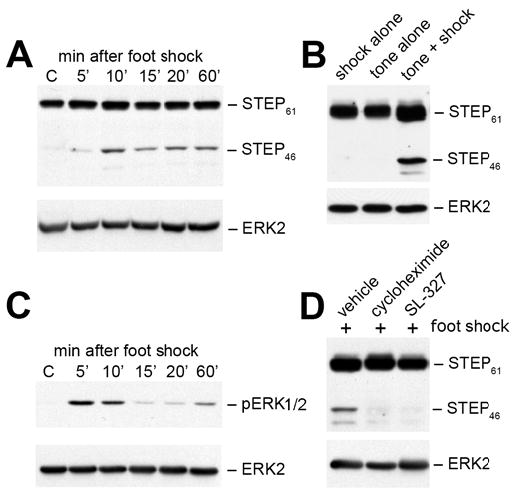
Results: STEP co-localizes with the ERKs within neurons of the lateral amygdala. A substrate-trapping STEP protein binds to the ERKs and prevents their nuclear translocation after glutamate stimulation in primary cell cultures. Administration of TAT-STEP into the lateral amygdala (LA) disrupts long-term potentiation (LTP) and selectively disrupts fear memory consolidation. Fear conditioning induces a biphasic activation of ERK1/2 in the LA with an initial activation within 5 minutes of training, a return to baseline levels by 15 minutes, and an increase again at 1 hour. In addition, fear conditioning results in the de novo translation of STEP. Inhibitors of ERK1/2 activation or of protein translation block the synthesis of STEP within the LA after fear conditioning.

Conclusions: Together, these data imply a role for STEP in experience-dependent plasticity and suggest that STEP modulates the activation of ERK1/2 during amygdala-dependent memory formation. The regulation of emotional memory by modulating STEP activity may represent a target for the treatment of psychiatric disorders such as posttraumatic stress disorder (PTSD), panic, and anxiety disorders.


Figures

Figure 1
Figure 1. STEP and ERK co-localize in the lateral amygdala
(A) Immunohistochemical analysis of ERK and STEP in the dorso-lateral amygdala (LAd), ventro-lateral amygdala (LAv), baso-lateral amygdala (BLA) and the central nucleus (CeN). Rat brain coronal sections were double-labeled with anti-ERK2 and anti-STEP antibodies. The ‘Merge’ column illustrates the co-localization of the two proteins in the same cell. (B) Tissue punches from the LA, CeN and the striatum were analyzed by SDS-polyacrylamide gel electrophoresis and western blots were probed with anti-STEP antibody to show the distribution of STEP isoforms in these regions. Note that STEP61 is the only isoform expressed in the LA.
Figure 2
Figure 2. Functional characterization of TAT-STEP in primary neuronal cultures
(A) Schematic diagram of TAT-STEP46 C-S indicating the positions of the TAT-peptide, six-histidine (His) and myc tags, the KIM domain, and the C-S mutation site within the phosphatase domain. (B) Immunocytochemical staining with anti-myc antibody and DAPI to characterize the transduction of TAT-STEP. (C) Neuron cultures were incubated with either TAT-myc or TAT-STEP, which were immunoprecipitated with anti-myc antibody, analyzed by SDS-PAGE, and probed with anti-ERK2 antibody (upper panels). The blots were then reprobed with anti-myc antibody (lower panels). (D) Immunocytochemical analysis using an antibody that recognizes phospho-ERK1/2 illustrates the cytoplasmic and nuclear distribution of pERK1/2 following glutamate treatment for 5 min, in the absence or presence of TAT-STEP. Arrows point to perinuclear pERK1/2.
Figure 3
Figure 3. TAT-STEP blocks induction of LTP in LA without affect basal synaptic transmission
(A) aCSF (control) or TAT-STEP (2 μM) was added to 300 μm amygdala slices and tissue processed with anti-myc antibody at 5’ and 10’ after start of incubation. (B) Left panel. Field EPSP (fEPSP) amplitude (mean ± SEM) is plotted every 1 min for control slices (n = 3 slices, closed circles), for a slices treated with 300 nM TAT-myc (n = 4 slices, open circles) and for slices treated with 300 nM TAT-STEP (n = 5, gray circles) that was applied during the period indicated by the bar. In the middle are averaged traces from control slices (upper panel) or before (a) application of either TAT-myc or TAT-STEP and (b) 50 min after application of TAT-myc (middle panel) or TAT-STEP (lower panel). On the right, histogram showing average NMDAR mediated fEPSPs before or during application of TAT-STEP (20 min after start of application). (C) Left panel. Field EPSP (fEPSP) amplitude (mean ± SEM) is plotted every 1 min for control slices (n = 6 slices, closed circles), for a slices treated with 300 nM TAT-myc (n = 6 slices, open circles) and for slices treated with 300 nM TAT-STEP (n = 7, gray circles) that was applied during the period indicated by the bar. Tetanus (HFS; three trains of 100 Hz stimuli, 1 sec duration, intertrain interval of 90 sec) is indicated by the arrowhead. On the right are averaged traces taken from control slices (upper panel) or before (a) application of either TAT-myc (middle panel) or TAT-STEP (lower panel) and (b) 50 min after tetanus. Six sweeps were averaged to produce the responses shown. Scale bar, 0.5 msec, 0.5 mV for B and C. (D) Representative, continuous field recordings illustrating responses during HFS trains in control, TAT-myc- or TAT-STEP-treated slices. Scale bar, 100 msec, 0.2 mV.
Figure 4
Figure 4. Effect of intra-amygdala infusion of TAT-STEP prior to fear conditioning
Rats were infused with TAT-STEP (n=6) or TAT-myc (control; n=7) 1 hr prior to training on Pavlovian fear conditioning (two pairings of 30 sec tone and 1 sec 1.5 mA shock). They were then assessed for fear memory formation at 1 hr (A, short term memory, STM) or 24 hr (B, long-term memory, LTM) after training. (A & B) Mean ± S.E. freezing in rats 30 sec prior to CS presentation (pre CS) and during the three tone presentations (CS). *p < 0.0001 relative to the TAT-myc control. (C) Scattergram demonstrating the distribution of freezing in TAT-myc and TAT-TAT-STEP in the LTM test. (D) Histological verification of cannula tip placements in rats infused with either TAT-STEP (▴) or TAT-myc (•).
Figure 5
Figure 5. Effect of post-session intra-amygdala infusion of TAT-STEP
Rats were infused with TAT-STEP (n=7) or TAT-myc (control; n=7) immediately following training on Pavlovian fear conditioning (two pairings of 30 sec tone and 1 sec 1.5 mA shock). They were then assessed for fear memory formation at 1 hr (A, short term memory, STM) or 24 hr (B, long-term memory, LTM) after training. (A & B) (A & B) Mean ± S.E. freezing in rats prior to the tone (pre CS) and the three tone presentations (CS). *p < 0.05, **p < 0.01, ***p < 0.001 relative to the TAT-myc control. (C) Scattergram demonstrating the distribution of freezing in TAT-myc and TAT-STEP in the LTM test. (D) Histological verification of cannula tip placements in rats infused with either TAT-STEP (▴) or TAT-myc (•).
Figure 6
Figure 6. Time kinetics of ERK1/2 activation in the lateral amygdala
Rats were presented with two pairs of tone and shock and processed for immunohistochemistry or immunoblotting. (A) Representative immunohistochemical staining of pERK1/2 in the amygdala at 0, 5, 10, 60, 120 and 360 min after fear conditioning. (B) Bar diagram showing pERK1/2 immunoreactive cells per 100 cells in serial sections through the LA (mean ± S.E., n = 5 sections per time point). *p < 0.05, **p < 0.01, ***p < 0.001 relative to control. (C) pERK1/2 immunoblot of tissue punches taken from the LA at 5, 10, 15, 20 and 60 min after fear conditioning training (upper panel). The blots were reprobed with total ERK2 antibody (lower panel). (D) Quantification of pERK1/2 levels from 3 separate experiments (***p < 0.001).
Figure 7
Figure 7. De novo synthesis of STEP in the lateral amygdala following fear conditioning
(A) Tissue punches taken from LA at 5, 10, 15, 20 and 60 min after fear conditioning or from control animals (no tone or shock, lane C) were analyzed by SDS-PAGE and processed for immunoblotting with anti-STEP (upper panel). (B) Rats were exposed to either shock or tone alone and punches were taken from the LA at 10 minutes, when STEP levels are at their maximum after fear conditioning (n = 3 independent trials with 3 rats per treatment). Note the increase in both STEP isoforms after fear conditioning. (C) pERK1/2 levels were determined after fear conditioning (upper panel). (D) Rats were injected intraperitoneally with DMSO (vehicle), cycloheximide (protein synthesis inhibitor) or SL327 (MEK inhibitor) 1 hr prior to fear conditioning. Tissue punches were again processed at 10 minutes for immunoblotting with anti-STEP antibody (upper panel). These experiments were repeated in triplicate with p<0.02 for both inhibitors. All blots were reprobed with anti-ERK2 antibody to allow for comparisons of loading levels (A, B, C & D, all lower panels).

Similar articles

Cited by

References

    1. Aarts M, Liu Y, Liu L, Besshoh S, Arundine M, Gurd JW, Wang YT, Salter MW, Tymianski M. Treatment of ischemic brain damage by perturbing NMDA receptor- PSD-95 protein interactions. Science. 2002;29:846–850. - PubMed
    1. Atkins CM, Selcher JC, Petraitis JJ, Trzaskos JM, Sweatt JD. The MAPK cascade is required for mammalian associative learning. Nat Neurosci. 1998;1:602–609. - PubMed
    1. Bauer EP, Schafe GE, LeDoux JE. NMDA receptors and L-type voltage-gated calcium channels contribute to long-term potentiation and different components of fear memory formation in the lateral amygdala. J Neuroscience. 2002;22:5239–5249. - PMC - PubMed
    1. Berman DE, Hazvi S, Rosenblum K, Seger R, Dudai Y. Specific and differential activation of mitogen-activated protein kinase cascades by unfamiliar taste in the insular cortex of the behaving rat. J Neurosci. 1998;18:10037–10044. - PMC - PubMed
    1. Blair HT, Schafe GE, Bauer EP, Rodrigues SM, LeDoux JE. Synaptic plasticity in the lateral amygdala: a cellular hypothesis of fear conditioning. Learn Mem. 2001;8:229–242. - PubMed

Publication types

MeSH terms