

Noninvasive corneal stromal collagen imaging using two-photon-generated second-harmonic signals
- PMID: 17081858
- PMCID: PMC1855208
- DOI: 10.1016/j.jcrs.2006.08.027
Noninvasive corneal stromal collagen imaging using two-photon-generated second-harmonic signals
Abstract
Purpose: To investigate the feasibility of using femtosecond-pulse lasers to produce second-harmonic generated (SHG) signals to noninvasively assess corneal stromal collagen organization.
Setting: The Eye Institute, University of California, Irvine, California, USA.
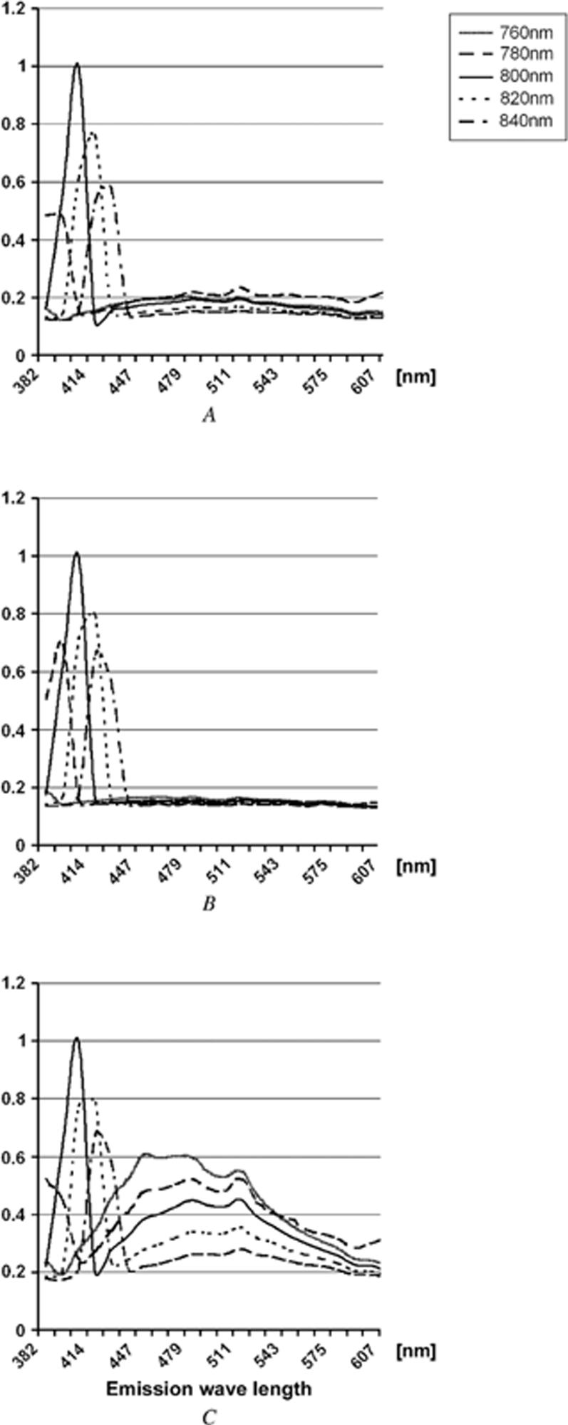
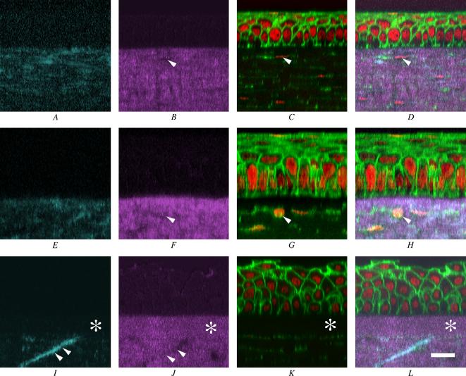
Methods: Mouse, rabbit, and human corneas were examined by two-photon confocal microscopy using a variable-wavelength femtosecond lasers to produce SHG signals. Two types were detected: forward scattered and backward scattered. Wavelength dependence of the SHG signal was confirmed by spectral separation using the 510 Meta (Zeiss). To verify the spatial relation between SHG signals and corneal cells, staining of cytoskeletons and nuclei was performed.
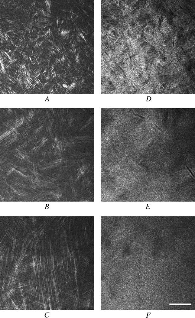
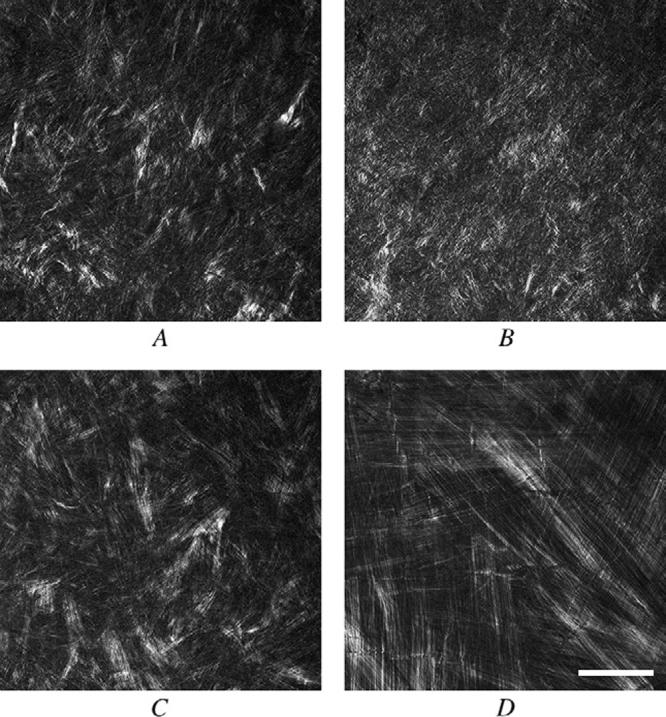
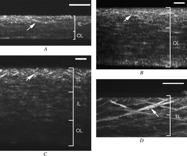
Results: Second-harmonic-generated signal intensity was strongest with an excitation wavelength of 800 nm for all 3 species. Second-harmonic-generated forward signals showed a distinct fibrillar pattern organized into bands suggesting lamellae, while backscattered SHG signals appeared more diffuse and indistinct. Reconstruction of SHG signals showed two patterns of lamellar organization: highly interwoven in the anterior stroma and orthogonally arranged in the posterior stroma. Unique to the human cornea was the presence of transverse, sutural lamellae that inserted into Bowman's layer, suggesting an anchoring function.
Conclusions: Using two-photon confocal microscopy to generate SHG signals from the corneal collagen provides a powerful new approach to noninvasively study corneal structure. Human corneas had a unique organizational pattern with sutural lamellae to provide important biomechanical support that was not present in mouse or rabbit corneas.
Figures
References
-
- Hamada R, Giraud J-P, Graf B, Pouliquen Y. Étude analytique et statistique des lamelles, des keratocytes, des fibrilles de collagéne de la région centrale de la cornée humaine normale. (Microscopie optique et électronique) Arch Ophtalmol Rev Gen Ophtalmol. 1972;32:563–570. - PubMed
-
- Komai Y, Ushiki T. The three-dimensional organization of collagen fibrils in the human cornea and sclera. Invest Ophthalmol Vis Sci. 1991;32:2244–2258. - PubMed
-
- Radner W, Zehetmayer M, Aufreiter R, Mallinger R. Interlacing and cross-angle distribution of collagen lamellae in the human cornea. Cornea. 1998;17:537–543. - PubMed
-
- Binder PS, Rock ME, Schmidt KC, Anderson JA. High-voltage electron microscopy of normal human cornea. Invest Ophthalmol Vis Sci. 1991;32:2234–2243. - PubMed
-
- Müller LJ, Pels L, Vrensen GFJM. Novel aspects of the ultrastructural organization of human corneal keratocytes. Invest Ophthalmol VisSci. 1995;36:2557–2567. - PubMed
Publication types
MeSH terms
Substances
Grants and funding
LinkOut - more resources
Full Text Sources
