

Measuring total corneal power before and after laser in situ keratomileusis with high-speed optical coherence tomography
- PMID: 17081867
- PMCID: PMC1808223
- DOI: 10.1016/j.jcrs.2006.04.046
Measuring total corneal power before and after laser in situ keratomileusis with high-speed optical coherence tomography
Abstract
Purpose: To measure total corneal power using optical coherence tomography (OCT).
Setting: Refractive surgery practices at 2 academic eye centers in Cleveland, Ohio, and Los Angeles, California, USA.
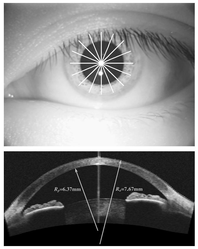
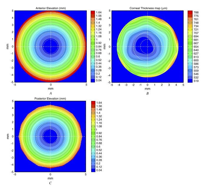
Methods: Thirty-two eyes of 17 patients having myopic laser in situ keratomileusis (LASIK) were enrolled in a prospective observational study. Manifest refraction, OCT, and Placido ring corneal topography with the Atlas 995 (Carl Zeiss Meditec, Inc.) were performed preoperatively and 3 months after laser in situ keratomileusis (LASIK). A high-speed (2000 axial scans/second) corneal and anterior segment OCT prototype was used. The total corneal power was calculated by summation of the anterior and posterior surface powers, and the value was compared with that determined by simulated keratometry. Two methods of measuring total corneal power were tested: the direct method, which used OCT to measure both corneal surfaces directly, and the hybrid method, which combined OCT with anterior corneal topography.
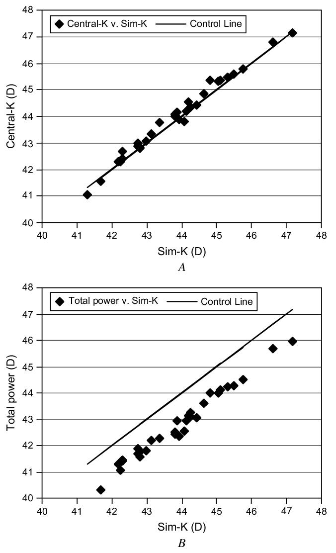
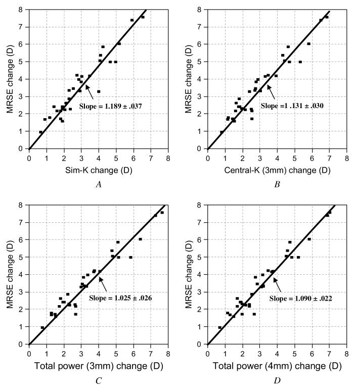
Results: The repeatability (pooled standard deviation) of measuring total corneal power using the hybrid method was 3 times better than that using the direct method. It was 0.23 diopter (D) before LASIK and 0.26 D after LASIK. Preoperative total power was 1.13 D (2.6%) lower than the simulated keratometry. Compared to the LASIK-induced change in spherical equivalent refraction, the change in total corneal power was equivalent, while the change in simulated keratometry power was significantly smaller (-18.8%) (P<.001).
Conclusions: Keratometry using the traditional index of 1.3375 overestimated the total power in preoperative corneas and underestimated LASIK-induced refractive change. Measuring both corneal surfaces using a combination of OCT and Placido ring topography provided a better measure of total corneal power that closely tracked the refractive change in post-LASIK eyes.
Figures
References
-
- Henson DB. Optometric Instrumentation. Butter-worths; London, Boston, MA: 1983.
-
- Rabbetts RB. Bennett and Rabbetts’ Clinical Visual Optics. 3rd ed. Butterworth-Heinemann; Oxford, Boston, MA: 1998.
-
- Huang D, Tang M, Shekhar R. Mathematical model of corneal surface smoothing after laser refractive surgery. Am J Ophthalmol. 2003;135:267–278. - PubMed
-
- Leyland M. Validation of Orbscan II posterior corneal curvature measurement for intraocular lens power calculation. Eye. 2004;18:357–360. - PubMed
-
- Seitz B, Langenbucher A, Nguyen NX, et al. Underestimation of intraocular lens power for cataract surgery after myopic photorefractive keratectomy. Ophthalmology. 1999;106:693–702. - PubMed
Publication types
MeSH terms
Grants and funding
LinkOut - more resources
Full Text Sources
Other Literature Sources
