. 2006 Nov 3;127(3):495-508.
doi: 10.1016/j.cell.2006.08.047.

Structural basis for the histone chaperone activity of Asf1

Affiliations

Structural basis for the histone chaperone activity of Asf1

Christine M English et al. Cell. .

Abstract

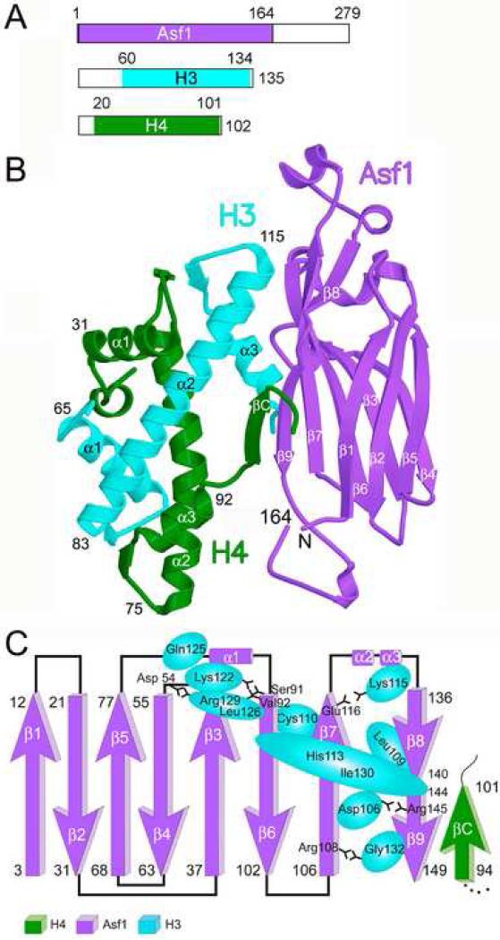
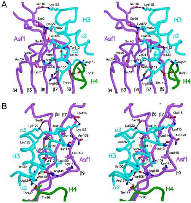
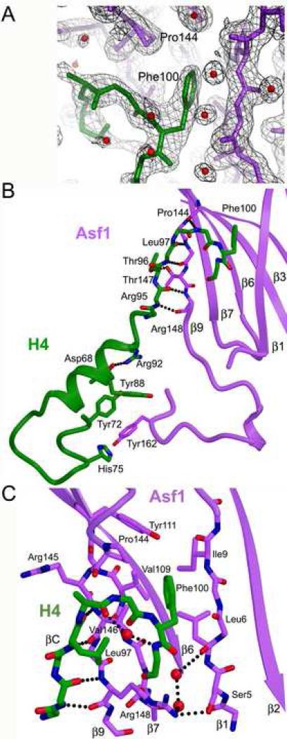
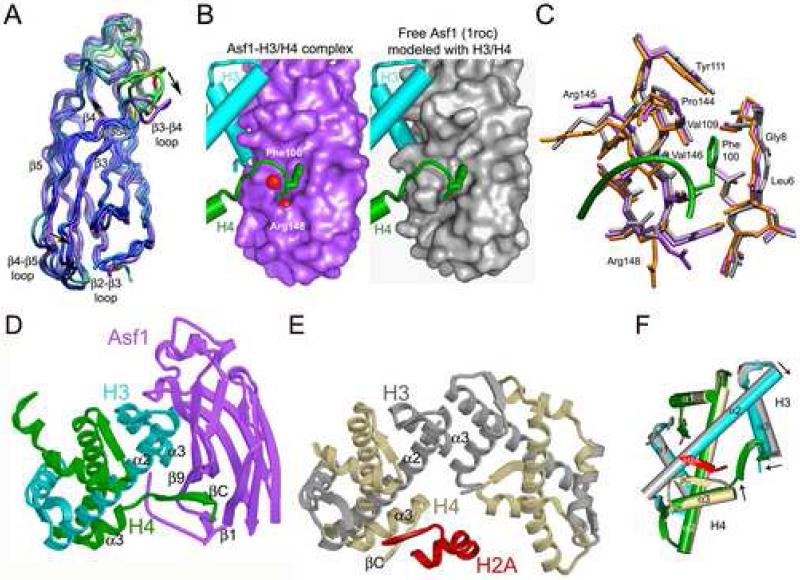
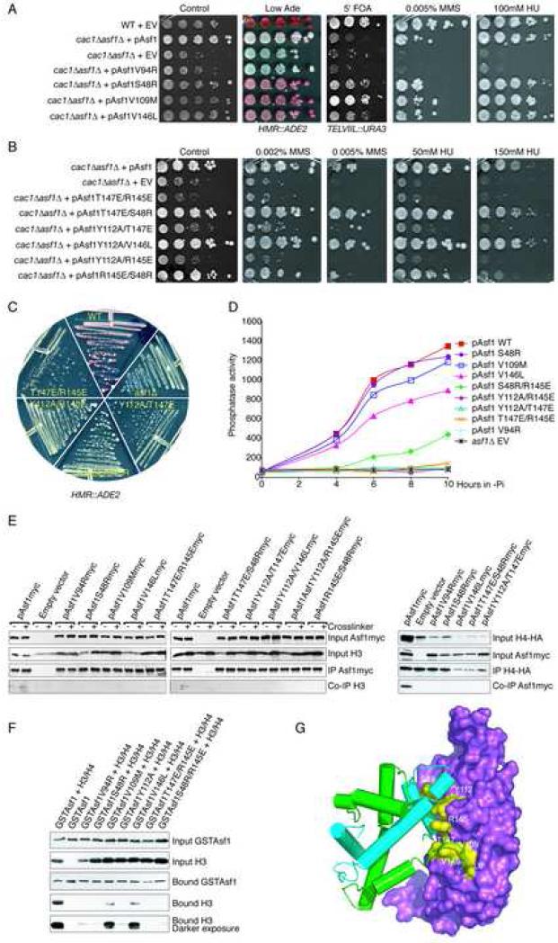
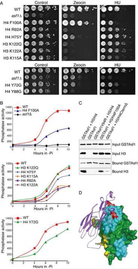
Anti-silencing function 1 (Asf1) is a highly conserved chaperone of histones H3/H4 that assembles or disassembles chromatin during transcription, replication, and repair. The structure of the globular domain of Asf1 bound to H3/H4 determined by X-ray crystallography to a resolution of 1.7 Angstroms shows how Asf1 binds the H3/H4 heterodimer, enveloping the C terminus of histone H3 and physically blocking formation of the H3/H4 heterotetramer. Unexpectedly, the C terminus of histone H4 that forms a mini-beta sheet with histone H2A in the nucleosome undergoes a major conformational change upon binding to Asf1 and adds a beta strand to the Asf1 beta sheet sandwich. Interactions with both H3 and H4 were required for Asf1 histone chaperone function in vivo and in vitro. The Asf1-H3/H4 structure suggests a "strand-capture" mechanism whereby the H4 tail acts as a lever to facilitate chromatin disassembly/assembly that may be used ubiquitously by histone chaperones.


Figures

Figure 1
Figure 1. Asf1-H3/H4 structure
A. Regions of the Asf1, H3 and H4 proteins that appear in this structure are shown as colored regions of the boxes representing the full-length proteins. B. The ribbon diagram shows the overall structure of the Asf1-H3/H4 complex, with Asf1 in violet, H3 in cyan, and H4 in green. The major secondary structure elements are labeled. C. Topology diagram of Asf1 with contacts between Asf1 and H3 mapped to indicate the extent of H3-Asf1 interactions. The amino acids of histone H3 that significantly contribute to the Asf1 interface are represented by the cyan ovals, whose sizes are proportional to the extent of the buried surface, and are labeled accordingly.
Figure 2
Figure 2. Interactions of Asf1 with histone H3
A. The stereodiagram shows the details of the interface between Asf1 and H3 focusing on the interactions mediated by alpha helix (α3) of histone H3. Asf1 is colored in violet, H3 in cyan, and H4 in green, with several residues labeled, and dotted lines to indicate inferred hydrogen bonds. B. The view in this stereodiagram is rotated approximately 120° clockwise about the vertical axis and 25° toward the viewer about the horizontal axis compared to panel A, to show the details of the interface between Asf1 and alpha helix (α2) of histone H3, colored as in A.
Figure 3
Figure 3. Interactions of H4 with Asf1
A. The electron density map (σA weighted 2Fo-Fc map contoured at 1σ) is shown with Asf1 in violet, solvent molecules in red, and H4 in green. B. The diagram illustrates the two regions of H4 that bind to Asf1. The side chains of H4 that interact directly with Asf1 are shown with other portions of the proteins shown in a ribbon representation. The figure is colored as in A, with oxygen atoms in red, nitrogen atoms in blue, and inferred hydrogen bonds are shown as dotted lines. C. Close-up view of the H4 C-terminal interaction with the core of Asf1. The view is zoomed in and rotated slightly clockwise about the vertical axis compared to panel B.
Figure 4
Figure 4. Asf1 and histones undergo structural changes to accommodate binding
A. The overlay diagram of Asf1 from this study (in violet) has the three other Asf1 structures superimposed. The free Asf1 models are colored according to r.m.s.d. value from red (most different) to blue (most similar). Arrows indicate the directions and positions of notable structural changes, and Asp54 is indicated. B. Surface representations of Asf1 in the current structure on the left and free Asf1 modeled with histones (from PDB 1roc (Daganzo et al., 2003)). H3 is in cyan, H4 is in green, and free Asf1 is in grey. Water molecules are shown as red spheres. C. The superimposed Asf1 proteins are from two Asf1 structures (1roc is in grey and 1wg3 is in orange) overlaid onto the Asf1-H3/H4 complex. D. Ribbon diagram of the Asf1-H3/H4 complex, with Asf1 colored in violet, H3 in cyan, and H4 in green. E. Ribbon diagram showing the histone H3/H4 heterotetramer from the nucleosome core particle (PDB ID 1k5x (Davey et al., 2002)) oriented so that the H3/H4 dimer on the left is superimposed with the H3/H4 dimer in panel A, with coloring of H3 in silver, H4 in tan, and H2A in red. F. Cartoon diagram showing the superposition of the H3/H4 dimer from the Asf1-H3/H4 complex onto one H3/H4 dimer with the C-terminus of H2A from the nucleosome, as in panels A and B. Black arrows indicate structural differences between the helices of the histones that occur in the different environments.
Figure 5
Figure 5. Disruption of Asf1 functions by mutations in the histone binding interfaces
A. Asf1 mutations that lead to enhanced transcriptional silencing. A wild type (“WT”) strain containing pRS314 (“EV”) and cac1Δasf1Δ containing pRS314 (“EV”) or the wild type or mutagenized pAsf1 plasmid (as indicated) were analyzed by 10 fold serial-dilution analysis onto the indicated plates. B. Asf1 mutations that inactivate Asf1. cac1Δasf1Δ containing pRS314 (“EV”), pAsf1 plasmid, or mutagenized pAsf1 (as indicated) were analyzed as in A. C. Analyses of growth and silencing in Asf1 mutants. D. Ability of asf1 mutants to activate the PHO5 gene. Phosphate was depleted from the media of strains cac1Δasf1Δ carrying the indicated plasmids at time 0, and samples were taken and assayed for acid phosphatase activity. E. Disruption of the Asf1-histone interaction by Asf1 mutations in yeast. Co-immunoprecipitation analysis (left panel) was performed using an anti-myc antisera from strain ROY1169 carrying the indicated plasmids. The inclusion of the DSP crosslinker is indicated by the “+” sign. The input and immunoprecipitation (“IP”) samples were western blotted as indicated. Co-immunoprecipitation analysis (right panel) was performed using anti-HA antisera from strain ROY1169 carrying the indicated plasmids and overexpressing HA-H4. F. Disruption of the Asf1-histone interaction by Asf1 mutations in vitro. The inputs of the indicated co-expressed proteins and the material bound to the Glutathione affinity column were western blotted, as indicated. G. Locations of the Asf1 substitutions that alter Asf1 function.
Figure 6
Figure 6. Disruption of Asf1 functions by mutations of H3/H4
A. Sensitivity of histone mutants to DNA damaging agents and replication stress, as described in Fig. 5A. B. Phosphatase activity was measured as described in Fig. 5D. C. The stability of the Asf1-H3/H4 complex is reduced by the F100A mutation or deletion of residues 92-102 of histone H4. The analysis was performed as described in Fig. 5F. D. Location of the H3 (in red) and H4 residues (in yellow) whose mutation alters Asf1 function.

Comment in

Similar articles

Cited by

References

    1. Adkins MW, Howar SR, Tyler JK. Chromatin disassembly mediated by the histone chaperone Asf1 is essential for transcriptional activation of the yeast PHO5 and PHO8 genes. Mol Cell. 2004;14:657–666. - PubMed
    1. Akey CW, Luger K. Histone chaperones and nucleosome assembly. Curr Opin Struct Biol. 2003;13:6–14. - PubMed
    1. Arents G, Burlingame RW, Wang BC, Love WE, Moudrianakis EN. The nucleosomal core histone octamer at 3.1 Å resolution: a tripartite protein assembly and a left-handed superhelix. Proc Natl Acad Sci U S A. 1991;88:10148–10152. - PMC - PubMed
    1. Bailey S. The CCP4 suite: programs for protein crystallography. Acta Crystallogr. 1994;D50:760–763. - PubMed
    1. Daganzo SM, Erzberger JP, Lam WM, Skordalakes E, Zhang R, Franco AA, Brill SJ, Adams PD, Berger JM, Kaufman PD. Structure and function of the conserved core of histone deposition protein Asf1. Curr Biol. 2003;13:2148–2158. - PubMed

Publication types

MeSH terms

Associated data