. 2006 Nov 3;24(3):331-9.
doi: 10.1016/j.molcel.2006.09.013.

The mechanisms of PML-nuclear body formation

Affiliations

The mechanisms of PML-nuclear body formation

Tian Huai Shen et al. Mol Cell. .

Erratum in

  • The Mechanisms of PML-Nuclear Body Formation.
    Shen TH, Lin HK, Scaglioni PP, Yung TM, Pandolfi PP. Shen TH, et al. Mol Cell. 2006 Dec 8;24(5):805. doi: 10.1016/j.molcel.2006.11.010. Epub 2006 Dec 7. Mol Cell. 2006. PMID: 29654784 No abstract available.

Abstract

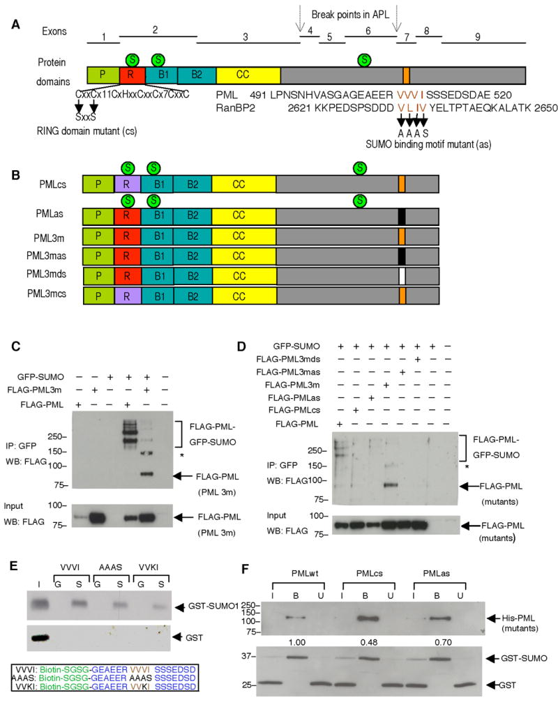
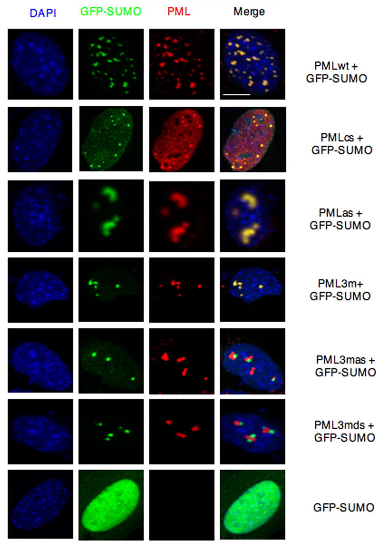
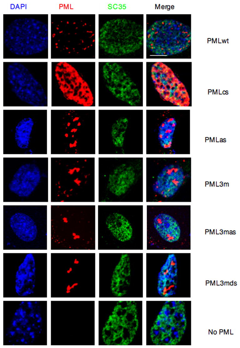
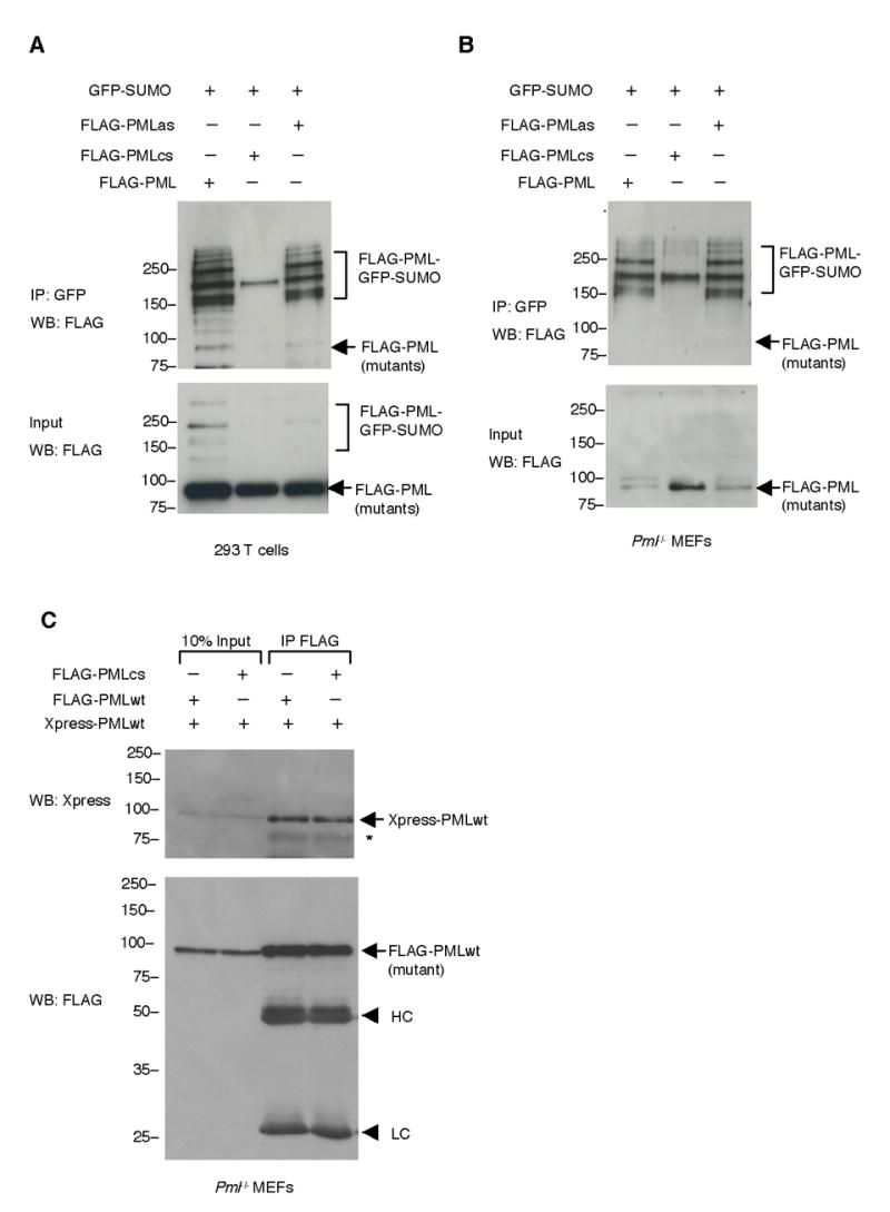
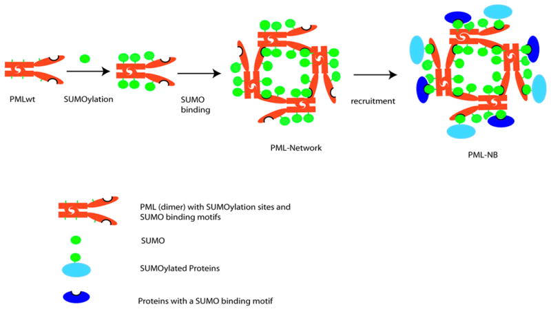
PML nuclear bodies (NBs) are nuclear structures that have been implicated in processes such as transcriptional regulation, genome stability, response to viral infection, apoptosis, and tumor suppression. PML has been found to be essential for the formation of the NBs, as these structures do not form in Pml null cells, although PML add back fully rescues their formation. However, the basis for such a structural role of PML is unknown. We demonstrate that PML contains a SUMO binding motif that is independent of its SUMOylation sites and is surprisingly necessary for PML-NB formation. We demonstrate that the PML RING domain is critical for PML SUMOylation and PML-NB formation. We propose a model for PML-NB formation whereby PML SUMOylation and noncovalent binding of PML to SUMOylated PML through the SUMO binding motif constitutes the nucleation event for subsequent recruitment of SUMOylated proteins and/or proteins containing SUMO binding motifs to the PML NBs.


Figures

Figure 1
Figure 1. PML Has a SUMO Binding Motif Independent of Its SUMOylation Sites
(A) Exon organization of the PML gene and the corresponding protein domains: proline rich domain (P), zinc binding boxes (B1, B2), coiled-coil domain (CC) and RING domain (R). The characteristic feature of the RING domain (Lorick et al., 1999), and two point mutations that are supposed to disrupt it, are shown below. C, cysteine, H, histidine, x, any amino acid. The numbers (11, 7) following × represent numbers of amino acids between the cysteines in PML RING domain. The putative SUMO binding motif of PML along with that of RanBP2 are aligned to reveal the consensus sequence, which is presumably composed of four bulk and hydrophobic amino acids (highlighted in brown). The VVVI in PML was mutated to AAAS in this study. The three SUMOylation sites (indicated by green circles labeled with an S), and the chromosomal break points (indicated by arrows) in acute promyelocytic leukemia (APL) are also shown. (B) Schematic presentation of PML mutants used in the study. RING domain mutant (PMLcs), SUMO binding motif mutant (PMLas), SUMOylation deficient mutant (PML3m), SUMOylation deficient and SUMO binding motif mutant (PML3mas), SUMOylation deficient and SUMO binding motif deletion mutant (PML3mds), SUMOylation deficient and RING domain mutant (PML3mcs) (C) SUMOylation deficient PML co-immunoprecipitates with GFP-SUMO1. FLAG-tagged PML or PML3m were transfected into immortalized Pml−/− MEFs with or without GFP-SUMO1. Top panel: Immunoprecipitation using anti-GFP antibodies and Western blot using anti-FLAG antibodies. Bottom panel: Western blot using anti-FLAG antibodies on 10% of protein inputs used for immunoprecipitation. (D) PML SUMO binding motif is required for SUMOylation deficient PML and SUMO co-immunoprecipitation. FLAG-tagged PML and its mutants described in (B) were analyzed for the SUMO-binding activities as in (C). Asterisk (*) indicates a cross-reacting band. Molecular weight markers (kDa) are indicated. (E) The SUMO binding motif (VVVI) in a synthetic PML-derived peptide is responsible for SUMO binding activity in vitro. Recombinant GST-SUMO1 or GST proteins were incubated with VVVI, AAAS, or VVKI peptides. The bound proteins were determined by Western blot by using antibodies against SUMO1 (upper panel), or GST (middle panel). I: input (20%), G: GST, S: GST-SUMO1. The lower panel shows peptide sequences synthesized. The biotin and linker amino acids are in green, PML sequence in blue, SUMO binding motif in brown and the mutant amino acids in the SUMO binding motif are in black. (F) PML SUMO binding motif mediates PML and SUMO interaction in vitro. Purified recombinant PMLwt, RING domain mutant (PMLcs), or SUMO binding motif mutant (PMLas) were incubated with purified GST and GST-SUMO1, the bound fractions (B) together with 10% of both input (I) and unbound (U) fractions were analyzed by SDS-PAGE and Western blot using antibodies against His (upper panel) or GST (lower panel). The numbers between panels are the ratios of GST-SUMO to PML. They are normalized to that of PMLwt, which is set to 1. Molecular weight markers (kDa) are indicated.
Figure 2
Figure 2. PML RING Domain and SUMO Binding Motif Are Essential for PML-NB Formation
Pml−/− immortalized MEFs were transfected with the indicated plasmids and analyzed by immunofluorescence. Representative confocal microscopy images are presented. Scale bar, 10 μm.
Figure 3
Figure 3. The Nuclear Structures Formed by PMLwt or Its Mutants Do Not Overlap with Nucleoli
Pml−/− immortalized MEFs were transfected with the indicated plasmids and analyzed by immunofluorescence. Representative confocal microscopy images are presented. Scale bar, 10 μm.
Figure 4
Figure 4. The Nuclear Structures Formed by PMLwt or Its Mutants Do Not Overlap with Splicing Speckles
Pml−/− immortalized MEFs were transfected with the indicated plasmids and analyzed by immunofluorescence. Representative confocal microscopy images are presented. Scale bar, 10 μm.
Figure 5
Figure 5. PML RING Domain Is Required for PML SUMOylation
(A) Mutation of the PML RING domain drastically decreases PML SUMOylation. 293T cells were transfected with indicated plasmids. Immunoprecipitation was carried out with an anti-GFP antibody and Western blot analysis with an anti-FLAG antibody (top panel). Ten percent of the lysates used for immunoprecipitation was also analyzed by Western blot (bottom panel) (B) Pml−/− MEFs were transfected as indicated and analyzed as in (A). (C) The RING domain mutant PML maintains the ability of hetero-dimerizing with wild type PML. Pml−/− MEFs were transfected as indicated. Cell lysates were immunoprecipitated with anti-FLAG antibodies. The immunoprecipitates and 10% of inputs used for immunoprecipitation were analyzed by Western blotting with anti-Xpress antibodies (top panel) and anti-FLAG antibodies (bottom panel). Asterisk (*) indicates an Xpress-PML degradation product. HC: Immunoglobulin heavy chains. LC: Immunoglobulin light chains. Molecular weight markers (kDa) are indicated.
Figure 6
Figure 6. A Model of PML-NB Formation
In mitosis, deSUMOylation of PML causes disassembly of PML-NBs. As a result, PML is in aggregate mediated by its RBCC motif (shown as a dimer). In interphase, PML is SUMOylated. Subsequently, the non-covalent interactions between two PML molecules mediated both by SUMO binding motif and RBCC motif promote the growth of concentered PML networks, in which nuclear body proteins that are SUMOylated and/or contain SUMO binding motifs will be recruited through SUMO moieties and SUMO binding motifs that are amply provided by PML. Although, based on this model, it is not clear how the electron dense core material in the center of the PML-NB is packaged during the NB biogenesis (not shown in the model), it is likely that PML SUMOylation is associated with the packaging process, and that the diameter of the NB is determined by the volume of the core material. Red rods: PML dimers. Green circles: SUMO. Light blue circle joined to green circle: SUMOylated proteins. Dark blue circle with indentation: proteins containing a SUMO binding motif

Similar articles

Cited by

References

    1. Bernardi R, Pandolfi PP. Role of PML and the PML-nuclear body in the control of programmed cell death. Oncogene. 2003;22:9048–9057. - PubMed
    1. Bernardi R, Scaglioni PP, Bergmann S, Horn HF, Vousden KH, Pandolfi PP. PML regulates p53 stability by sequestering Mdm2 to the nucleolus. Nat Cell Biol 2004 - PubMed
    1. Boddy MN, Howe K, Etkin LD, Solomon E, Freemont PS. PIC 1, a novel ubiquitin-like protein which interacts with the PML component of a multiprotein complex that is disrupted in acute promyelocytic leukaemia. Oncogene. 1996;13:971–982. - PubMed
    1. Boisvert FM, Hendzel MJ, Bazett-Jones DP. Promyelocytic leukemia (PML) nuclear bodies are protein structures that do not accumulate RNA. J Cell Biol. 2000;148:283–292. - PMC - PubMed
    1. Dellaire G, Eskiw CH, Dehghani H, Ching RW, Bazett-Jones DP. Mitotic accumulations of PML protein contribute to the re-establishment of PML nuclear bodies in G1. J Cell Sci. 2006;119:1034–1042. - PubMed

Publication types

MeSH terms