. 2006 Nov 3;24(3):397-408.
doi: 10.1016/j.molcel.2006.09.015.

A dynamic mechanism for AKAP binding to RII isoforms of cAMP-dependent protein kinase

Affiliations

A dynamic mechanism for AKAP binding to RII isoforms of cAMP-dependent protein kinase

Francis S Kinderman et al. Mol Cell. .

Abstract

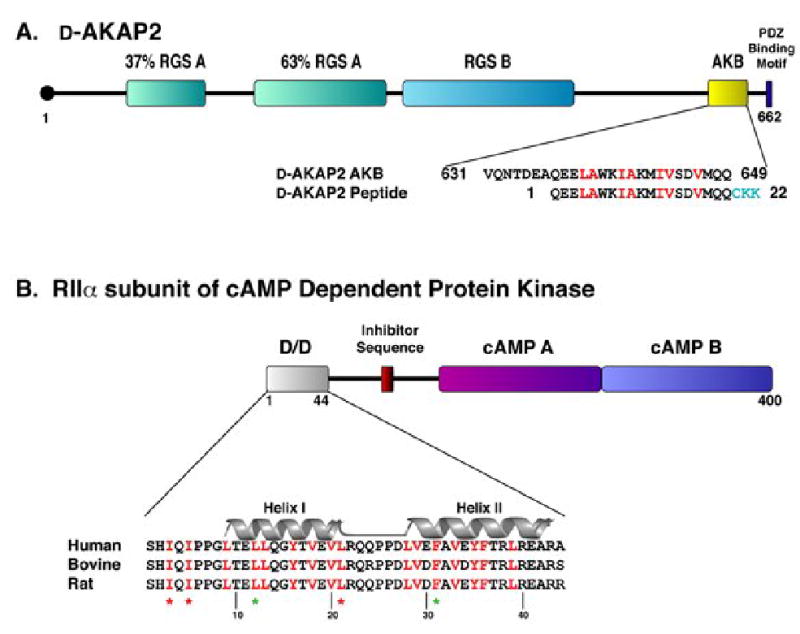
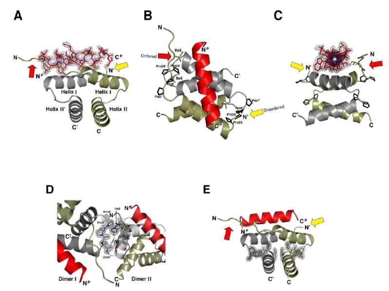
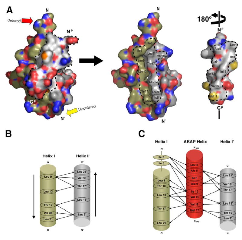
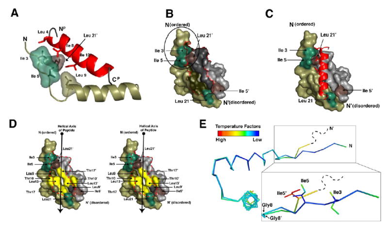
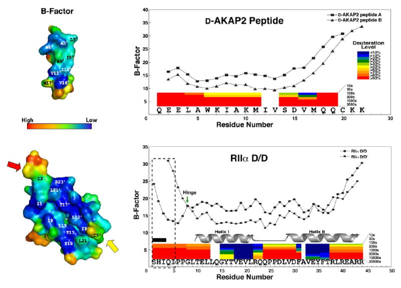
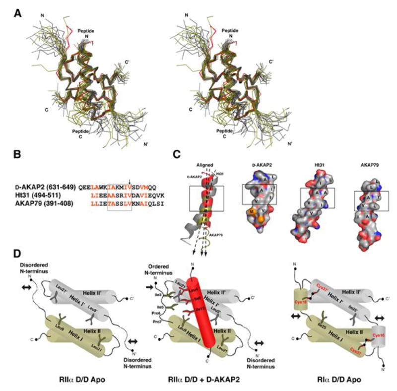
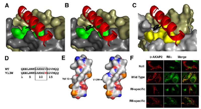
A kinase-anchoring proteins (AKAPs) target PKA to specific microdomains by using an amphipathic helix that docks to N-terminal dimerization and docking (D/D) domains of PKA regulatory (R) subunits. To understand specificity, we solved the crystal structure of the helical motif from D-AKAP2, a dual-specific AKAP, bound to the RIIalpha D/D domain. The 1.6 Angstrom structure reveals how this dynamic, hydrophobic docking site is assembled. A stable, hydrophobic docking groove is formed by the helical interface of two RIIalpha protomers. The flexible N terminus of one protomer is then recruited to the site, anchored to the peptide through two essential isoleucines. The other N terminus is disordered. This asymmetry provides greater possibilities for AKAP docking. Although there is strong discrimination against RIalpha in the N terminus of the AKAP helix, the hydrophobic groove discriminates against RIIalpha. RIalpha, with a cavity in the groove, can accept a bulky tryptophan, whereas RIIalpha requires valine.


Figures

Figure 1
Figure 1. Amphipathic helices mediate D-AKAP2 interaction with RIIα D/D domain
(A) Full length D-AKAP2 contains two putative RGS domains, a PDZ binding motif, and the A Kinase Binding (AKB) Domain, which forms an amphipathic helix for binding the D/D domain. This fragment corresponds to residues 623–649 (numbering according to Swiss-Prot entry O8845). For our studies, a 22-residue peptide was synthesized, including residues 631–649 of D-AKAP2 and three extra residues (shown in turquoise) on the C-terminus for functional purposes. For simplicity, we have numbered the peptide 1–22. Residues highlighted in red correspond to the surface of the helix involved in docking to the D/D domain of RIα and RIIα (Burns-Hamuro et al., 2005). (B) The dimerization/ docking domain resides at the N-terminus of the RIIα subunit, which also includes an inhibitor sequence that binds the active site of the PKA catalytic subunit and two cAMP binding domains. Each protomer of the D/D domain forms a helix-turn-helix motif (Helix I and Helix II are labeled). The conserved hydrophobic, dimer interface is highlighted in red. Residues essential for dimerization are denoted by a green star and residues that are essential for AKAP binding, but do not effect dimerization are denoted by a red star (Hausken et al., 1994; Li and Rubin, 1995).
Figure 2
Figure 2. High resolution crystal structure depicts RIIα D/D domain complexed with the D-AKAP2 peptide
(A-C) The crystal structure of the RIIαD/D:D-AKAP2 complex is displayed at three different angles. The two protomers of the D/D domain, RIIαD/D and RIIαD/D’, are displayed in tan and grey, respectively. The D-AKAP2 peptide, shown in red, is bound to Helix I and Helix I’ of the RIIα D/D dimer. Prolines 6R, 7R, 25R, and 26R, which stablize the loops between helices, as well as, contribute to crystal packing, are highlighted. (D) The two molecules in the asymmetric unit are associated by contacts involving Gln24R’, Pro25R’, and Pro25R’ of Dimer I and His2R and Gln4R of Dimer II. (E) The hydrophobic core of the D/D domain is stabilized by several aromatic side chains extending from all four helices.
Figure 3
Figure 3. Complementary hydrophobic surfaces on RIIα D/D domain and D-AKAP2 mediate a tight interaction between helices
(A) The D-AKAP2 peptide is removed from the interface and rotated 180° in order to see the buried interacting surfaces displayed by the peptide and the RIIα D/D domain. The hydrophobic residues of the peptide form a ridge required for PKA binding. RIIα D/D domain displays a complementary hydrophobic surface, formed by Helix I and I’, that allows the peptide’s hydrophobic ridge to dock. Dotted lines outline the residues of both peptide and the D/D domain that pack together in the hydrophobic interface. (B) Helix I and Helix I’ interact in an antiparallel manner to form the stable binding site for AKAP docking. Arrows represent the hydrophobic interactions between the two helices. (C) Specific side chain interactions between residues of the D/D domain and peptide were elucidated and listed.
Figure 4
Figure 4. RIIα D/D domain forms an asymmetric pocket upon D-AKAP2 binding
(A) Ile3R, Ile5R, Leu9R and Leu21R’ make critical contacts with peptide residues Leu4P, Ala5P, Ile8P, and Ile12P. These residues are highlighted to show their proximity to each other and the importance of the branching of the isoleucine side chains. (B and C) The surface of RIIα D/D domain is displayed alone and with the AKAP. The Ile3R, Ile5R, Leu21R are shown in green. Ile5R’ and Leu21R’ are shown in brown. These residues are highlighted to illustrate the two potential docking sites created Ile3R, Ile5R and Leu21R’ and Ile5R’ and Leu21R. The N-terminus of RIIαD/D, including Ile3R and Ile5R (circled in B) is ordered in the structure and cluster with Leu21R’ to form a hydrophobic site that interacts with a number of side chains from the peptide. However, the N-terminal extension of RIIαD/D’ is disordered in our crystal, so it cannot be seen in our model. (D) The hydrophobic groove of the RIIα D/D domain is illustrated with a stereo-representation of the D/D domain surface. Residues that line this groove and contribute to AKAP docking are highlighted. Residues colored in yellow represent the center of the groove, which are important for binding specificity. (E) The two protomers from the D/D domain are aligned and colored according to B-factor to show the asymmetry more precisely. The RIIαD/D’ protomer is disordered at the N-terminus beyond residue 4 and residue 5 is relatively dynamic according to B-factors. However, this region is well ordered on the protomer that contacts the N-terminus of the peptide. The position of Ile5R and Ile5R’ is different due to changes around Gly8R and Gly8R’.
Figure 5
Figure 5. The B-factors of the crystal structure correlate with H/D exchange protection data
B-factors of both RIIα D/D domain and D-AKAP2 peptide are shown. We see a sharp rise in B-factor at the C-terminus of the peptide, which corresponds with a high deuteration rates demonstrated by previous H/DMS experiments (Burns-Hamuro et al., 2005). The colored bars represent the deuteration levels of peptides from D-AKAP2 AKB (631–649) in complex with RIIα D/D domain and the peptides of RIIαD/D domain in the presence of the peptide. In addition, we see elevation in B-factors at the termini of the RIIα D/D domain, which corresponds to high deuteration rates as well. The flexible N-terminus (1–5) is boxed to show the differences between the two protomers. RIIαD/D’ exhibits higher B-factors for residues 4 and 5 and residues 1–3 are disordered (labeled by the black box), suggesting great flexibility for this region. Gly 8 is marked with an arrow, because it may act as a hinge for the flexible N-terminus. The helix I and helix II are highly protected when bound to the AKAP.
Figure 6
Figure 6. Crystal and NMR structures reveal conserved features for AKAP docking to RIIα and provide insight into RI binding
(A) Both Dimer I and Dimer II of the crystal structure (in red) are aligned with NMR structures of RIIα D/D bound to Ht31 (gray) and AKAP79 (olive) (Newlon et al., 2001). The structures are aligned based purely on Helix I and Helix I’. The AKAP peptides differ in helical register. (B) The sequences of D-AKAP2, Ht31, AKAP79 involved in RIIα binding are aligned according to the structural alignment. The two turns of the helix that are critical for binding the D/D domain are shown in the box. The arrow points to Val13P, which is important for RI vs. RII specificity. (C). Despite the diversity of sequence these AKAPs display a hydrophobic ridge that interacts with the D/D of RIIα. The two turns of the helices that are crucial for binding are boxed for each. Ht31 and AKAP79 peptides are each taken from one structure in an ensemble. The C-terminus of AKAP79 is not illustrated as a ribbon because it does not form a well ordered helix in the NMR structures. The axis of each peptide helix is shown to demonstrate that the D/D domain can accommodate different orientations of the AKAP helix. (D) A cartoon representation of the D/D domain of RIIα subunits demonstrates the presence of a stable core and two flexible N-terminal tails. Upon binding to an AKAP, one of these tails is stabilized. RI subunits possess two conserved cysteines (Cys16R and Cys37R), which can stabilize the N-1 helix when disulfide bonded. Cys16R and Cys37R’ are located in analogous positions to Ile3R and Leu21R’ of RIIα (Banky et al., 2003), which are essential for binding. Crystal structures of RIα D/D may detail the specific involvement of these residues in AKAP docking.
Figure 7
Figure 7. AKAP specificity for RII subunits of PKA can be toggled by Val13P mutations
(A) Val13P, highlighted in black, binds to the surface of the D/D domain at the center of the hydrophobic groove comprised of Thr10R, Leu13R, Thr17R and Leu13R’, which are highlighted in green. (B) Mutating Val13P to Trp eliminates binding to the RIIα D/D domain, as shown previously (Burns-Hamuro et al., 2003). Modeling this mutation into the D-AKAP2 in our structure causes steric clashes with the D/D domain; therefore, we believe this mutation disrupts the hydrophobic packing within the interface. (C) By modeling our structure with the NMR structure of the RIα D/D domain (Banky et al., 2003), we observe a pocket in the RIα D/D domain that can accommodate the V13W mutation. This is consistent with peptide array data. Qln26R, Leu29R, Ile33R, and Leu29R’ form the surface highlighted in yellow can accommodate the Trp mutation in RIα. (D) The sequence of the V13W mutation is shown above. This is located in the segment of the peptide that is critical for binding the D/D domains. (E) The surfaces of the wild type and mutant peptides are quite different because the Trp protrudes from the peptide surface. (F) The specificity of this mutation is demonstrated in vivo. Hela cells were transfected with wild-type, null, RI or RII specific versions of the D-AKAP2 peptide fused to a mitochondrial targeting sequence. Localization of these constructs is shown in the column on the left. RIIα was stained with fluorescent antibodies, which is shown in the middle column. Each AKAP construct was targeted to the mitochondria; however, only the wild type and RII-specific constructs successfully redirected the RIIα away from the golgi and to the mitochondria.

References

    1. Alto NM, Soderling SH, Hoshi N, Langeberg LK, Fayos R, Jennings PA, Scott JD. Bioinformatic design of A-kinase anchoring protein-in silico: a potent and selective peptide antagonist of type II protein kinase A anchoring. Proc Natl Acad Sci U S A. 2003;100:4445–4450. - PMC - PubMed
    1. Banky P, Roy M, Newlon MG, Morikis D, Haste NM, Taylor SS, Jennings PA. Related protein-protein interaction modules present drastically different surface topographies despite a conserved helical platform. J Mol Biol. 2003;330:1117–1129. - PubMed
    1. Burns LL, Canaves JM, Pennypacker JK, Blumenthal DK, Taylor SS. Isoform specific differences in binding of a dual-specificity A-kinase anchoring protein to type I and type II regulatory subunits of PKA. Biochemistry. 2003;42:5754–5763. - PubMed
    1. Burns-Hamuro LL, Hamuro Y, Kim JS, Sigala P, Fayos R, Stranz DD, Jennings PA, Taylor SS, Woods VL., Jr Distinct interaction modes of an AKAP bound to two regulatory subunit isoforms of protein kinase A revealed by amide hydrogen/deuterium exchange. Protein Sci. 2005;14:2982–2992. - PMC - PubMed
    1. Burns-Hamuro LL, Ma Y, Kammerer S, Reineke U, Self C, Cook C, Olson GL, Cantor CR, Braun A, Taylor SS. Designing isoform-specific peptide disruptors of protein kinase A localization. Proc Natl Acad Sci U S A. 2003;100:4072–4077. - PMC - PubMed

Publication types

MeSH terms

Associated data