. 2006 Nov 3;24(3):433-43.
doi: 10.1016/j.molcel.2006.09.010.

Deregulated replication licensing causes DNA fragmentation consistent with head-to-tail fork collision

Affiliations

Deregulated replication licensing causes DNA fragmentation consistent with head-to-tail fork collision

Iain F Davidson et al. Mol Cell. .

Abstract

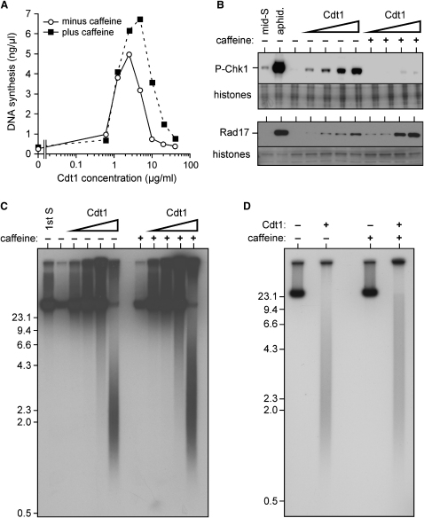
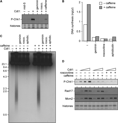
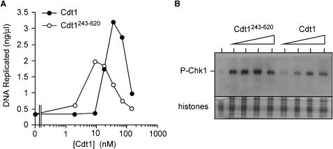
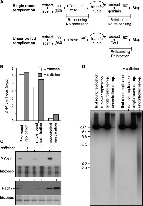
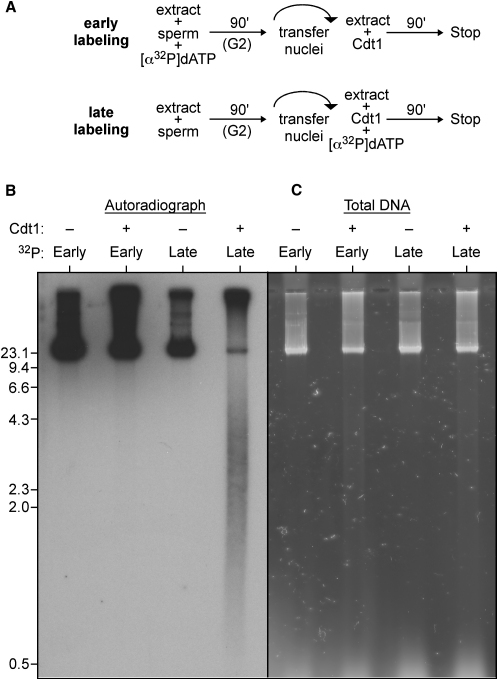
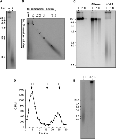
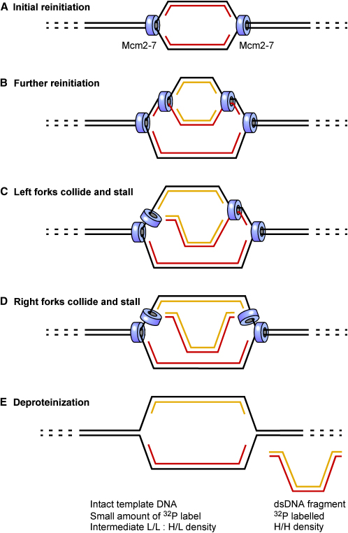
Correct regulation of the replication licensing system ensures that no DNA is rereplicated in a single cell cycle. When the licensing protein Cdt1 is overexpressed in G2 phase of the cell cycle, replication origins are relicensed and the DNA is rereplicated. At the same time, checkpoint pathways are activated that block further cell cycle progression. We have studied the consequence of deregulating the licensing system by adding recombinant Cdt1 to Xenopus egg extracts. We show that Cdt1 induces checkpoint activation and the appearance of small fragments of double-stranded DNA. DNA fragmentation and strong checkpoint activation are dependent on uncontrolled rereplication and do not occur after a single coordinated round of rereplication. The DNA fragments are composed exclusively of rereplicated DNA. The unusual characteristics of these fragments suggest that they result from head-to-tail collision (rear ending) of replication forks chasing one another along the same DNA template.


Figures

Figure 1
Figure 1
Addition of Cdt1 to Egg Extract Causes Rereplication, DNA Damage, and Checkpoint Activation (A–C) Sperm nuclei were incubated in interphase extract for 90 min; extract was then supplemented with different concentrations of Cdt1 plus or minus 5 mM caffeine and incubated for a further 90 min. (A) Sperm nuclei were added at 5 ng DNA/μl extract. [α32P]dATP was added along with Cdt1. After 90 min, rereplication was measured by 32P incorporation. (B) Nuclei were immunoblotted for phosphorylated Chk1 (upper panel); chromatin was immunoblotted for Rad17 (lower panel). Final concentrations of Cdt1 were 2.5, 5, 10, or 20 μg/ml. As controls, nuclei were isolated after 40 min sperm incubation by using either untreated extract (mid S) or extract supplemented with 40 μM aphidicolin (aphid.). (C) [α32P]dATP was added along with Cdt1. After 90 min, DNA was isolated, separated by neutral agarose gel electrophoresis, and autoradiographed. Final concentrations of Cdt1 were 2.5, 5, 10, or 20 μg/ml. As control, [α32P]dATP was added along with the sperm, and DNA was isolated after 90 min (first S). (D) Sperm nuclei were incubated in interphase extract supplemented with [α32P]dATP ±20 μg/ml Cdt1 and 5 mM caffeine. After 90 min, DNA was isolated, separated by neutral agarose gel electrophoresis, and autoradiographed. Molecular weight markers (kb) are shown to the side.
Figure 2
Figure 2
Cdt1-Induced DNA Damage and Checkpoint Activation Requires Rereplication Sperm nuclei were incubated in interphase extract for 90 min to allow a single round of replication; extract was then supplemented with different concentrations of Cdt1 plus or minus caffeine, geminin, roscovitine (2 mM), aphidicolin (100 μM), or [α32P]dATP and incubated for 90 min. (A) Nuclei were isolated and immunoblotted for phosphorylated Chk1. As control, sperm nuclei were isolated after 40 min (prior to Cdt1 addition) when they were in mid S phase. Coomassie-stained histones provide a loading control. Cdt1 was added at 20 μg/ml. (B) [α32P]dATP was added along with 20 μg/ml Cdt1. After 90 min, 32P incorporation into DNA was measured (rereplicative DNA synthesis). (C) [α32P]dATP was added along with sperm nuclei, and after 90 min, 20 μg/ml Cdt1 plus or minus inhibitors was added. After a further 90 min, DNA was isolated, separated by neutral agarose gel electrophoresis, and autoradiographed. Molecular weight markers (kb) are shown to the side. (D) Nuclei were isolated and immunoblotted for phosphorylated Chk1 (upper panel), or chromatin was isolated and immunoblotted for Rad17 or Mcm2 (lower panel). Coomassie-stained histones provide a loading control. Final concentrations of Cdt1 were 1.25, 5, or 10 μg/ml.
Figure 3
Figure 3
Geminin Titration Is Not Required to Cause Checkpoint Activation Sperm nuclei were incubated in interphase extract (5 ng DNA/μl) for 90 min to allow a single round of replication; extract was then supplemented with different concentrations of full-length Cdt1 or Cdt1243–620 and incubated for a further 90 min. Cdt1 constructs were added at 2, 9, 18, 35, 71, 141, and 283 nM, equivalent to 0.16, 0.63, 1.25, 2.5, 5, 10, and 20 μg/ml full-length Cdt1. (A) [α32P]dATP was added along with the Cdt1. After 90 min, 32P incorporation into DNA, representing rereplicative DNA synthesis, was measured. (B) Nuclei were isolated and immunoblotted for phosphorylated Chk1. Cdt1 constructs were added at 35, 71, 141, and 283 nM. Coomassie-stained histones provide a loading control.
Figure 4
Figure 4
Multiple Rounds of Rereplication Are Required for Cdt1-Induced DNA Damage and Checkpoint Activation (A) Protocol for a single round of rereplication (upper panel) or uncontrolled rereplication (lower panel). (B) [α32P]dATP plus or minus caffeine was added to the second extract. As control, roscovitine (0.5 mM) was added to the first extract along with the sperm nuclei (first round replication). (C) Nuclei were immunoblotted for phospho-Chk1 (upper panel), or chromatin was immunoblotted for Rad17 (lower panel). As control, nuclei and chromatin were isolated after incubation for 90 min in the first extract. Coomassie-stained histones provide a loading control. (D) [α32P]dATP plus or minus caffeine was added to the second extract. DNA was isolated, separated by neutral gel electrophoresis, and autoradiographed. As control, roscovitine (0.5 mM) was added to the first extract along with the sperm nuclei (first round replication). In a second control, Cdt1 was added to neither extract (run over replication).
Figure 5
Figure 5
Template DNA Remains Intact during Uncontrolled Rereplication (A) Early labeling protocol (upper panel): only DNA synthesized during the first S phase is labeled with [α32P]dATP before uncontrolled rereplication is induced by Cdt1. Late labeling protocol (lower panel): only rereplicated DNA is labeled with [α32P]dATP during uncontrolled rereplication induced by Cdt1. (B and C) DNA was subjected to early- and late-labeling protocols as in (A). DNA was separated on neutral agarose gels and either autoradiographed (B) or stained with Sybr Safe to show total DNA (C). Molecular weight markers (kb) are shown to the side.
Figure 6
Figure 6
Damage Caused by Cdt1 Is Consistent with Head-to-Tail Fork Collision Sperm nuclei were incubated in interphase extract for 90 min to allow a single round of replication; extract was then supplemented with 20 μg/ml Cdt1 plus [α32P]dATP and incubated for a further 90 min. (A) DNA was isolated and incubated plus or minus AluI for 3 hr. Samples were separated by neutral agarose gel electrophoresis and then autoradiographed. (B) DNA was isolated and separated by 2D neutral:alkaline gel electrophoresis. The migration of double-stranded molecular weight markers in the first (neutral) dimension is shown along the top. The migration of double-stranded markers on a parallel 2D electrophoresis is indicated by crosses. (C) Chromatin was pelleted; DNA was isolated from both the pellet fraction (P) and the supernatant fraction (S), and neutral agarose gel electrophoresis was performed. An identical sample was diluted in buffer but not centrifuged to provide total level of DNA (T). As control, sperm nuclei were replicated in the presence of [α32P]dATP and then incubated plus or minus micrococcal nuclease (MNase) before being pelleted. (D and E) BrdUTP was added to extract along with Cdt1. After 90 min, DNA was isolated and separated by CsCl equilibrium gradient centrifugation. 32P activity across the gradient is shown in (C). HH, HL, and LL show the expected position of heavy/heavy, heavy/light, and light/light DNA, respectively. DNA from fractions 1–10 (HH) and 19–27 (HL/LL) was pooled, separated on a neutral agarose gel, and autoradiographed. Molecular weight markers (kb) are shown to the side.
Figure 7
Figure 7
Model for the Creation of DNA Fragments by Head-to-Tail Fork Collision A small segment of chromosomal DNA containing a replication origin is shown. Mcm2-7 is indicated by blue cylinders. (A) Reinitiation at the replication origin forms a replication bubble. (B) A further initiation event forms a second replication bubble whose forks are chasing the forks in the first bubble. (C) The left-moving forks undergo head-to-tail collision, shutting them both down. (D) The right-moving forks undergo head-to-tail collision, shutting them both down. (E) When replication fork proteins are removed from the DNA, a small DNA fragment is released from the chromosomal DNA.

References

    1. Annunziato A.T., Seale R.L. Histone deacetylation is required for the maturation of newly replicated chromatin. J. Biol. Chem. 1983;258:12675–12684. - PubMed
    1. Archambault V., Ikui A.E., Drapkin B.J., Cross F.R. Disruption of mechanisms that prevent rereplication triggers a DNA damage response. Mol. Cell. Biol. 2005;25:6707–6721. - PMC - PubMed
    1. Arias E.E., Walter J.C. Replication-dependent destruction of Cdt1 limits DNA replication to a single round per cell cycle in Xenopus egg extracts. Genes Dev. 2005;19:114–126. - PMC - PubMed
    1. Blow J.J., Dutta A. Preventing re-replication of chromosomal DNA. Nat. Rev. Mol. Cell Biol. 2005;6:476–486. - PMC - PubMed
    1. Blow J.J., Laskey R.A. A role for the nuclear envelope in controlling DNA replication within the cell cycle. Nature. 1988;332:546–548. - PubMed

Publication types