. 2007 Jan 15;578(Pt 2):471-9.
doi: 10.1113/jphysiol.2006.123588. Epub 2006 Nov 2.

Cellular mechanisms of burst firing-mediated long-term depression in rat neocortical pyramidal cells

Affiliations

Cellular mechanisms of burst firing-mediated long-term depression in rat neocortical pyramidal cells

Antonny Czarnecki et al. J Physiol. .

Abstract

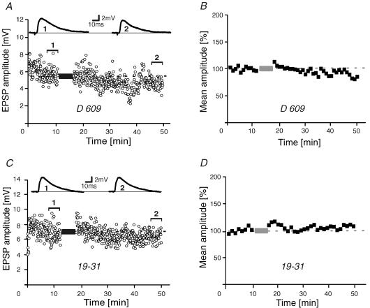
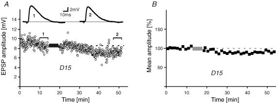
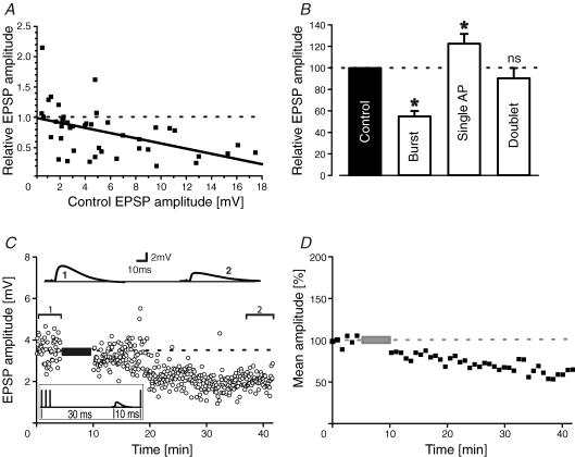
During wakefulness and sleep, neurons in the neocortex emit action potentials tonically or in rhythmic bursts, respectively. However, the role of synchronized discharge patterns is largely unknown. We have recently shown that pairings of excitatory postsynaptic potentials (EPSPs) and action potential bursts or single spikes lead to long-term depression (burst-LTD) or long-term potentiation, respectively. In this study, we elucidate the cellular mechanisms of burst-LTD and characterize its functional properties. Whole-cell patch-clamp recordings were obtained from layer V pyramidal cells in somatosensory cortex of juvenile rats in vitro and composite EPSPs and EPSCs were evoked extracellularly in layers II/III. Repetitive burst-pairings led to a long-lasting depression of EPSPs and EPSCs that was blocked by inhibitors of metabotropic glutamate group 1 receptors, phospholipase C, protein kinase C (PKC) and calcium release from the endoplasmic reticulum, and that required an intact machinery for endocytosis. Thus, burst-LTD is induced via a Ca2+- and phosphatidylinositol-dependent activation of PKC and expressed through phosphorylation-triggered endocytosis of AMPA receptors. Functionally, burst-LTD is inversely related to EPSP size and bursts dominate single spikes in determining the sign of synaptic plasticity. Thus burst-firing constitutes a signal by which coincident synaptic inputs are proportionally downsized. Overall, our data thus suggest a mechanism by which synaptic weights can be reconfigured during non-rapid eye movement sleep.


Figures

Figure 1
Figure 1. Burst-LTD of EPSPs/EPSCs
A, single-cell EPSP/EPSC amplitude–time series and sample traces in control (1) and after burst-conditioning (2). B, mean time course of normalized EPSP/EPSC time-averaged (1 min) amplitudes (n = 11). C, sample EPSPs and amplitude–time series of a pairing experiment in the presence of the type 1 metabotropic glutamate receptor-specific antagonist 1-aminoindan-1,5-dicarboxylic acid (AIDA; 0.2 mm). D, population data of all AIDA experiments (n = 7). Dashed lines and bars represent average values in control and the pairing period, respectively.
Figure 2
Figure 2. Induction of burst-LTD
A and B, EPSP amplitude–time series and sample traces (A) and normalized time-averaged (1 min) population data (B) of burst-pairing experiments in the presence of the phospholipase C inhibitor O-(octahydro-4,7-methano-1H-inden-5-yl) carbonopotassium dithioate (D609) added to the patch pipette (30 μm; n = 6). C and D, similar experiments during intracellular application of the protein kinase C pseudosubstrate 19-31 (0.5 μm; n = 8). Dashed lines and bars represent average control values and the pairing period, respectively.
Figure 3
Figure 3. Burst-LTD and intracellular Ca2+
A and B, EPSP amplitude–time series with sample traces (A) and normalized population data (B) of burst-pairing experiments during intracellular application of BAPTA (30 mm, n = 8). C and D, EPSP amplitude–time series with sample traces (C) and normalized 1 min time-averaged population data (D) of burst-pairing experiments in the presence of the inositol 1,4,5-trisphosphate receptor blocker heparin added to the pipette solution (1 mg ml−1, n = 7). E and F, comparable experiments in the presence of ruthenium red (0.2 mm) to block ryanodine receptors (n = 6). Dashed lines and bars represent average control values and the pairing period, respectively.
Figure 5
Figure 5. Summary graph of pharmacology of burst-LTD
A and B, histograms of EPSP amplitudes after burst-conditioning. A, antagonists that successfully prevented burst-LTD. B, inhibitors that did not interfere with burst-LTD. Dotted and dashed line, average control and burst-conditioned EPSP amplitude, respectively. AIDA, 1-aminoindan-1,5-dicarboxylic acid; AM-251, N-(piperidin-1-yl)-5-(4-iodophenyl)-1-(2,4-dichlorophenyl)-4-methyl-1H-pyrazole-3-carboxamide; BAPTA, 1,2-bis(o-aminophenoxy)ethane-N,N,N′,N′-tetraacetic acid; CDC, cinnamyl 3,4,-dihydroxy-α-cyanocinnamate; CN, calcineurin autoinhibitory peptide (ITSFEEAKGLDRINERMPPR); D15, PPPQVPSRPNRAPPG; D609, O-(octahydro-4,7-methano-1H-inden-5-yl) carbonopotassium dithioate; 19-31, RFARKGALRQKNV; Hep, heparin; Nif, nifedipine; L-NNA, NG.-nitro-l-arginine; RR, ruthenium red; ext, extracellularly applied; int, intracellularly applied.
Figure 4
Figure 4. Expression of burst-LTD
A and B, burst-LTD is blocked after intracellular application of the endocytosis inhibitor peptide D15 (1 mm). EPSP amplitude–time series in an individual experiment (A) and 1 min time-averaged population data (B) (n = 7). Dashed lines and bars represent average control values and the pairing period, respectively.
Figure 6
Figure 6. Burst and single spikes
A, summary graph of relative EPSP size after conditioning versus the initial EPSP amplitude. A straight line was fitted to the data points by linear regression (r = 0.49; P < 0.0001). B, summary histogram of pairing experiments with bursts (three to four spikes), single action potentials and spike doublets (*P < 0.05; ns, not significant). C, sample traces and EPSP amplitude–time series of an experiment where EPSPs were simultaneously paired with a −20 ms burst and a +10 ms single action potential. D, 1 min averaged time course of normalized EPSP amplitudes with concomitant burst–single spike pairings (n = 6 cells).

References

    1. Anwyl R. Metabotropic glutamate receptors: electrophysiological properties and role in plasticity. Brain Res Rev. 1999;366:151–158. - PubMed
    1. Artola A, Singer W. Long-term depression of excitatory synaptic transmission and its relationship to long-term potentiation. Trends Neurosci. 1993;16:480–487. - PubMed
    1. Batchelor AM, Garthwaite J. Frequency detection and temporally dispersed synaptic signal association through a metabotropic glutamate receptor pathway. Nature. 1997;385:74–77. - PubMed
    1. Birtoli B, Ulrich D. Firing mode-dependent synaptic plasticity in rat neocortical pyramidal neurons. J Neurosci. 2004;24:4935–4940. - PMC - PubMed
    1. Charpak S, Gähwiler B. Glutamate mediates a slow synaptic repsonse in hippocampal slices cultures. Proc R Soc Lond B Biol Sci. 1991;243:221–226. - PubMed

Publication types

MeSH terms

LinkOut - more resources