. 2007 Jan;27(1):42-8.
doi: 10.1161/01.ATV.0000251500.94478.18. Epub 2006 Nov 2.

Nox4 is required for maintenance of the differentiated vascular smooth muscle cell phenotype

Affiliations

Nox4 is required for maintenance of the differentiated vascular smooth muscle cell phenotype

Roza E Clempus et al. Arterioscler Thromb Vasc Biol. 2007 Jan.

Erratum in

  • Arterioscler Thromb Vasc Biol. 2007 Feb;27(2):e8. Schmidt, Harald H H [added]

Abstract

Objective: The mechanisms responsible for maintaining the differentiated phenotype of adult vascular smooth muscle cells (VSMCs) are incompletely understood. Reactive oxygen species (ROS) have been implicated in VSMC differentiation, but the responsible sources are unknown. In this study, we investigated the role of Nox1 and Nox4-derived ROS in this process.

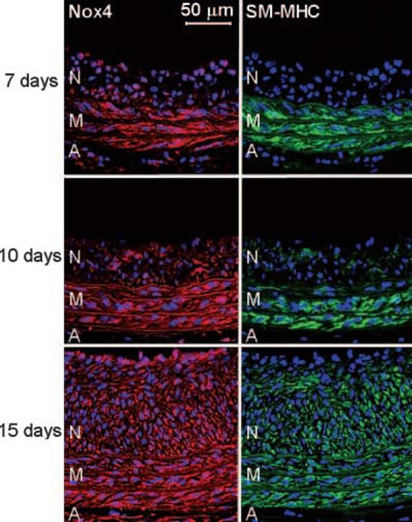
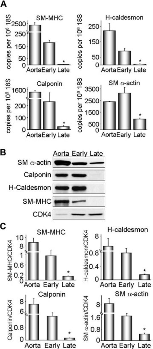
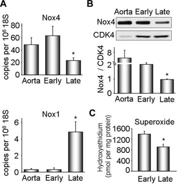
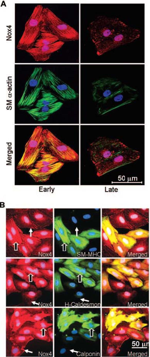
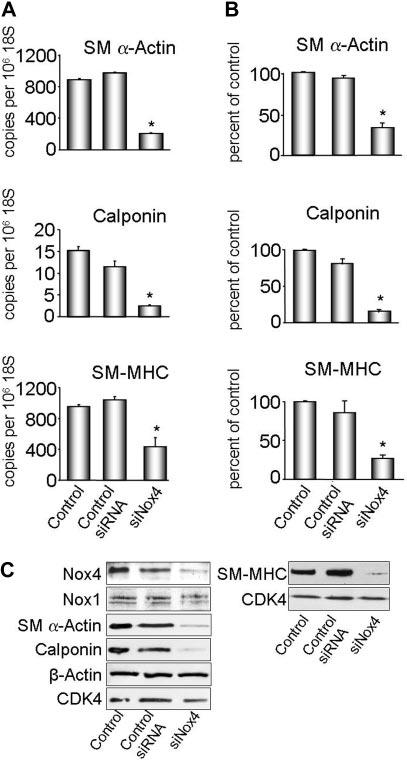
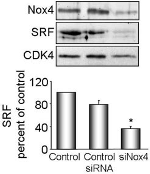
Methods and results: Primary VSMCs were used to study the relationship between Nox homologues and differentiation markers such as smooth muscle alpha-actin (SM alpha-actin), smooth muscle myosin heavy chain (SM-MHC), heavy caldesmon, and calponin. We found that Nox4 and differentiation marker genes were downregulated from passage 1 to passage 6 to 12, whereas Nox1 was gradually upregulated. Nox4 co-localized with SM alpha-actin-based stress fibers in differentiated VSMC, and moved into focal adhesions in de-differentiated cells. siRNA against nox4 reduced NADPH-driven superoxide production in serum-deprived VSMCs and downregulated SM-alpha actin, SM-MHC, and calponin, as well as SM-alpha actin stress fibers. Nox1 depletion did not decrease these parameters.

Conclusions: Nox4-derived ROS are critical to the maintenance of the differentiated phenotype of VSMCs. These findings highlight the importance of identifying the specific source of ROS involved in particular cellular functions when designing therapeutic interventions.


Figures

Figure 1
Figure 1
Nox4 correlates with SM-MHC differentiation markers in vivo. Immunofluorescence labeling using antibodies to Nox4 (red) and SM-MHC (green) of rat carotid arteries sections harvested at indicated time points after balloon injury. Cell nuclei are stained blue with DAPI. Lumen is to the top of each panel. N=neointima; M=media; A=adventitia. Elastic laminae appear light red in the left panels or green in the right panels. Bar=50 μm.
Figure 2
Figure 2
Characterization of VSMC differentiation state in vitro. Protein and RNA were extracted from early (1–2) and late (6–13) passage VSMCs. A, SM-MHC, H-caldesmon, calponin, and SM α-actin cDNA levels were measured by quantitative real-time polymerase chain reaction. Data were normalized to 106 copies 18S cDNA. Whole aorta cDNA was used as a positive control. Results are expressed as mean± SEM (n=4 to 6). *P<0.01 compared with early passage. B, Western blot analysis of differentiation marker protein expression. Cyclin-dependent kinase 4 (CDK4) was used as a loading control. Each differentiation marker gene was normalized to CDK4. C, Quantification of protein expression for 4 to 6 experiments. Results are expressed as mean± SEM. *P=0.01 compared with early passage.
Figure 3
Figure 3
Nox1 and Nox4 expression and superoxide production in early and late passage VSMCs. Protein and RNA were extracted from early (1–2) and late (6 –13) passage VSMCs. A. Nox4 and Nox1 cDNAs were measured by quantitative real-time polymerase chain reaction. Data are normalized to 106 copies 18S cDNA. Whole aorta cDNA was used as a positive control. Results are expressed as mean±SEM (n=6). *P<0.01 compared with early passage. B, Representative Western blot analysis of Nox4 protein expression (upper panel). CDK4 was used as a loading control. Lower panel, Quantification of protein expression for 6 experiments. Nox4 was normalized to CDK4. Results are expressed as mean± SEM. *P<0.01 compared with early passage. C, Superoxide production in quiescent early and late passage VSMCs, measured with dihydroethidium–high-performance liquid chromatography. Results are expressed as mean±SEM (n=3) and are normalized to protein.
Figure 4
Figure 4
Differential localization of Nox4 in early and late passage VSMCs. A, Immunofluorescence labeling using specific antibodies to Nox4 (red) and SM α -actin (green). Nuclei are stained with DAPI (blue). Optical sectioning of the same cells shows a stress fiber distribution of Nox4 in early passage cells, and a focal adhesion pattern in late passage cells. Nox4 appears to colocalize with SM μ-actin in early passage (yellow color in bottom left panel), whereas there is no colocalization in the late passage cells (bottom right panel). Images are representative of at least four separate experiments. Bar=50 μm. B, Immunofluorescent cytochemistry for Nox4 (red), differentiation markers (green): SM-MHC, H-caldesmon, and calponin, and nuclei (blue) in early passage VSMCs. Nox4 appears along stress fibers only in the most differentiated cells (outlined arrows), whereas in cells expressing lower levels of differentiation markers, Nox4 moves to focal adhesions (plain arrows). Note: all cells express SM α-actin (not shown).
Figure 5
Figure 5
Nox4 is necessary for expression of differentiation markers. VSMCs were transfected with siNox4 or control siRNA, and harvested for RNA or protein. A, SM α-actin, calponin and SM-MHC cDNA levels were measured by quantitative real-time polymerase chain reaction. Data are normalized to 106 copies 18S cDNA. Results are expressed as mean±SEM (n=3, n=2 for SM-MHC). *P<0.01 for SM α-actin and calponin and P<0.05 for SM-MHC compared with control siRNA. B, Quantification of protein expression (n=3). Results are normalized to CDK4 and are expressed as mean±SEM. *P<0.05 compared with control siRNA. C, Western blot analysis of Nox1, Nox4, and differentiation marker protein expression. CDK4 was used as a loading control, and β-actin as a nondifferentiation marker protein.
Figure 6
Figure 6
Nox4 regulates SRF expression. VSMCs were transfected with siNox4 or control siRNA, and harvested for protein. Nox4 and SRF were measured by Western analysis. Protein expression was quantified by densitometry (n=4). Results are normalized to CDK4 and are expressed as mean± SEM. *P<0.001 compared with control siRNA.

Comment in

References

    1. Owens GK, Kumar MS, Wamhoff BR. Molecular regulation of vascular smooth muscle cell differentiation in development and disease. Physiol Rev. 2004;84:767–801. - PubMed
    1. Rovner AS, Murphy RA, Owens GK. Expression of smooth muscle and nonmuscle myosin heavy chains in cultured vascular smooth muscle cells. J Biol Chem. 1986;261:14740–14745. - PubMed
    1. Taniyama Y, Griendling KK. Reactive oxygen species in the vasculature: molecular and cellular mechanisms. Hypertension. 2003;42:1075–1081. - PubMed
    1. Su B, Mitra S, Gregg H, Flavahan S, Chotani MA, Clark KR, Goldschmidt-Clermont PJ, Flavahan NA. Redox regulation of vascular smooth muscle cell differentiation. Circ Res. 2001;89:39–46. - PubMed
    1. Jin L, Ying Z, Webb RC. Activation of Rho/Rho kinase signaling pathway by reactive oxygen species in rat aorta. Am J Physiol Heart Circ Physiol. 2004;287:H1495–H1500. - PubMed

Publication types

MeSH terms